. 2023 Oct 26;186(22):4818-4833.e25.
doi: 10.1016/j.cell.2023.09.007. Epub 2023 Oct 6.

Vertebrate-class-specific binding modes of the alphavirus receptor MXRA8

Affiliations

Vertebrate-class-specific binding modes of the alphavirus receptor MXRA8

Ofer Zimmerman et al. Cell. .

Abstract

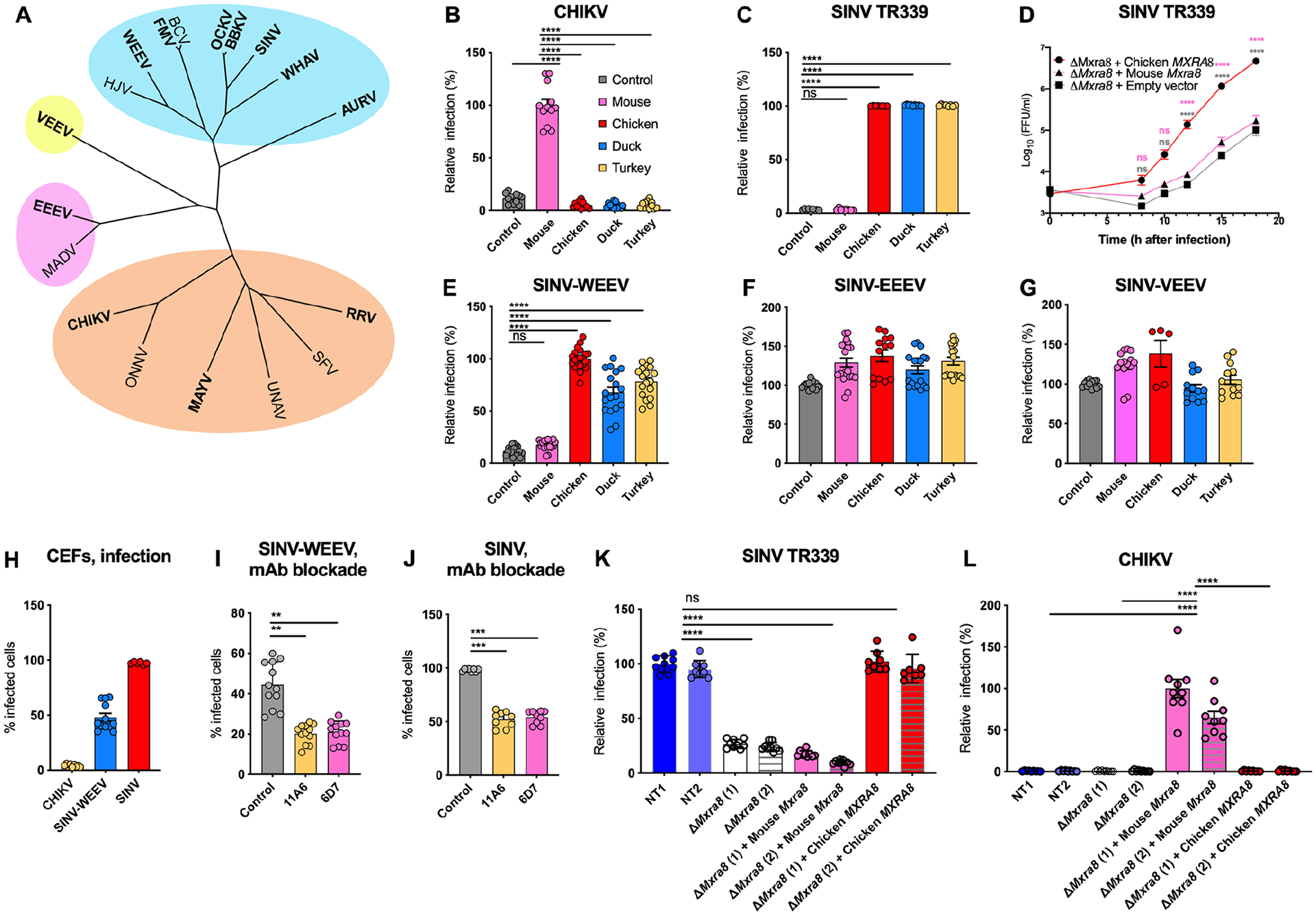
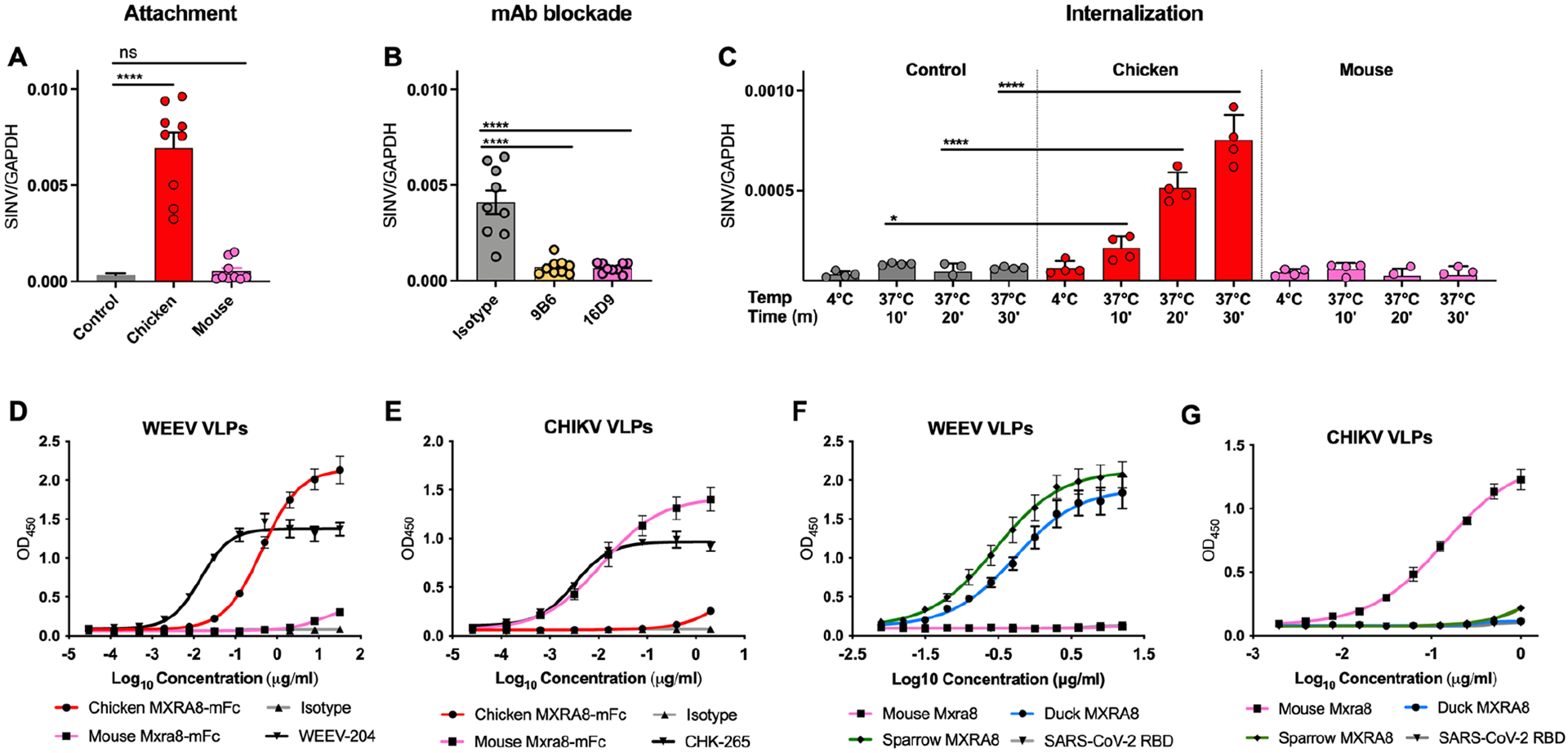
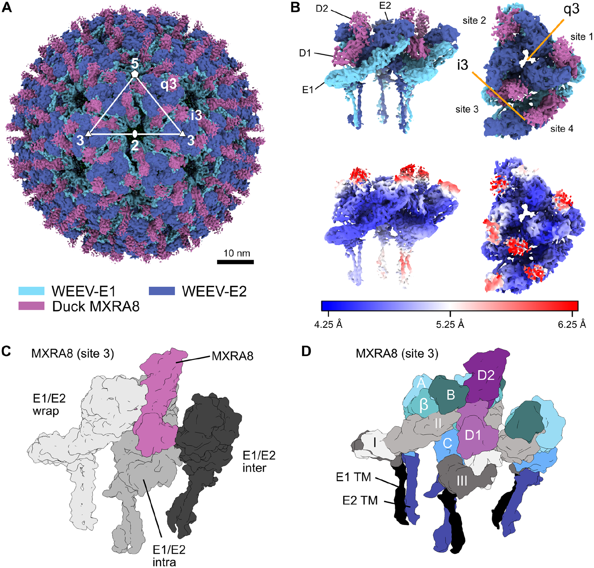
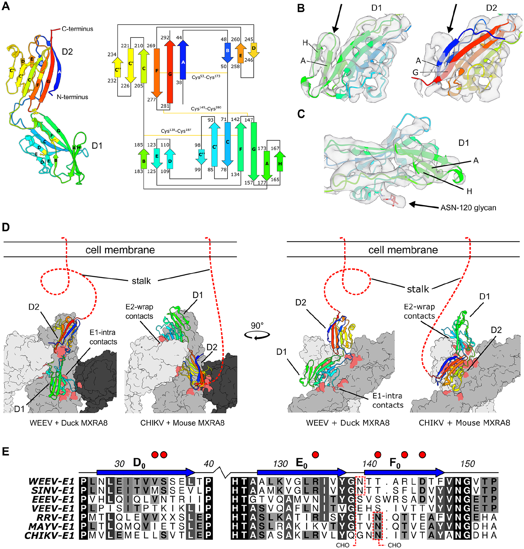
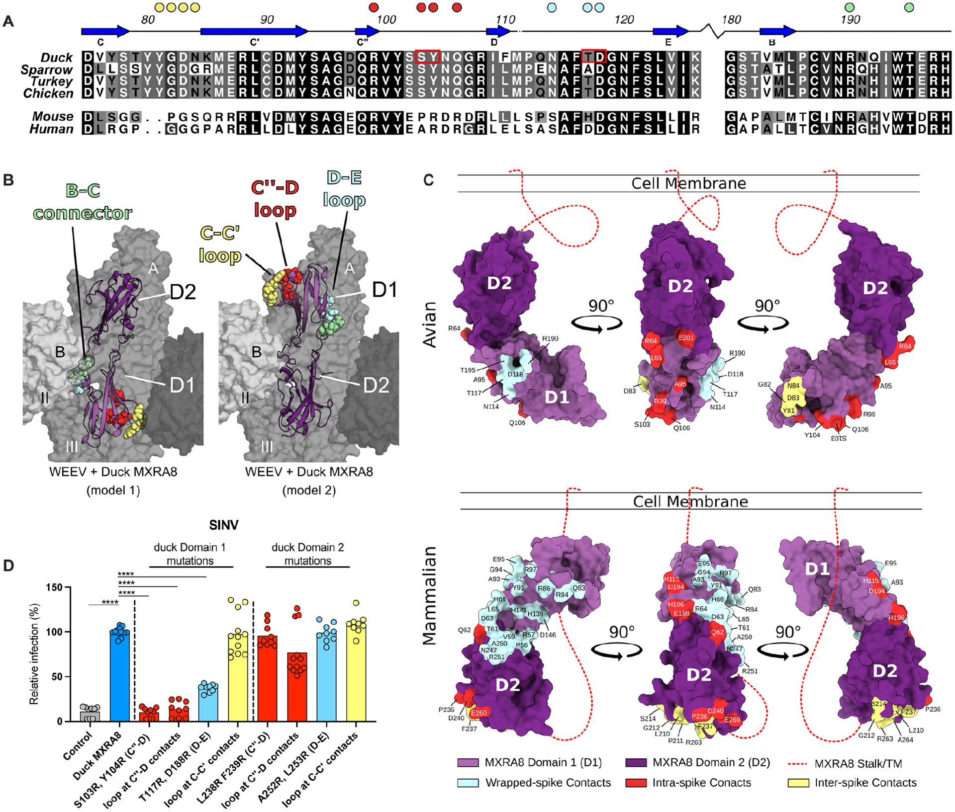
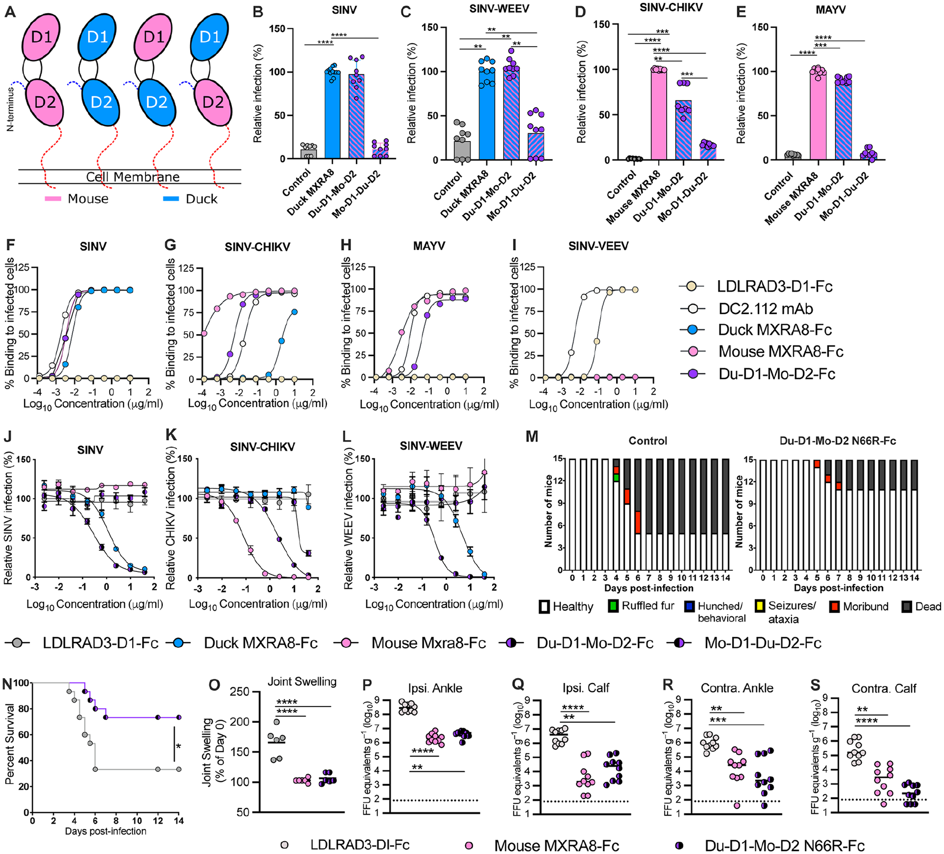
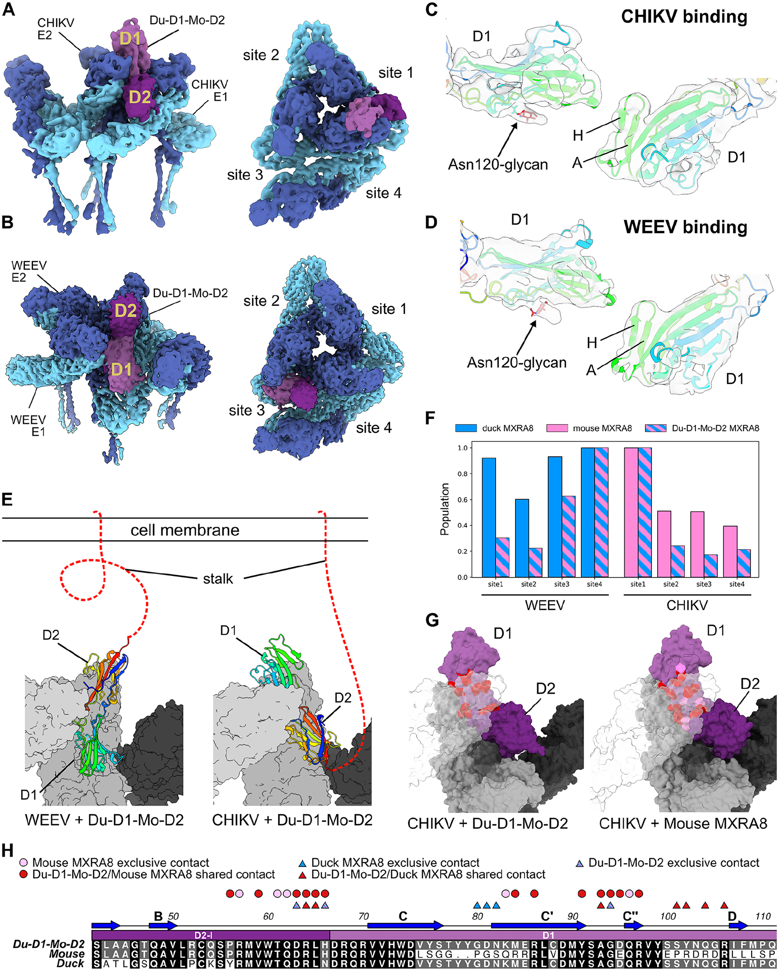
MXRA8 is a receptor for chikungunya (CHIKV) and other arthritogenic alphaviruses with mammalian hosts. However, mammalian MXRA8 does not bind to alphaviruses that infect humans and have avian reservoirs. Here, we show that avian, but not mammalian, MXRA8 can act as a receptor for Sindbis, western equine encephalitis (WEEV), and related alphaviruses with avian reservoirs. Structural analysis of duck MXRA8 complexed with WEEV reveals an inverted binding mode compared with mammalian MXRA8 bound to CHIKV. Whereas both domains of mammalian MXRA8 bind CHIKV E1 and E2, only domain 1 of avian MXRA8 engages WEEV E1, and no appreciable contacts are made with WEEV E2. Using these results, we generated a chimeric avian-mammalian MXRA8 decoy-receptor that neutralizes infection of multiple alphaviruses from distinct antigenic groups in vitro and in vivo. Thus, different alphaviruses can bind MXRA8 encoded by different vertebrate classes with distinct engagement modes, which enables development of broad-spectrum inhibitors.

Keywords: alphavirus; birds; cryoelectron microscopy; evolution; inhibitor; mammals; pathogenesis; receptor; species; structure; tropism.


Conflict of interest statement

Declaration of interests M.S.D. is a consultant to or member of a Scientific Advisory Board for Inbios, Ocugen, Vir Biotechnology, Topspin Therapeutics, and Moderna. The Diamond laboratory has received unrelated funding support in sponsored research agreements from Emergent BioSolutions, Moderna, Generate Biomedicines, Vir Biotechnology, and Immunome. D.H.F. is a founder of Courier Therapeutics and his laboratory has received unrelated funding support from Emergent BioSolutions and Mallinckrodt Pharmaceuticals. J.M.E. is an employee of Vir Biotechnology.

Figures

Figure 1.
Figure 1.. Avian MXRA8 is required for optimal infection of SINV and WEEV alphaviruses.
A. A phylogenetic tree based on alphavirus E1 and E2 protein sequences with complexes clustered in blue (WEEV complex), orange (SFV complex), purple (EEEV complex), and yellow (VEEV complex). Viruses in this study are bolded. B-C. ΔMxra8 3T3 cells complemented with mouse (pink), chicken (red), duck (blue) or turkey (yellow) MXRA8 or empty vector control (gray) were inoculated with CHIKV 181/25 (B) or SINV TR339 (C) and stained for CHIKV or SINV antigen (4 and 3 experiments in triplicate, respectively). Infection is normalized to levels detected with cells expressing mouse (B) or chicken (C) MXRA8. D. Multi-step growth curves with SINV TR339 in ΔMxra8 3T3 cells complemented with chicken or mouse MXRA8 or empty vector control. Virus was titrated by focus-forming unit (FFU) assay (4 experiments, duplicate). E-G. ΔMxra8 3T3 cells complemented with mouse, chicken, duck or turkey MXRA8 or empty vector control were inoculated with SINV-WEEV-GFP (E), SINV-EEEV-GFP (F) or SINV-VEEV-GFP (G). Infection was assessed by GFP expression (4 to 7 experiments, triplicate). H. CEFs were inoculated with CHIKV 181/25, SINV-WEEV-GFP, or SINV TR339 and stained for viral antigen. I-J. CEFs were pre-incubated with anti-chicken Mxra8 or an isotype control mAb and inoculated with SINV-WEEV-GFP (I) or SINV TR339 (J). Infection was assessed by GFP expression or E1 staining and compared between infected (red) and non-infected cells (blue) (3 experiments in triplicate). K-L. ΔMxra8 chicken fibroblasts complemented with chicken or mouse MXRA8 (2 independent clones of each shown) or empty vector control and non-targeted (NT1 and NT2) CEFs were inoculated with SINV TR339 (K), CHIKV 181/25 (L). Infection was assessed by flow cytometry, derived from the percent and mean fluorescence intensity of the infected cells, and normalized to the non-targeting control NT1 (for SINV) or ΔMxra8 (1) + Mouse Mxra8 (for CHIKV) (3 experiments, in triplicate). B, C, E, I, J, K and L: one-way ANOVA with Dunnett’s post-test; mean ± standard deviation (SD). D: Two-way ANOVA with Tukey post-test; mean ± SD. ns, non-significant; *, P < 0.05; **, P < 0.01; ****, P < 0.0001). See also Fig S1 S2, S3, S4, and Table S1.
Figure 2.
Figure 2.. Avian MXRA8 modulates SINV attachment and internalization and binds to WEEV VLPs.
A-C. SINV was incubated with ΔMxra8 3T3 cells (control), chicken Mxra8-complemented ΔMxra8 3T3 cells, or mouse Mxra8-complemented ΔMxra8 3T3 cells at 4 °C without (A) or with (B) pre-incubation with mouse-anti-chicken MXRA8 mAbs. Bound virions were quantified by measuring viral RNA levels and Gapdh levels via qRT–PCR. (C) After removal of unbound virus, the temperature was shifted to 37 °C to allow internalization. Intracellular RNA (SINV and Gapdh) levels were measured by qRT–PCR. (3 experiments, triplicate). D-E. Binding of chicken MXRA8–Fc and murine MXRA8-Fc to CHIKV (D) or WEEV (E) VLPs by ELISA (WEEV positive control, WEEV-204; CHIKV positive control, CHK-265; negative control, anti-HCV H77.39; CHIKV VLP ELISA: 2 experiments, triplicate; WEEV VLP ELISA: 2 experiments, duplicate). F-G. Binding of WEEV VLPs (F) and CHIKV VLPs (G) to duck, sparrow, or mouse MXRA8 by ELISA. SARS-CoV-2 receptor-binding domain served as a negative control (3 experiments, duplicate). A-C: one-way ANOVA with Dunnett’s post-test; mean ± SD. *, P < 0.05; ****, P < 0.0001).
Figure 3.
Figure 3.. Cryo-EM reconstruction of duck MXRA8 bound to WEEV.
A. Cryo-EM density map of WEEV-VLP bound to duck MXRA8 at 4.74 Å resolution. WEEV-E1, WEEV-E2, and MXRA8 are colored as light-blue, dark-blue, and violet, respectively. Rotational symmetries along the 2-fold, 3-fold, and 5-fold axes are displayed with white numbers. B. Map of WEEV-VLP bound to duck MXRA8. Asymmetric unit contains the entire quasi 3-fold spike (q3), and a single icosahedral spike (i3) E1-E2 heterodimer. Shown are two views of the asymmetric unit, a side view (left, parallel with the viral membrane) and a top-down view (right, perpendicular with the viral membrane). Local resolution of map is colored from blue (4.25 Å) to white (5.25 Å) to red (6.25 Å). Density maps are viewed at contour level = 0.26 (0.96σ). C. Surface diagram of MXRA8 at site 3, detailing the three unique viral E1-E2 heterodimer contacts, wrapped (light gray), intraspike (gray), and interspike (dark gray). D. Surface diagram of MXRA8 at site 3 and interacting E1-E2 heterodimers, termed: E1-E2-wrapped, E1-E2-intraspike, or E1-E2-interspike. MXRA8 D1: light magenta; MXRA8 D2: dark magenta; E1 domain I: light gray; E1 domain II: medium-gray; E1 domain III: dark-gray; E1-TM (black); E2 A domain: light-cyan; E2 β-linker: medium-cyan; E2 B domain: dark-cyan; E2 C domain: medium-blue; E2-TM: dark-blue. See also Fig S5, S6, and Tables S2–S4.
Figure 4.
Figure 4.. Duck and mouse MXRA8 use a domain-inverted binding paradigm to engage WEEV and CHIKV.
A. Ribbon and topology diagram of duck MXRA8 with β-strands labeled with standard Ig-like fold conventions. The β-strands are colored by a rainbow and are depicted as a cartoon with residue number start/stop locations. Disulfide bonds between cysteines are depicted with a yellow line. B. Cryo-EM density of the duck MXRA8 domain in the binding-groove, modeled as either D1 or D2. D1 has an extra β-strand (strand H) that better fits the density, whereas D2 lacks this β-strand and leaves this density unfilled. Density maps are viewed at contour level = 0.42 (1.56σ). C. Cryo-EM map of duck D1 with clear density in the region expected for N-linked glycan on Asn-120. Density map is viewed at contour level = 0.42 (1.56σ). D. Differential binding modes for avian and mammalian MXRA8 binding to WEEV and CHIKV, respectively. Avian MXRA8 D1 is distal to the cell membrane and binds E1-intraspike heterodimer, whereas mammalian MXRA8 D1 is proximal to the cell and binds E2-wrapped heterodimer. E. Multi-sequence alignment of WEE- and SF-complex E1 proteins with E1-intraspike duck MXRA8 D1 contacts (depicted with red circles). N-linked glycosylation sites are shown with dotted red boxes. Sequence similarities are colored from white to black, with black being most similar. E1 β-strands are labeled as described in Voss et al. See also Fig S7–S9.
Figure 5.
Figure 5.. Assessment of the MXRA8 binding model
A. Sequence alignment of D1 of MXRA8 at regions that contact WEEV E1. Similar sequence identities are colored by most variable (white) to conserved (black). β-strands are labeled based on topology in Fig 4A. Filled circles above the alignment denote avian MXRA8 contact residues in the C-C’ loop (yellow), C”-D loop (red), D-E loop (cyan), and B-C connector (pale green) are denoted by circles. B. Structure of avian MXRA8 in two possible binding modes, non-flipped (model 1, left) and flipped (model 2, mouse/human-like, right), with contact residues in either D1 or D2 shown as space filling spheres in the C-C’ loop (yellow), C”-D loop (red), D-E loop (cyan), and B-C connector (pale green). C. Binding sites for WEEV and CHIKV on D1 (light purple) and D2 (dark purple) of MXRA8. Structurally defined binding sites and amino acid contact residues are colored according to the viral E2-E1 heterodimer engaged: wrapped (cyan), intraspike (red), or interspike (yellow). Stalk attaching MXRA8 to the cell membrane is represented as a red dashed line and is used to denote mode of binding, with mammalian MXRA8 adopting a flipped orientation relative to avian MXRA8. Avian and mammalian MXRA8 engage E2-E1 heterodimers most proximal to viral membrane with D1 and D2, respectively. D. SINV-GFP infection in ΔMxra8 3T3 cells complemented with wild-type (blue) and indicated mutants (light blue, yellow, or red) duck MXRA8 (3 experiments, triplicate; normalized to wild-type duck MXRA8 infection. Infection and GFP fluorescence were analyzed by flow cytometry. D: one-way ANOVA with Dunnett’s post-test; mean ± standard deviation (SD). ****, P < 0.0001). See also Fig S10.
Figure 6.
Figure 6.. Chimeric avian-mammalian MXRA8 interacts with both WEE and SF complex alphaviruses.
A. Schematic of chimeric MXRA8 proteins used to assess binding modes. In addition to mouse or duck MXRA8, chimeras shown are Du-D1-Mo-D2 and Mo-D1-Du-D2. B-C. SINV-GFP (B) and SINV-WEEV-GFP (C) infection in ΔMxra8 3T3 cells complemented with wild-type duck (blue) and chimeric (blue/pink) duck-mouse MXRA8 (3 experiments in triplicate; normalized to wild-type duck MXRA8 infection. Infection and GFP fluorescence were analyzed by flow cytometry. D-E. SINV-CHIKV (LR 2006 strain) (D) and MAYV (E) infection of ΔMxra8 3T3 cells complemented with an empty vector, or wild-type mouse and chimeric duck-mouse MXRA8 (3 experiments in triplicate; normalized to wild-type mouse MXRA8 infection). Infection was analyzed by flow cytometry by evaluating viral antigen staining with mAbs (CHIKV or MAYV). F-I. Staining of the surface of Vero cells infected with SINV-TR339 (F), SINV-CHIKV (LR 2006 strain), (G) MAYV (H), or SINV-VEEV (I) after incubation with serially diluted mouse MXRA8-Fc, duck MXRA8-Fc, Du-D1-Mo-D2 MXRA8-Fc, LDLRAD3-D1-Fc proteins or cross-reactive anti-E1 mAb (DC2.112, positive control)) control. Data are expressed as the percentage of infected cells that bound positively to the indicated proteins by flow cytometry (representative of 2 experiments). J-L. Neutralization of SINV TRR399 (J) SINV-WEEV (K) or SINV-CHIKV (L) (all three viruses expressing eGFP) infection by duck MXRA8-Fc, mouse MXRA8-Fc, Du-D1-Mo D2-MXRA8-Fc, Mo-D1-Du-D2-MXRA8-Fc, or LDLRAD3-D1-Fc control (3 experiments, duplicates). Infection was analyzed by flow cytometry (GFP expression) and normalized to levels after incubation with LDLRAD3-D1-Fc protein. M-N. Clinical disease (M) and survival (N) of 4-week-old female CD-1 mice inoculated with 103 PFU of WEEV (McMillan strain) mixed with Du-D1-Mo D2 N66R-MXRA8-Fc or LDLRAD3-D1-Fc. O-S. Foot swelling (O) and viral RNA levels in indicated tissues (P-S) of 4-week-old male C57BL/6J mice at 72 h after inoculation with 103 FFU of CHIKV (La Reunion 2006) mixed with 50 μg of mouse Mxra8-Fc, Du-D1-Mo-D2-N66R-MXRA8-Fc, or LDLRAD3-D1-Fc (O: 2 experiments n = 6; P-S: 2 experiments n = 10). B-E and O: one-way ANOVA with Dunnett’s post-test; mean ± standard deviation (SD). N: Log rank test. P-S: Kruskal-Wallis ANOVA with Dunn’s post-test. **, P < 0.01; ***, P < 0.001; ****, P < 0.0001. See also Fig S11, S12, and Table S5.
Figure 7.
Figure 7.. Cryo-EM structure of Du-D1-Mo-D2 MXRA8 chimera bound to CHIKV VLP.
A-B. Cryo-EM density of Du-D1-Mo-D2 bound to CHIKV (A) or WEEV (B) VLPs. Shown are two views of a single asymmetric unit, with E1 (light blue), E2 (dark blue), D1 of Du-D1-Mo-D2 (light purple), and D2 of Du-D1-Mo-D2 (dark purple) at site 1. C-D. Atomic models of Du-D1-Mo-D2 MXRA8 with experimental density highlighting the symmetry breaking N-linked glycan at Asn120 and β-strands A and H in D1 when binding to CHIKV (C) or WEEV (D). E. Atomic models of Du-D1-Mo-D2 MXRA8 binding to WEEV (left) or CHIKV (right) highlighting the flipped orientations. Surfaces are shown for the wrapped E1-E2 heterodimer (light gray), intra E1-E2 heterodimer (gray), and inter E1-E2 heterodimer (dark gray). Du-D1-Mo-D2 is depicted as a ribbon and is rainbow-colored from the N- to C-terminus. Stalk attaching MXRA8 to the cell membrane is represented as a red dashed line. F. MXRA8 site occupancies for duck MXRA8 (light blue), mouse MXRA8 (pink), and Du-D1-Mo-D2 (blue and pink stripes) bound to WEEV (left) or CHIKV (right). G. Molecular models of Du-D1-Mo-D2 and mouse MXRA8 bound to CHIKV. Shown are wrapped E1-E2 heterodimer (light gray, transparent), intraspike E1-E2 heterodimer (gray), interspike E1-E2 heterodimer, MXRA8 D1 (light purple), and MXRA8 D2 (dark purple). Observed contact residues in both Du-D1-Mo-D2 and mouse MXRA8 are colored dark red, and contacts observed only for mouse MXRA8 are colored pink. H. Sequences of Du-D1-Mo-D2, mouse, and duck MXRA8, highlighting conserved contacts between Du-D1-Mo-D2 and mouse MXRA8 to CHIKV (red circles), contacts only in mouse MXRA8 to CHIKV (pink circles), conserved contacts between Du-D1-Mo-D2 and duck MXRA8 to WEEV (red triangles), contacts only in duck MXRA8 to WEEV (blue triangles), and contacts only in Du-D1-Mo-D2 to WEEV (blue and pink stripped triangles). See also Fig S5, S11, and Tables S2–S4.

Similar articles

Cited by

References

    1. Weaver SCS, D.W. (2011). Alphavirus Infections. In Tropical Infectious Diseases: Principles, Pathogens and Practice, D.H.W. Guerrant Richard L., Weller Peter F., ed. (Saunders WB), pp. 519–524. 10.1016/B978-0-7020-3935-5.00078-1. - DOI
    1. Garmashova N, Gorchakov R, Volkova E, Paessler S, Frolova E, and Frolov I (2007). The Old World and New World alphaviruses use different virus-specific proteins for induction of transcriptional shutoff. J Virol 81, 2472–2484. JVI.02073–06 [pii]10.1128/JVI.02073-06. - DOI - PMC - PubMed
    1. Weaver SC, Kang W, Shirako Y, Rumenapf T, Strauss EG, and Strauss JH (1997). Recombinational history and molecular evolution of western equine encephalomyelitis complex alphaviruses. J Virol 71, 613–623. 10.1128/JVI.71.1.613-623.1997. - DOI - PMC - PubMed
    1. Hahn CS, Lustig S, Strauss EG, and Strauss JH (1988). Western equine encephalitis virus is a recombinant virus. Proc Natl Acad Sci U S A 85, 5997–6001. 10.1073/pnas.85.16.5997. - DOI - PMC - PubMed
    1. Strauss JHSEG (1997). Recombination in Alphaviruses. Seminars in VIROLOGY 8,, 85–94, VI9701151044.

Publication types

LinkOut - more resources