

Successful conservative treatment for left ventricular free wall rupture after acute myocardial infarction
- PMID: 37805478
- PMCID: PMC10560420
- DOI: 10.1186/s13019-023-02397-w
Successful conservative treatment for left ventricular free wall rupture after acute myocardial infarction
Abstract
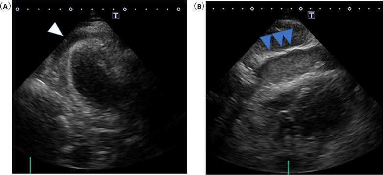
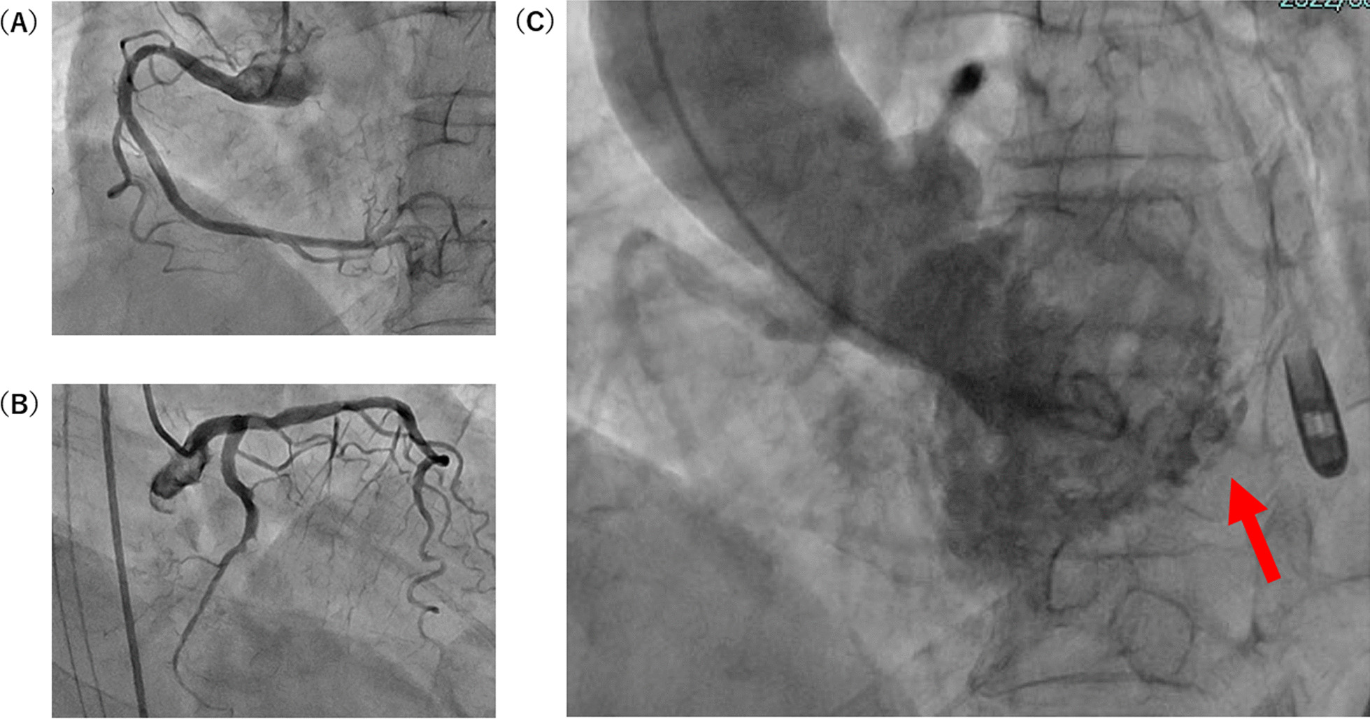
Left ventricular free wall rupture (LVFWR) is a rare but fatal complication of acute myocardial infarction (AMI). An 81-year-old female patient with several cardiovascular risk factors presented to the emergency department with symptoms of developing a chronic stomachache and cold sweat. An echocardiograph showed wall motion abnormalities from the lateral to posterior wall, as well as pericardial effusion containing clots of up to 17 mm in the posterior wall that indicated LVFWR after AMI. Although she was conscious after being brought to the initial care unit, she suddenly lost consciousness and fell into electromechanical dissociation (EMD). Endotracheal intubation was immediately initiated and her pericardial drainage and intra aortic balloon pump (IABP) placement, and hemodynamics recovered. Although she had 100% obstruction in the left circumflex artery (LCX) #12 on coronary angiography (CAG), she was discharged to the Intensive Care Unit (ICU) without percutaneous coronary intervention (PCI). Conservative treatment such as intubation, sedation, pericardiocentesis and strict blood pressure management as well as treatment by IABP long-term support led to the patient being uneventfully discharged after 60 days.
Keywords: Acute myocardial infarction (AMI); Cardiac tamponade; Cardiogenic shock; Conservative treatment; Left ventricular free wall rupture (LVFWR).
© 2023. BioMed Central Ltd., part of Springer Nature.
Conflict of interest statement
The authors declare that they have no competing interests.
Figures

Similar articles
-
Intraprocedural left ventricular free wall rupture diagnosed by left ventriculogram in a patient with infero-posterior myocardial infarction and severe aortic stenosis.BMC Cardiovasc Disord. 2016 Jun 6;16:126. doi: 10.1186/s12872-016-0302-7. BMC Cardiovasc Disord. 2016. PMID: 27266264 Free PMC article.
-
Survival after left ventricular free wall rupture following acute myocardial infarction by conservative treatment.Am J Emerg Med. 2021 Jan;39:21-23. doi: 10.1016/j.ajem.2020.08.035. Epub 2020 Aug 15. Am J Emerg Med. 2021. PMID: 32829991
-
Use of intra-aortic balloon pump support for oozing-type cardiac rupture after acute myocardial infarction.Am J Emerg Med. 2016 Jan;34(1):120.e1-3. doi: 10.1016/j.ajem.2015.05.054. Epub 2015 Jun 14. Am J Emerg Med. 2016. PMID: 26145582
-
[Reperfusion therapy and mechanical circulatory support in patients in cardiogenic shock].Herz. 1999 Oct;24(6):448-64. doi: 10.1007/BF03044431. Herz. 1999. PMID: 10546149 Review. German.
-
Subacute left ventricular free-wall rupture in early course of acute myocardial infarction. Clinical report of two cases and review of the literature.G Ital Cardiol. 1999 Feb;29(2):163-70. G Ital Cardiol. 1999. PMID: 10088074 Review.
References
-
- Becker RC, Hochman JS, Cannon CP, Spencer FA, Ball SP, Rizzo MJ, et al. Fatal cardiac rupture among patients treated with thrombolytic agents and adjunctive thrombin antagonists: observations from the Thrombolysis and Thrombin Inhibition in Myocardial Infarction 9 Study. J Am Coll Cardiol. 1999;33(2):479–487. doi: 10.1016/S0735-1097(98)00582-8. - DOI - PubMed
Publication types
MeSH terms
LinkOut - more resources
Full Text Sources
Medical
Miscellaneous
