. 2023 Oct 7;18(1):6.
doi: 10.1186/s13064-023-00174-y.

Drosophila CASK regulates brain size and neuronal morphogenesis, providing a genetic model of postnatal microcephaly suitable for drug discovery

Affiliations

Drosophila CASK regulates brain size and neuronal morphogenesis, providing a genetic model of postnatal microcephaly suitable for drug discovery

Judith A Tello et al. Neural Dev. .

Abstract

Background: CASK-related neurodevelopmental disorders are untreatable. Affected children show variable severity, with microcephaly, intellectual disability (ID), and short stature as common features. X-linked human CASK shows dosage sensitivity with haploinsufficiency in females. CASK protein has multiple domains, binding partners, and proposed functions at synapses and in the nucleus. Human and Drosophila CASK show high amino-acid-sequence similarity in all functional domains. Flies homozygous for a hypomorphic CASK mutation (∆18) have motor and cognitive deficits. A Drosophila genetic model of CASK-related disorders could have great scientific and translational value.

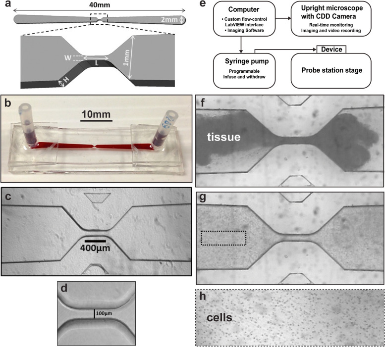
Methods: We assessed the effects of CASK loss of function on morphological phenotypes in Drosophila using established genetic, histological, and primary neuronal culture approaches. NeuronMetrics software was used to quantify neurite-arbor morphology. Standard nonparametric statistics methods were supplemented by linear mixed effects modeling in some cases. Microfluidic devices of varied dimensions were fabricated and numerous fluid-flow parameters were used to induce oscillatory stress fields on CNS tissue. Dissociation into viable neurons and neurite outgrowth in vitro were assessed.

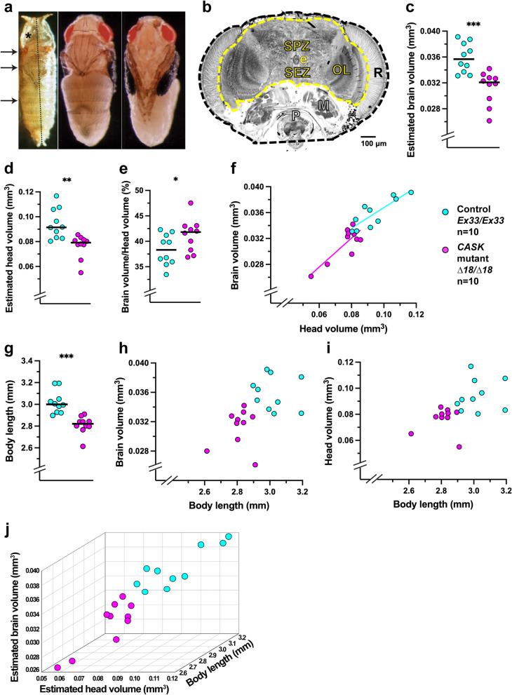
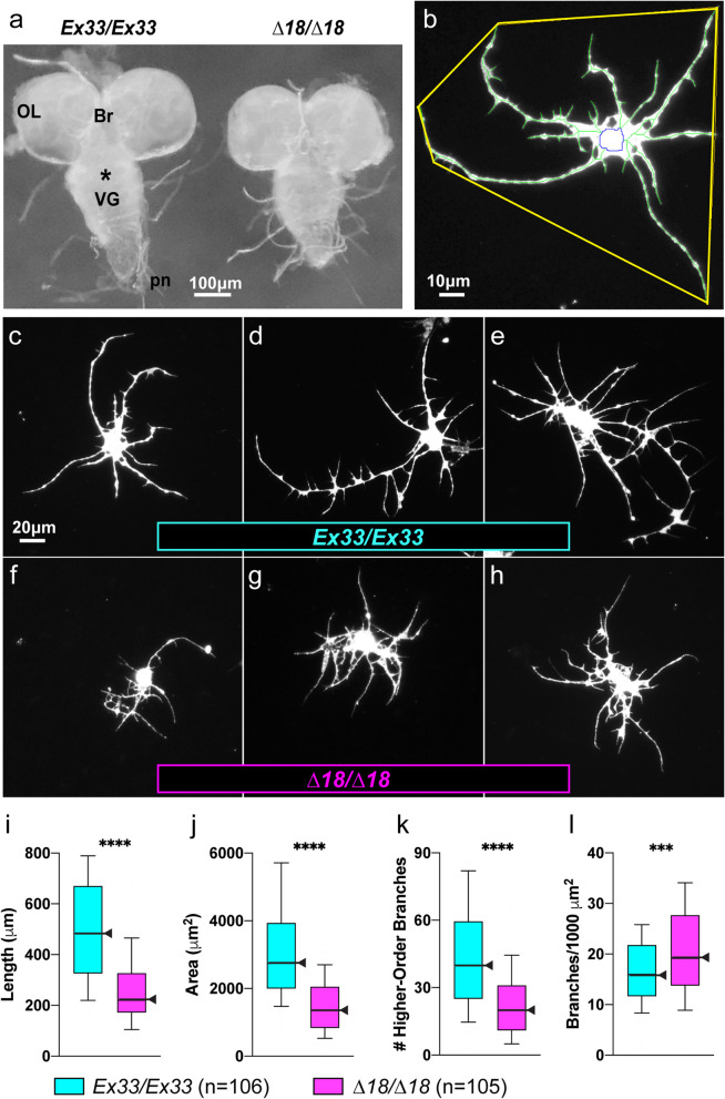
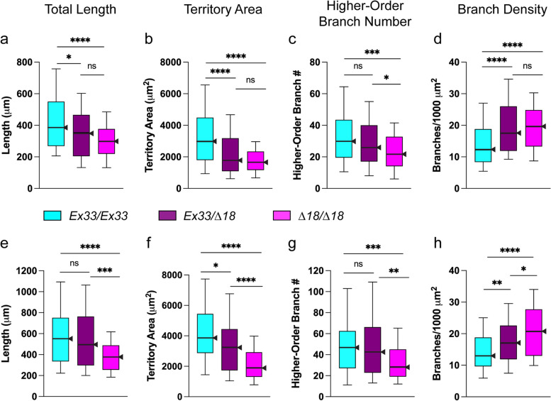
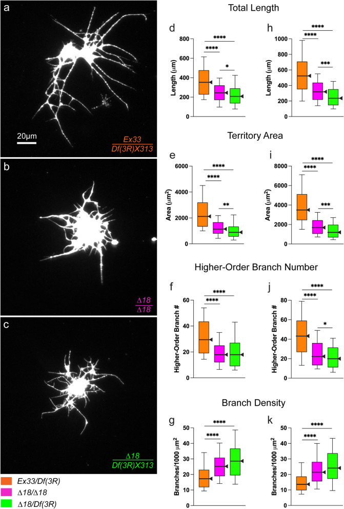
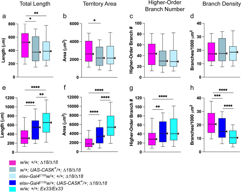
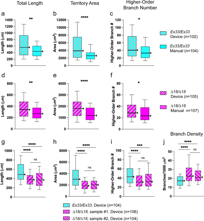
Results: We demonstrated that ∆18 homozygous flies have small brains, small heads, and short bodies. When neurons from developing CASK-mutant CNS were cultured in vitro, they grew small neurite arbors with a distinctive, quantifiable "bushy" morphology that was significantly rescued by transgenic CASK+. As in humans, the bushy phenotype showed dosage-sensitive severity. To overcome the limitations of manual tissue trituration for neuronal culture, we optimized the design and operation of a microfluidic system for standardized, automated dissociation of CNS tissue into individual viable neurons. Neurons from CASK-mutant CNS dissociated in the microfluidic system recapitulate the bushy morphology. Moreover, for any given genotype, device-dissociated neurons grew larger arbors than did manually dissociated neurons. This automated dissociation method is also effective for rodent CNS.

Conclusions: These biological and engineering advances set the stage for drug discovery using the Drosophila model of CASK-related disorders. The bushy phenotype provides a cell-based assay for compound screening. Nearly a dozen genes encoding CASK-binding proteins or transcriptional targets also have brain-development mutant phenotypes, including ID. Hence, drugs that improve CASK phenotypes might also benefit children with disorders due to mutant CASK partners.

Keywords: Haploinsufficiency; Immunostaining; Intellectual disability; Microfluidics; Neurite arbor; Neurogenetics; Primary neuronal culture; Short stature.


Conflict of interest statement

The authors declare no competing interests.

Figures

Fig. 1
Fig. 1
Drosophila CASK gene, proteins, and genetic reagents. a Color-coded schematic, drawn to scale, showing identical organization of CASK protein domains in H. sapiens and D. melanogaster. Amino-acid (aa) sequence similarity (%) as indicated; identities are CaMK-like 68%; L27A 31%; L27B 45%; PDZ 82%; SH3 67%; GUK 70%. The white boxes between SH3 and GUK are the "hook" motif. b Cytogenetic and molecular maps of CASK (FlyBase ID FBgn0013759; https://flybase.org/reports/FBgn0013759, last accessed 6 February 2023). Region 93F10-12 on right arm of chromosome 3 [86], reproduced in [91], centromere to the left. CASK gene span: genomic DNA map 21,783 – 21,822 kb (Fly Base, FB2022_06); scale at top right applies to all maps in (b)-(d). Protein orientation in (a) is flipped 180° relative to transcription direction. CASK (blue), with two transcriptional start sites (left-pointing arrows), and tsl (green) genes. P-element EY07081 insertion site in first intron. Two overlapping chromosomal deficiencies delete CASK DNA, with breakpoints estimated by restriction mapping [96]. c CASK alleles [142]. Imprecise excision of EY07081 generated CASK mutation, ∆18, with deletion of exons 1 and 2, and insertion of small piece of roo transposon at the breakpoint. Precise excision yielded Ex33, which serves as the control allele. (d) Endogenous and engineered CASK transcripts. Boxes represent exons; color scheme for encoded protein domains as in (a), gray for 5'- and 3'-untranslated regions. CASK-β is the full-length transcript; internal promoter generates short transcript, CASK-α, which is missing the 5' exons. The UAS-CASK+ 10.20 construct expresses full-length wild-type CASK cDNA under UAS control
Fig. 2
Fig. 2
The morphological phenotype of CASK mutants involves head, brain, and body. a Photomicrographs of Drosophila pharate adults, anterior at the top. Left, Canton-S wildtype within the puparium (also called "pupal case"), dorsal view. Vertical dotted line represents body length. Arrows show the centers of the head, thorax, and abdomen (top to bottom, respectively). Asterisk marks the air space anterior to the animal. Center and right, dorsal and ventral views, respectively, of a female genetic control, dissected from the puparium but still wrapped by the transparent pupal cuticle. Adapted from [41], with permission. b Photomicrograph of a histological section through the head of a CASK-control (Ex13/Ex13) pharate adult, frontal plane. Scale bar as indicated. Colorized and thickened dashed outlines overlay the perimeters of the CNS (yellow) and head (dark blue), traced using cellSens software. e, esophagus; M, muscles; OL, optic lobe; P, proboscis; R, retina; SEZ, subesophageal zone; SPZ, supraesophageal zone. (c-j) Abnormal morphology of CASK Δ18/Δ18 mutants compared with Ex33/Ex13 controls. Each circle represents a single fly; color legend shown to the right of panel f. Horizontal black lines are medians. Significance levels: *, p < 0.05; **, p < 0.005; ***, p < 0.0005. In CASK mutants, c estimated brain volume was reduced (p = 0.0002); d estimated head volume was reduced (p = 0.0007); e the percentage of head volume occupied by the brain was increased (p = 0.0288); f scatter plot of head volume vs. brain volume shows a positive linear relationship in both mutants and controls; (g) body length was reduced (p < 0.0001); h-i scatter plots of body length vs. brain volume or head volume show distinct distributions of mutant and control data and no strong indication of linear relationships; j in a 3D scatter plot of brain volume, head volume, and body length, the data from the two genotypes are nonoverlapping, which was confirmed by rotating the graph
Fig. 3
Fig. 3
The “bushy” phenotype of CASK-mutant neurite arbors in vitro. a The CASK-mutant larval CNS is reduced in size. Stereomicroscopic view of freshly dissected, unfixed whole-CNS explants from wandering third-instar female larvae in buffered saline, dorsal side up, anterior to the top. CASK control (Ex33/Ex33; left) alongside mutant (∆18/∆18; right). OL, optic lobe; Br, brain; *, subesophageal zone; VG, ventral ganglia; pn, peripheral nerves. Scale bar = 100 µm. b-h Photomicrographs of cultured larval CNS neuron, immunofluorescently labeled for a neuronal membrane marker, after 3 div (60X magnification). b NeuronMetrics software output. The color-coded lines have been thickened to improve visibility: yellow polygon, territory; blue, central portion of the neuronal cell body; green, branches of the skeletonized neurite arbor. Scale bar = 10 µm. c-h Scale bar = 20 μm. Neurons representing the ~ 25th (c, f), ~ 50th (d, g), and ~ 75th (e, h) percentiles for each of three arbor-size parameters, total neurite length, territory area, and branch density. c-e CASK control, Ex33/Ex33. fh CASK mutant, ∆18/∆18. (i-l) The “bushy” phenotype: CASK-mutant neurite arbors are reduced in size with excessive branch density. Quantification of neurite-arbor morphology, depicted as box-plot distributions, comparing 3 div cultures of CASK-control (Ex33/Ex33; n = 106 neurons; aqua) and CASK-mutant (Δ18/∆18; n = 105 neurons; magenta) larval CNS neurons. Center lines and arrowheads represent the 50th percentile. Top and bottom of each box represent the 75th and 25th percentiles, respectively. The upper and lower whiskers represent the 90th and 10th percentiles, respectively. Significance levels: ***, p < 0.0005; ****, p < 0.00005. Total neurite length (i), territory area (j), and higher-order branch number (k) were all reduced in CASK-mutant neurons, whereas branch density (l) was increased. These data are from one of 9 independent experiments; see Additional file: Table A3 for analysis across all experiments
Fig. 4
Fig. 4
Analysis of ∆18 heterozygous neurons shows the bushy phenotype is neither strictly recessive nor dominant. Data from two independent experiments (a-d and eh, respectively), each with parallel 3-div cultures of larval CNS neurons from CASK control (Ex33/Ex33; aqua), CASK mutant (Δ18/∆18; magenta), and heterozygote (Δ18/Ex33; burgundy). Box-plot distributions depicted as in Fig. 3, with n = 101–104 neurons from each genotype in each experiment. a, e Total neurite length; b, f territory area; c, g higher-order branch number; and d, h branch density. For two key characteristic features of the bushy phenotype, decreased territory area and increased branch density, ∆18 was dominant (b, d) or semi-dominant (f, h). Decreased total neurite length showed opposite results in the two experiments (a, e) and decreased higher-order branch number appeared to be recessive (c, g). Significance levels: *, p < 0.05; ***, p < 0.0005; ****, p < 0.00005
Fig. 5
Fig. 5
Chromosomal deficiencies of CASK reveal dosage sensitivity of the bushy phenotype. For two deficiencies, 3 div larval CNS cultured neurons were compared among three genotypes: CASK homozygous mutant (Δ18/Δ18), CASK hemizygous mutant (∆18/Df), and control allele over deficiency (Ex33/Df). a-c Photomicrographs of representative neurons, immunostained for neuronal membrane marker, from near the median for each of three parameters: total neurite length, territory area and branch density. 60X magnification; scale bar = 20 μm. a Ex33/Df(3R)X313. b-c Both Δ18/Δ18 and Δ18/ Df(3R)X313 neurons show bushy neurite arbors. d-k CASK hemizygous mutant neurons are more severely affected on most parameters. Quantification of neurite-arbor morphology; box-plot distributions depicted as in Fig. 3. Significance levels: *, p < 0.05; ***, p < 0.0005; ****, p < 0.00005. d-g Ex33/Df(3R)X307 (n = 104; orange) compared with Δ18/∆18 (n = 106; magenta) and Δ18/ Df(3R)X307 (n = 106; green). hk Ex33/Df(3R)X313 (n = 106; orange), compared with Δ18/∆18 (n = 105; magenta) and Δ18/ Df(3R)X313 (n = 106; green)
Fig. 6
Fig. 6
Transgenic expression of CASK+ significantly improves the bushy phenotype. Neurite-arbor morphology parameters from 3 div larval CNS neuronal cultures. Box-plot distributions depicted as in Fig. 3. Significance levels: *, p < 0.05; **, p < 0.005; ***, p < 0.0005; ****, p < 0.00005. a-d Neither individual transgene, UAS-controlled wild-type full-length CASK cDNA or the neuronal driver, elav-Gal4C155, improved the bushy phenotype of CASK-mutant (Δ18/Δ18) neurons. Three genotypes were compared: w/w; + ; Δ18/Δ18 (n = 104; magenta); w/ + ; UAS-CASK+/ + ; ∆18/∆18 (n = 104; gray); and elav-Gal4C155 w/ + ; + ; ∆18/∆18 (n = 105; light blue). a Both transgenes caused modest reductions in total neurite length, i.e., worsening the phenotype. b Territory area was mildly reduced by UAS-CASK+ only. c Higher-order branch number was not affected by either transgene. d Branch density, the most distinctive feature of the bushy phenotype, was not affected by either transgene. eh Driving expression of transgenic CASK+ in neurons significantly improved all four parameters of the bushy phenotype. Three genotypes were compared: w/w; + ; Δ18/Δ18 (n = 105; magenta); elav-Gal4C155, w/ + ; UAS-CASK+/ + ; Δ18/Δ18 (n = 104; bright blue); and w/w; + ; Ex33/Ex33 (CASK control; n = 105; aqua). e Total neurite length. f Territory area. g Higher-order branch number. h Branch density. Strong but incomplete transgenic rescue is consistent with the dosage-sensitive nature of the bushy phenotype
Fig. 7
Fig. 7
A microfluidic system for dissociation of CNS into viable neurons. a Schematic drawing of the microdevice, top-down view, with central portion magnified and shown in oblique lateral view. Tissue dissociation takes place in the narrow central orifice. Length (L), width (W), and height (H) were varied during optimization. b Photograph of single-channel microfluidic device, lateral oblique view: ports extending upward from the distal ends provide inlet/outlet access and connection to the pump. The channel has been filled with red dye to make it easily visible. c, d Photomicrographs of the central part of the channel, top-down views. c As the main channel segments approach the central orifice, the walls narrow at 45° angles. d High-magnification view of the orifice. Note the smoothness of the channel walls. The 100-µm-wide channels were not needed or used in the experiments reported here. e A block diagram illustrating the main components of the experimental system. The microfluidic device sits on the stage of a Signatone probe station equipped with an upright compound microscope and computer-controlled CCD camera. One device port is connected by tubing to a computer-controlled syringe pump that drives oscillatory flow within the channel. fh Individual frames of video acquired during the dissociation of an enzyme-treated piece of rat E18 hippocampus in a microfluidic device. f Intact tissue is driven by the pump through the central orifice with dimensions L = 400 µm, W = 70 µm. g Following oscillatory cycles of flow-induced shear stress (flow rate 50 µl/sec, infusion volume 12.5 µl, 4 Hz), the tissue has been dissociated into cell clusters and single cells. Dotted rectangle indicates approximate size of the area shown in (h) at a later time at higher magnification. h Additional cycles have completed the dissociation into single cells which were collected and plated for culture
Fig. 8
Fig. 8
Device-dissociated neurons extend larger arbors and manifest the CASK-LOF bushy phenotype in vitro. Device dissociations were performed with flow parameters 50 µl/sec, infusion volume 10.5 µl, 4.8 Hz, mean cycle number1444. Neurite-arbor size parameters from 3 div cultures, comparing methods of dissociation and genotypes. Box-plot distributions depicted as in Fig. 3. Significance levels: *, p < 0.05; **, p < 0.005; ***, p < 0.0005; ****, p < 0.00005; ns, not significant. a-f Device vs. manual dissociation. a-c Ex33/Ex33. Device dimensions: channel height 500 µm; orifice length 400 µm; orifice width 60 µm. d-f ∆18/∆18. Device dimensions: channel height 450 µm; orifice length 400 µm; orifice width 50 µm. For both genotypes, total neurite length (a, d), territory area (b, e), and higher-order branch number (c-f) were significantly higher for microfluidic device-dissociated neurons. g-j Comparison of device-dissociated neurons from control (Ex33/Ex33; one sample) and CASK mutant (∆18/∆18; two samples) larval CNS. Device dimensions: channel height 450 µm; orifice length 400 µm; orifice width 50 µl. g Total neurite length. h Territory area. Higher-order branches. j Branch density. Device-dissociated CASK-mutant neurons extend bushy neurite arbors, replicating the phenotype seen in manual cultures. In addition, two cultures prepared after serial device-based dissociation show marked consistency of neurite-arbor parameters

Similar articles

References

    1. Anonymous, editors. Geneticist seeks engineer: must like flies and worms. Nat Methods. 2007;4(6):463. https://www.nature.com/articles/nmeth0607-463. - PubMed
    1. Al-Ali H, Blackmore M, Bixby JL, Lemmon VP. High content screening with primary neurons. 2013 [updated 2014 Oct 1]. In: Markossian S, Grossman A, Brimacombe K, et al., editors. Assay Guidance Manual [Internet]. Bethesda (MD): Eli Lilly & Company and the National Center for Advancing Translational Sciences. NCBI Bookshelf ID: NBK169433. https://www.ncbi.nlm.nih.gov/books/NBK169433/. Accessed 20 Aug 2023.
    1. Al-Mofty S, Elsayed M, Ali H, Ahmed O, Altayyeb A, Wahby A, et al. A microfluidic platform for dissociating clinical scale tissue samples into single cells. Biomed Microdevices. 2021;23:10. - PubMed
    1. Alburquerque-González B, Bernabé-García M, Montoro-García S, Bernabé-García Á, Rodrigues PC, Ruiz Sanz J, et al. New role of the antidepressant imipramine as a Fascin1 inhibitor in colorectal cancer cells. Exp Mol Med. 2020;52:281–292. - PMC - PubMed
    1. Aliaghaei M, Haun JB. Optimization of mechanical tissue dissociation using an integrated microfluidic device for improved generation of single cells following digestion. Front Bioeng Biotechnol. 2022;10:841046. - PMC - PubMed

Publication types

Substances