. 2023 Oct 7;14(10):654.
doi: 10.1038/s41419-023-06176-2.

Endothelial cell-derived RSPO3 activates Gαi1/3-Erk signaling and protects neurons from ischemia/reperfusion injury

Affiliations

Endothelial cell-derived RSPO3 activates Gαi1/3-Erk signaling and protects neurons from ischemia/reperfusion injury

Ting-Tao Liu et al. Cell Death Dis. .

Abstract

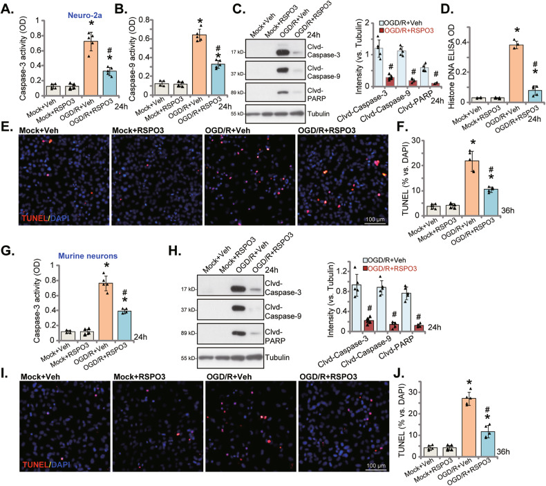
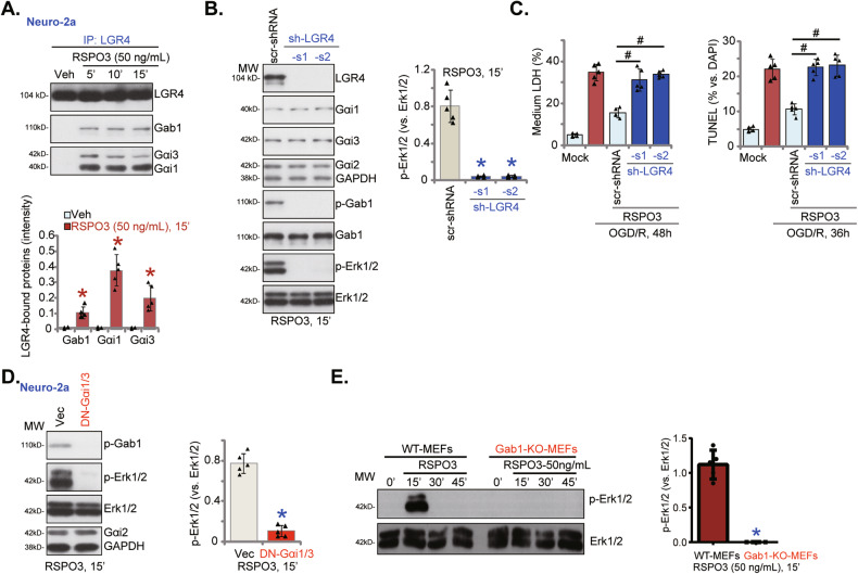
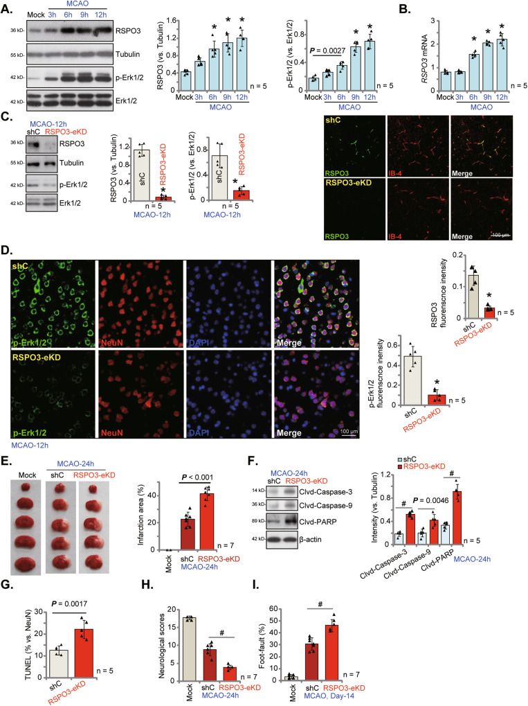
The current study explores the potential function and the underlying mechanisms of endothelial cell-derived R-spondin 3 (RSPO3) neuroprotection against ischemia/reperfusion-induced neuronal cell injury. In both neuronal cells (Neuro-2a) and primary murine cortical neurons, pretreatment with RSPO3 ameliorated oxygen and glucose deprivation (OGD)/re-oxygenation (OGD/R)-induced neuronal cell death and oxidative injury. In neurons RSPO3 activated the Akt, Erk and β-Catenin signaling cascade, but only Erk inhibitors reversed RSPO3-induced neuroprotection against OGD/R. In mouse embryonic fibroblasts (MEFs) and neuronal cells, RSPO3-induced LGR4-Gab1-Gαi1/3 association was required for Erk activation, and either silencing or knockout of Gαi1 and Gαi3 abolished RSPO3-induced neuroprotection. In mice, middle cerebral artery occlusion (MCAO) increased RSPO3 expression and Erk activation in ischemic penumbra brain tissues. Endothelial knockdown or knockout of RSPO3 inhibited Erk activation in the ischemic penumbra brain tissues and increased MCAO-induced cerebral ischemic injury in mice. Conversely, endothelial overexpression of RSPO3 ameliorated MCAO-induced cerebral ischemic injury. We conclude that RSPO3 activates Gαi1/3-Erk signaling to protect neuronal cells from ischemia/reperfusion injury.


Conflict of interest statement

The authors declare no competing interests.

Figures

Fig. 1
Fig. 1. RSPO3 ameliorates OGD/R-induced neuronal cell death.
Neuro-2a neuronal cells (A, B), the primary murine cortical neurons (C, D) were pretreated with RSPO3 (at 50 ng/mL, expert for A, B) or the vehicle control (“Veh”) for 30 min, followed by oxygen glucose deprivation (OGD) for 4 h and then re-oxygenation (OGD/R) for 48 h, viability and cell death were tested by CCK-8 (A, C) and medium LDH releasing (B, D) assays, respectively. “Mock” stands for the mock treatment (norm-oxygenated medium with glucose). Data were presented as mean ± standard deviation (SD, n = 5). *P < 0.001 vs. “Mock” cells with Veh treatment. #P < 0.001 vs. OGD/R with “Veh” pretreatment. Each experiment was repeated five times and similar results were obtained.
Fig. 2
Fig. 2. RSPO3 ameliorates OGD/R-induced neuronal cell apoptosis.
Neuro-2a neuronal cells (AF) or the primary murine cortical neurons (GI) were pretreated with RSPO3 (at 50 ng/mL) or the vehicle control (“Veh”) for 30 min, followed by oxygen glucose deprivation (OGD) for 4 h and then re-oxygenation (OGD/R) for applied time periods, the caspase-3/9 activities (A, B, G) were tested; Expression of listed proteins in total cell lysates were shown (C, H); Histone-bound DNA contents were measured (D). Cell apoptosis was examined by TUNEL staining (E, F, I, J). Data were presented as mean ± standard deviation (SD, n = 5). *P < 0.001 vs. “Mock” cells. #P < 0.001 vs. OGD/R with “Veh” pretreatment. Each experiment was repeated five times and similar results were obtained. Scale bar = 100 μm.
Fig. 3
Fig. 3. RSPO3 ameliorates OGD/R-induced oxidative injury in neuronal cells.
Neuro-2a neuronal cells (AE) or the primary murine cortical neurons (FH) were pretreated with RSPO3 (at 50 ng/mL) or the vehicle control (“Veh”) for 30 min, followed by oxygen glucose deprivation (OGD) for 4 h and then re-oxygenation (OGD/R) for 24 h, mitochondrial depolarization (A, F), ROS production (B, C, G), lipid peroxidation (D) and ssDNA contents (E, H) were tested by the described assays. “Mock” stands for the mock treatment (norm-oxygenated medium with glucose). Data were presented as mean ± standard deviation (SD, n = 5). *P < 0.001 vs. “Mock +Veh” group. #P < 0.001 vs. OGD/R with “Veh” pretreatment. Each experiment was repeated five times and similar results were obtained. Scale bar = 100 μm.
Fig. 4
Fig. 4. RSPO3-induced neuroprotection against OGD/R is abolished after Erk1/2 inhibition.
Neuro-2a neuronal cells (A) or the primary murine cortical neurons (B) were treated with RSPO3 (at 50 ng/mL) or the vehicle control (“Veh”) for indicated time periods, and expression of listed proteins was shown. Neuro-2a neuronal cells were pretreated with PD98059 or U0126 (10 μM, for 30 min), followed by RSPO3 (50 ng/mL) treatment for 30 min, cells were then maintained under oxygen glucose deprivation (OGD) for 4 h and then re-oxygenation (“OGD/R”) for 48 h, cell viability and death were tested by CCK-8 (C) and medium LDH release (D) assays, respectively. “Mock” stands for the mock treatment (norm-oxygenated medium with glucose). Data were presented as mean ± standard deviation (SD, n = 5). *P < 0.001 vs. “Veh” treatment (A, B). #P < 0.001 (C, D). Each experiment was repeated five times and similar results were obtained.
Fig. 5
Fig. 5. Gαi1 and Gαi3 are required for RSPO3-induced Erk activation and neuroprotection against OGD/R.
Wild-type (WT), Gαi1/3 double knockout (DKO), Gαi1, Gαi2 or Gαi3 single knockout (SKO) mouse embryonic fibroblasts (MEFs) were treated with RSPO3 (at 50 ng/mL) and cultivated for indicated time periods, expression of listed proteins was shown (A, B). Neuro-2a cells, with the Gαi1 shRNA plus the Gαi3 shRNA (“Gαi1/3-DshRNA”), the scramble control shRNA (“scr-shRNA”), were established and expression of listed genes and proteins was shown (C, D); Cells were treated with RSPO3 (50 ng/mL) or vehicle control and cultured for 15 min, expression of listed proteins was tested (E). Gαi1/3-DshRNA or scr-shRNA Neuro-2a cells were pretreated with RSPO3 (50 ng/mL) for 30 min, cells were then maintained under oxygen glucose deprivation (OGD) for 4 h and then re-oxygenation (“OGD/R”) for the applied time periods, cell viability, death and apoptosis were tested by CCK-8 (F), medium LDH release (G) and nuclear TUNEL staining (H) assays, respectively. “Mock” stands for the mock treatment (norm-oxygenated medium with glucose). Data were presented as mean ± standard deviation (SD, n = 5). *P < 0.001 vs. “WT” MEFs/“scr-shRNA”. #P < 0.001. Each experiment was repeated five times and similar results were obtained.
Fig. 6
Fig. 6. RSPO3 induces LGR4-Gab1-Gαi1/3 association, required for downstream Erk activation in neuronal cells.
Neuro-2a cells were treated with RSPO3 (50 ng/mL) and cultivated for 5–15 min, LGR4 association with Gab1 and Gαi1/3 was examined by co-immunoprecipitation (“Co-IP”) assays (A); Neuro-2a cells, stably expressing the lentiviral LGR4 shRNA (sh-LGR4-s1 and sh-LGR4-s2, representing two different sequences) or the scramble control shRNA (“scr-shRNA”), were treated with RSPO3 (50 ng/mL) and cultured for 15 min, expression of listed proteins was tested (B); Alternatively, cells were pretreated with RSPO3 (50 ng/mL) for 30 min, cells were then maintained under oxygen glucose deprivation (OGD) for 4 h and then re-oxygenation (“OGD/R”) for the applied time periods, cell death and apoptosis were tested by medium LDH release and nuclear TUNEL staining assays, respectively (C). Neuro-2a cells, stably expressing the dominant negative (DN)-Gαi1 plus DN-Gαi3 (“DN-Gαi1/3”) or the empty vector (“Vec”), were treated with RSPO3 (50 ng/mL) and cultured for 15 min, expression of listed proteins was shown (D). Wild-type (WT) and Gab1 knockout (KO) mouse embryonic fibroblasts (MEFs) were treated with RSPO3 (50 ng/mL) and cultivated for 15–45 min, expression of listed proteins was shown (E). Data were presented as mean ± standard deviation (SD, n = 5). *P < 0.001 vs. “Veh”/“scr-shRNA”/“Vec”/“WT MEFs”. #P < 0.001 (C). Each experiment was repeated five times and similar results were obtained.
Fig. 7
Fig. 7. Endothelial knockdown of RSPO3 intensifies MCAO-caused cerebral ischemic injury.
C57BL/6 J mice were subject to MCAO procedure for applied time periods, and the ischemic penumbra brain regions were isolated. Expression of listed genes and proteins in the brain tissues were tested (A, B). RSPO3 shRNA-expressing adenovirus (RSPO3 shRNA-TIE1-AAV5, “RSPO3-eKD”) or the scramble control shRNA-expressing adenovirus (TIE1-AAV5, “shC”) were injected to the lateral ventricle of the mice; After 20 days, the mice were subject to MCAO procedure, and the ischemic penumbra brain regions were isolated and expression of listed proteins was tested (C, F). The ischemic penumbra brain slides were subject to designated fluorescence staining and representative images were shown (C, the right panel, D). TTC staining was employed to stain the ischemic region and results were quantified (E). The brain slides were also subjected to TUNEL/NeuN staining and TUNEL-positive nuclei ratio was recorded (G). Other mice were subject to the behavior tests, the neurological scores were recorded (H, at 24 h) and foot-fault tests (I, at Day-14) were performed. Data were presented as mean ± standard deviation (SD). *P < 0.001 vs. “Mock”/“shC” group. #P < 0.001. In each group there were five/seven mice (n = 5/7). Scale bar = 100 μm.
Fig. 8
Fig. 8. Endothelial conditional knockout of RSPO3 exacerbates MCAO-induced cerebral ischemic injury.
AAV5-FLEX-CRISPR/Cas9-RSPO3-KO was injected into the lateral ventricle of the TIE1-DIO-Cre C57 mice (4-week old, male). After 20 days, RSPO3 endothelial conditional knockout (RSPO3-eCKO) mice were established. RSPO3-eCKO mice and TIE1-DIO-Cre control mice were subject to MCAO procedure. After indicated time periods, the ischemic penumbra brain regions were isolated and expression of listed proteins was tested (A). TTC staining was employed to stain the ischemic region and results were quantified (B). Other mice were subject to the behavior tests, the neurological scores were recorded (C, at 24 h) and foot-fault tests (D, at Day-14) were carried out. Data were presented as mean ± standard deviation (SD). *P < 0.001 vs. “TIE1-DIO-Cre” group (A). #P < 0.001 (BD). In each group there were five/seven mice (n = 5/7).
Fig. 9
Fig. 9. Endothelial RSPO3 overexpression ameliorates MCAO-induced cerebral ischemic injury.
The RSPO3-overexpressing adenovirus with TIE1 promoter region (RSPO3 cDNA-TIE1-AAV5) was injected to mouse lateral ventricle (“RSPO3-nOE” group); The control group mice were injected with the TIE1-AAV5 empty vector adenovirus (“Vec”); After 20 days, MCAO was applied to the mice. After indicated time periods, the ischemic penumbra brain regions were isolated and expression of listed proteins in the brain tissues was measured (A, D). The ischemic penumbra brain slides were subject to designated fluorescence staining and representative images were shown (B). The ischemic region was stained by TTC and results were quantified (C). Mice were also subject to the behavior tests, the neurological scores were recorded (E, at 24 h) and foot-fault tests (F, at Day-14) were performed. Data were presented as mean ± standard deviation (SD). #P < 0.001. In each group there were five/seven mice (n = 5/7). Scale bar = 100 μm.
Fig. 10
Fig. 10
The proposed signaling cascade of the present study.

Similar articles

Cited by

References

    1. Campbell BCV, Khatri P. Stroke. Lancet. 2020;396:129–42. doi: 10.1016/S0140-6736(20)31179-X. - DOI - PubMed
    1. Pandian JD, Gall SL, Kate MP, Silva GS, Akinyemi RO, Ovbiagele BI, et al. Prevention of stroke: a global perspective. Lancet. 2018;392:1269–78. doi: 10.1016/S0140-6736(18)31269-8. - DOI - PubMed
    1. Shahjouei S, Sadighi A, Chaudhary D, Li J, Abedi V, Holland N, et al. A 5-Decade Analysis of Incidence Trends of Ischemic Stroke After Transient Ischemic Attack: A Systematic Review and Meta-analysis. JAMA Neurol. 2021;78:77–87. doi: 10.1001/jamaneurol.2020.3627. - DOI - PMC - PubMed
    1. Mendelson SJ, Prabhakaran S. Diagnosis and Management of Transient Ischemic Attack and Acute Ischemic Stroke: A Review. JAMA. 2021;325:1088–98. doi: 10.1001/jama.2020.26867. - DOI - PubMed
    1. Campbell BCV, De Silva DA, Macleod MR, Coutts SB, Schwamm LH, Davis SM, et al. Ischaemic stroke. Nat Rev Dis Prim. 2019;5:70. doi: 10.1038/s41572-019-0118-8. - DOI - PubMed

Publication types