. 2023 Nov;32(11):e4802.
doi: 10.1002/pro.4802.

On the regulation of human D-aspartate oxidase

Affiliations

On the regulation of human D-aspartate oxidase

Valentina Rabattoni et al. Protein Sci. 2023 Nov.

Abstract

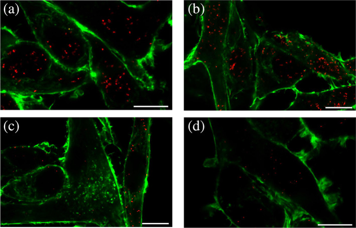
The human flavoenzyme D-aspartate oxidase (hDASPO) controls the level of D-aspartate in the brain, a molecule acting as an agonist of NMDA receptors and modulator of AMPA and mGlu5 receptors. hDASPO-induced D-aspartate degradation prevents age-dependent deterioration of brain functions and is related to psychiatric disorders such as schizophrenia and autism. Notwithstanding this crucial role, less is known about hDASPO regulation. Here, we report that hDASPO is nitrosylated in vitro, while no evidence of sulfhydration and phosphorylation is apparent: nitrosylation affects the activity of the human flavoenzyme to a limited extent. Furthermore, hDASPO interacts with the primate-specific protein pLG72 (a well-known negative chaperone of D-amino acid oxidase, the enzyme deputed to D-serine degradation in the human brain), yielding a ~114 kDa complex, with a micromolar dissociation constant, promoting the flavoenzyme inactivation. At the cellular level, pLG72 and hDASPO generate a cytosolic complex: the expression of pLG72 negatively affects the hDASPO level by reducing its half-life. We propose that pLG72 binding may represent a protective mechanism aimed at avoiding cytotoxicity due to H2 O2 produced by the hDASPO enzymatic degradation of D-aspartate, especially before the final targeting to peroxisomes.

Keywords: D-aspartate; flavooxidase; neurotransmission; pLG72; post-translational modification; protein-protein interaction.


Conflict of interest statement

The authors declare no conflict of interest.

Figures

FIGURE 1
FIGURE 1
In silico analysis of cysteine and tryptophan residues in hDASPO. (a) Position, exposure to the solvent and amino acidic environment of cysteine residues in hDASPO. (Top) Solvent accessible surface as calculated for the hDASPO monomer (pdb code 6RKF). Cysteine residues are represented as spheres (carbon atoms in cyan, sulfur atoms in yellow); the FAD cofactor is represented as sticks (yellow). The backbone is shown as a cartoon. Protein surface is colored by surface proximity with cysteine residues (cyan = cysteine residues located within 5 Å from the surface; orange = cysteine residues located at a distance higher than 15 Å from the surface). Solvent accessible surface has been calculated based on a solvent radius of 1.4 Å. (Bottom) Analysis of the environment of selected cysteine residues, represented as spheres (carbon atoms in cyan, sulfur atoms in yellow, nitrogen atoms in blue, and oxygen atoms in red): the surrounding residues are depicted as sticks and the van der Waals surface is shown. Hydrophobic residues are colored in orange, while charged residues are colored by element (carbon atoms in magenta, nitrogen atoms in blue, and oxygen atoms in red). (b) Frequency of sequence conservation among mammalian DASPOs. Weblogo representation of conserved residues identified by the alignment of selected regions of DASPO sequences from Homo sapiens, Mus musculus, Rattus norvegicus, Sus scrofa, Bos taurus, Cavia porcellus, Macaca fascicularis, and Pongo abelii. The x‐axis identifies the amino acid positions (the annotated numbering refers to the human enzyme) and the height of symbols is proportional to the degree of conservation of each single residue; cysteine residues are reported in red. Figure prepared using WebLogo (https://weblogo.berkeley.edu/logo.cgi). (c) Tryptophan residues in hDASPO: solvent‐exposed tryptophans (showing an exposed surface >20 Å2) were considered surface residues (dark brown). FAD cofactor is represented as yellow sticks. FAD, flavin adenine dinucleotide.
FIGURE 2
FIGURE 2
Analysis of in vitro S‐nitrosylation and S‐sulfhydration of hDASPO holoenzyme and apoprotein forms. Non‐reducing SDS‐PAGE analysis of recombinant hDASPO (7.5 or 15 μg) following in vitro S‐nitrosylation (panel a) or S‐sulfhydration (panel b). Top: fluorescence switch assay (image acquisition was performed upon excitation of the fluorescent probe). Bottom: Coomassie blue proteins staining. Mixtures in which the NO donor GSNO was replaced with GSH, or the sulfide donor NaHS was omitted were analyzed as negative controls; whereas positive controls (CTRL) were protein samples in which all cysteine residues were labeled by Alexa Fluor 350 C5 Maleimide (by omitting the starting blocking step during the fluorescence switch assay). GSH, reduced glutathione; GSNO, S‐nitrosoglutathione.
FIGURE 3
FIGURE 3
Effect of in vitro nitrosylation on hDASPO structural and functional properties. (a) Comparison of far‐UV CD spectra of holo‐ and apoprotein forms of hDASPO following nitrosylation (blue and green lines, respectively) with respect to controls (black and brown lines, respectively). (b) Titration of hDASPO apoprotein reacted with DTNB or GSNO with increasing amounts of FAD (the arrow indicates the spectral changes recorded at increasing cofactor concentration). (c) Analysis of FAD binding to hDASPO apoprotein following the reaction with DTNB or GSNO (as reported in panel b) assessed as quenching of protein fluorescence at ~340 nm. Values are expressed as a percentage of the total change. Blue: control mixture in the absence of DTNB or GSNO; red: mixture containing 100 μM DTNB or 500 μM GSNO. Insets show a closer view in the 0–1 μM FAD concentration range. (d) Time course of the residual activity of hDASPO in the presence (red) or absence (blue) of 500 μM GSNO at 25°C. Inset: relative activity of hDASPO after 1 hour of incubation at different GSNO concentrations (0–1 mM range). Data are the mean ± SD (n = 5) and are expressed relatively to the activity recorded before the incubation (=100%). DTNB, 5,5′‐dithiobis(2‐nitrobenzoic acid); GSNO, S‐nitrosoglutathione.
FIGURE 4
FIGURE 4
Effect of in vitro sulfhydration on hDASPO structural and functional properties. (a) Far‐UV CD spectra of holo‐ and apoprotein forms of hDASPO in the presence of 5 mM NaHS (blue and green lines, respectively) compared to controls (black and brown lines, respectively). (b, c) Titration of 5 μM hDASPO apoprotein with increasing amounts of FAD after sulfhydration by 5 mM NaHS: (b) spectral changes (the arrow indicates the changes recorded at increasing cofactor concentration); (c) fluorescence intensity values at 342 nm expressed as percentage of the total change. Blue: control mixture without the sulfide donor; red: mixture containing 5 mM NaHS. Inset shows a closer view in the 0–1 μM FAD concentration range. (d) Effect of sulfhydration on hDASPO residual activity at 25 °C. Blue: control; red: mixture containing 5 mM NaHS. Inset: relative activity of hDASPO after 1 h of incubation at different concentrations of NaHS. Data are the mean ± SD (n = 3–6) and are expressed relative to the activity measured without NaHS (=100%).
FIGURE 5
FIGURE 5
Effect on hDASPO properties by binding of pLG72 variants. (a, b) SEC analysis of hDASPO binding to R30 (a) and R30K (b) pLG72 variants. Protein mixtures containing 25 nmol of hDASPO and different amounts of pLG72 variants corresponding to 0.5–3 molar ratios were incubated at 4 °C for 10 min and centrifuged before column injection. Dotted curves: 25 nmol hDASPO; dashed curves: 25 nmol pLG72. The recorded elution profiles showed multiple peaks corresponding to: hDASPO (at ~15.5 mL), pLG72 (at ~8.0 and ~14.2 mL), and hDASPO–pLG72 protein complex (at 13.0 ± 0.2 mL). Insets: effect of hDASPO:pLG72 molar ratio on the area of the 13 mL peak (~114 kDa), corresponding to the hDASPO complexes with the R30 and R30K pLG72 variants, and indicated by the arrowheads in the chromatograms. (c) SDS‐PAGE analysis of the SEC fractions. SM: sample mixtures containing hDASPO (25 nmoles) and R30 or R30K  pLG72 variants (50 nmoles). Bands corresponding to hDASPO (15 μg) and pLG72 variants (7 μg) were detected at 39 and 18 kDa, respectively. About 30 μL of the indicated SEC fractions (a–c) was also analyzed. (d) Effect on hDASPO activity of pLG72 variants. Sample mixtures were prepared by adding a fixed amount of recombinant hDASPO (0.4 nmol) with increasing amounts of R30 (black bars) or R30K (gray bars) pLG72 variants and incubated for 30 min at 25 °C. hDASPO residual activity was reported as percentage with respect to the initial value. Data are reported as mean ± SE (n = 3).
FIGURE 6
FIGURE 6
Investigation of hDASPO–pLG72 interaction at the cellular level. (a–c) U87 cells expressing pLG72‐ECFP were transiently transfected with a plasmid encoding hDASPO and were fixed at 24 (a), 48 (b), and 72 h (c) after transfection. The hDASPO–pLG72 interaction was detected via the Duolink PLA technique using mouse anti‐FLAG and rabbit anti‐pLG72 antibodies. (d) Negative control was prepared by performing the assay on non‐transfected U87 cells. Red: proximity spots indicating protein interaction. Green: cytoskeleton stained with Phalloidin CruzFluor™ 488. Scale bar = 10 μm.

References

    1. Abe K, Kimura H. The possible role of hydrogen sulfide as an endogenous neuromodulator. J Neurosci. 1996;16:1066–1071. - PMC - PubMed
    1. Cappelletti P, Campomenosi P, Pollegioni L, Sacchi S. The degradation (by distinct pathways) of human D‐amino acid oxidase and its interacting partner pLG72‐two key proteins in D‐serine catabolism in the brain. FEBS J. 2014;281:708–723. - PubMed
    1. Chumakov I, Blumenfeld M, Guerassimenko O, Cavarec L, Palicio M, Abderrahim H, et al. Genetic and physiological data implicating the new human gene G72 and the gene for D‐amino acid oxidase in schizophrenia. Proc Natl Acad Sci U S A. 2002;99:13675–13680. - PMC - PubMed
    1. Cristino L, Luongo L, Squillace M, Paolone G, Mango D, Piccinin S, et al. D‐Aspartate oxidase influences glutamatergic system homeostasis in mammalian brain. Neurobiol Aging. 2015;36:1890–1902. - PubMed
    1. Errico F, D'Argenio V, Sforazzini F, Iasevoli F, Squillace M, Guerri G, et al. A role for D‐aspartate oxidase in schizophrenia and in schizophrenia‐related symptoms induced by phencyclidine in mice. Transl Psychiatry. 2015;5:e512. - PMC - PubMed

Publication types