

Improving risk stratification of indeterminate adnexal masses on MRI: What imaging features help predict malignancy in O-RADS MRI 4 lesions?
- PMID: 37806193
- PMCID: PMC11186047
- DOI: 10.1016/j.ejrad.2023.111122
Improving risk stratification of indeterminate adnexal masses on MRI: What imaging features help predict malignancy in O-RADS MRI 4 lesions?
Abstract
Purpose: Ovarian-Adnexal Reporting and Data System (O-RADS) MRI uses a 5-point scale to establish malignancy risk in sonographically-indeterminate adnexal masses. The management of O-RADS MRI score 4 lesions is challenging, as the prevalence of malignancy is widely variable (5-90%). We assessed imaging features that may sub-stratify O-RADS MRI 4 lesions into malignant and benign subgroups.
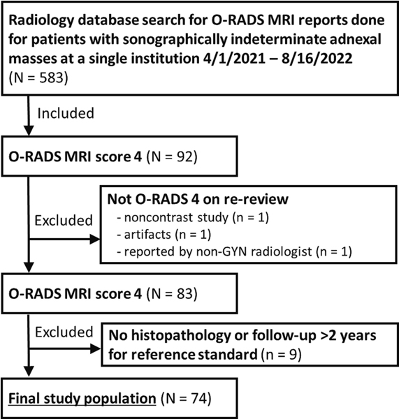
Method: Retrospective single-institution study of women with O-RADS MRI score of 4 adnexal masses between April 2021-August 2022. Imaging findings were assessed independently by 2 radiologists according to the O-RADS lexicon white paper. MRI and clinical findingswere compared between malignant and benign adnexal masses, and inter-reader agreement was calculated.
Results: Seventy-four women (median age 52 years, IQR 36-61) were included. On pathology, 41 (55.4%) adnexal masses were malignant. Patients with malignant masses were younger (p = 0.02) with higher CA-125 levels (p = 0.03). Size of solid tissue was greater in malignant masses (p = 0.01-0.04). Papillary projections and larger solid portion were more common in malignant lesions; irregular septations and predominantly solid composition were more frequent in benign lesions (p < 0.01). Solid tissue of malignant lesions was more often hyperintense on T2-weighted and diffusion-weighted imaging (p ≤ 0.03). Other imaging findings were not significantly different (p = 0.09-0.77). Inter-reader agreement was excellent-good for most features (ICC = 0. 662-0.950; k = 0. 650-0.860).
Conclusion: Various MRI and clinical features differed between malignant and benign O-RADS MRI score 4 adnexal masses. O-RADS MRI 4 lesions may be sub-stratified (high vs low risk) based on solid tissue characteristics and CA-125 levels.
Keywords: Adnexal mass; MRI; Malignancy; O-RADS; Ovary.
Copyright © 2023 Elsevier B.V. All rights reserved.
Conflict of interest statement
Declaration of Competing Interest The authors declare the following financial interests/personal relationships which may be considered as potential competing interests: This research was funded at the institutional level in part through the NIH/NCI Cancer Center Support Grant P30 CA008748. Sungmin Woo was supported by the National Academy of Medicine of the National Academy of Sciences under award number 20000013477 for this project. The content is solely the responsibility of the authors and does not necessarily represent the official views of the National Academy of Medicine or the National Academy of Sciences. The other authors do not have anything to disclose. None of the above entities had any role in study design; in the collection, analysis and interpretation of data; in the writing of the report; and in the decision to submit the article for publication.
Figures
Similar articles
-
Value of MRI-Based Ovarian-Adnexal Reporting and Data System for the Diagnosis of Adnexal Masses.Zhongguo Yi Xue Ke Xue Yuan Xue Bao. 2024 Dec;46(6):909-917. doi: 10.3881/j.issn.1000-503X.16176. Zhongguo Yi Xue Ke Xue Yuan Xue Bao. 2024. PMID: 39773510
-
Added Value of Quantitative Analysis of Diffusion-Weighted Imaging in Ovarian-Adnexal Reporting and Data System Magnetic Resonance Imaging.J Magn Reson Imaging. 2022 Jul;56(1):158-170. doi: 10.1002/jmri.28003. Epub 2021 Nov 19. J Magn Reson Imaging. 2022. PMID: 34797013
-
Does Combing O-RADS US and CA-125 Improve Diagnostic Accuracy in Assessing Adnexal Malignancy Risk in Women With Different Menopausal Status?J Ultrasound Med. 2023 Feb;42(3):675-685. doi: 10.1002/jum.16065. Epub 2022 Jul 26. J Ultrasound Med. 2023. PMID: 35880406
-
O-RADS MRI: A Systematic Review and Meta-Analysis of Diagnostic Performance and Category-wise Malignancy Rates.Radiology. 2023 Apr;307(1):e220795. doi: 10.1148/radiol.220795. Epub 2022 Nov 22. Radiology. 2023. PMID: 36413127
-
O-RADS MRI SCORE: An Essential First-Step Tool for the Characterization of Adnexal Masses.J Magn Reson Imaging. 2024 Mar;59(3):720-736. doi: 10.1002/jmri.28947. Epub 2023 Aug 7. J Magn Reson Imaging. 2024. PMID: 37550825 Review.
Cited by
-
Node-RADS: a systematic review and meta-analysis of diagnostic performance, category-wise malignancy rates, and inter-observer reliability.Eur Radiol. 2025 May;35(5):2723-2735. doi: 10.1007/s00330-024-11160-1. Epub 2024 Nov 6. Eur Radiol. 2025. PMID: 39505734 Free PMC article.
-
Developing a deep learning model for predicting ovarian cancer in Ovarian-Adnexal Reporting and Data System Ultrasound (O-RADS US) Category 4 lesions: A multicenter study.J Cancer Res Clin Oncol. 2024 Jul 9;150(7):346. doi: 10.1007/s00432-024-05872-6. J Cancer Res Clin Oncol. 2024. PMID: 38981916 Free PMC article.
-
Efficacy of a combination of O-RADS, CEUS, and CA125, in identification of ovary-adnexal malignant lesions.Am J Cancer Res. 2025 Feb 15;15(2):631-642. doi: 10.62347/JHJJ1149. eCollection 2025. Am J Cancer Res. 2025. PMID: 40084369 Free PMC article.
-
Diagnostic performance of a modified O-RADS classification system for adnexal lesions incorporating clinical features.Abdom Radiol (NY). 2025 Feb;50(2):953-965. doi: 10.1007/s00261-024-04538-8. Epub 2024 Aug 20. Abdom Radiol (NY). 2025. PMID: 39164457
-
Navigating Ovarian-Adnexal Reporting and Data System Magnetic Resonance Imaging (O-RADS MRI): A Review of Its Evolution, Current Advances, and Persistent Challenges in Ovarian Imaging.Cureus. 2025 Jun 25;17(6):e86717. doi: 10.7759/cureus.86717. eCollection 2025 Jun. Cureus. 2025. PMID: 40718198 Free PMC article. Review.
References
-
- Froyman W, Landolfo C, De Cock B, Wynants L, Sladkevicius P, Testa AC, et al. Risk of complications in patients with conservatively managed ovarian tumours (IOTA5): a 2-year interim analysis of a multicentre, prospective, cohort study. Lancet Oncol. 2019. Mar;20(3):448–58. - PubMed
-
- ACR-O-RADS-MRI-Lexicon. https://www.acr.org/Clinical-Resources/Reporting-and-Data-Systems/O-Rads.... Accessed June 14, 2023.
-
- Thomassin-Naggara I, Poncelet E, Jalaguier-Coudray A, Guerra A, Fournier LS, Stojanovic S, et al. Ovarian-Adnexal Reporting Data System Magnetic Resonance Imaging (O-RADS MRI) Score for Risk Stratification of Sonographically Indeterminate Adnexal Masses. JAMA Netw Open. 2020. Jan 24;3(1):e1919896. - PMC - PubMed
-
- Rizzo S, Cozzi A, Dolciami M, Del Grande F, Scarano AL, Papadia A, et al. O-RADS MRI: A Systematic Review and Meta-Analysis of Diagnostic Performance and Category-wise Malignancy Rates. Radiology. 2023. Apr;307(1):e220795. - PubMed
MeSH terms
Substances
Grants and funding
LinkOut - more resources
Full Text Sources
Medical
Research Materials
Miscellaneous
