Review
. 2023 Dec:78:101817.
doi: 10.1016/j.molmet.2023.101817. Epub 2023 Oct 6.

Sensory spinal interoceptive pathways and energy balance regulation

Affiliations
Review

Sensory spinal interoceptive pathways and energy balance regulation

Heike Münzberg et al. Mol Metab. 2023 Dec.

Abstract

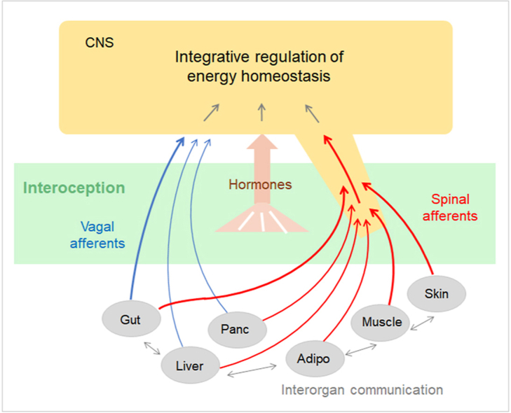
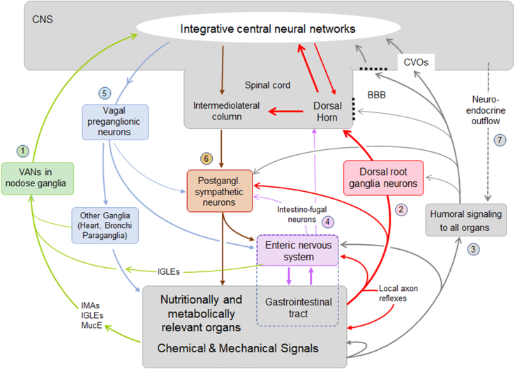
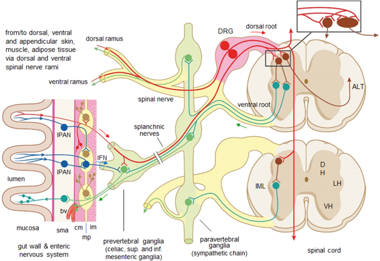
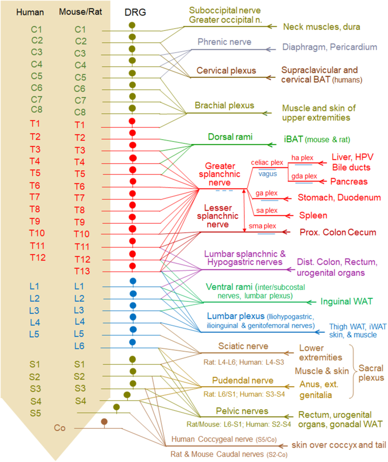
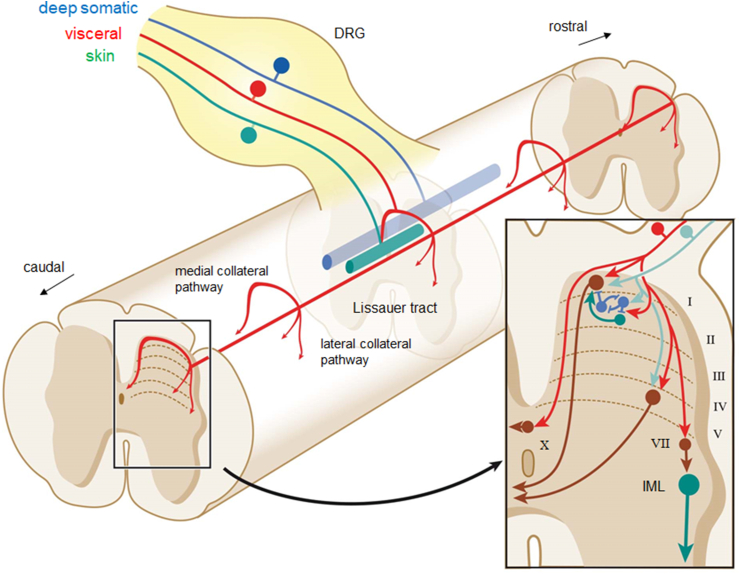
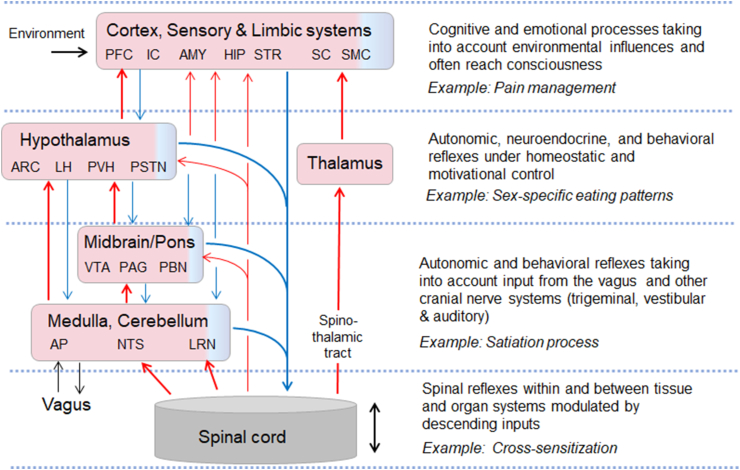
Interoception plays an important role in homeostatic regulation of energy intake and metabolism. Major interoceptive pathways include gut-to-brain and adipose tissue-to brain signaling via vagal sensory nerves and hormones, such as leptin. However, signaling via spinal sensory neurons is rapidly emerging as an additional important signaling pathway. Here we provide an in-depth review of the known anatomy and functions of spinal sensory pathways and discuss potential mechanisms relevant for energy balance homeostasis in health and disease. Because sensory innervation by dorsal root ganglia (DRG) neurons goes far beyond vagally innervated viscera and includes adipose tissue, skeletal muscle, and skin, it is in a position to provide much more complete metabolic information to the brain. Molecular and anatomical identification of function specific DRG neurons will be important steps in designing pharmacological and neuromodulation approaches to affect energy balance regulation in disease states such as obesity, diabetes, and cancer.

Keywords: Diabetes; Energy expenditure; Food intake; Gut-brain communication; Interoception; Interorgan communication; Obesity; Sensory nerves; Spinal cord.


Conflict of interest statement

Declaration of competing interest The authors declare the following financial interests/personal relationships which may be considered as potential competing interests: Heike Muenzberg reports financial support was provided by National Institute of Health. Hans-Rudolf Berthoud reports financial support was provided by National Institute of Health.

Figures

Image 1
Graphical abstract
Figure 1
Figure 1
Schematic diagram showing signaling pathways between the brain and peripheral organs potentially contributing to the regulation of energy balance. Communication in both directions is accomplished by three major pathways, vagal, spinal, and humoral. Communication from specific peripheral organs/tissues such as the gut and other associated visceral organs (but not cutaneous and subcutaneous tissues such as subcutaneous fat) to the brain, is accomplished by vagal afferent neurons (VANs, 1). All peripheral organs/tissues communicate with the brain through dorsal root ganglia neurons and the spinal cord (2), and through the general blood circulation (3). Signals generated in DRGs as well as humoral signals can also communicate with the enteric nervous system, postganglionic neurons, and the spinal cord via short loops. In addition to DRGs, the gut can also communicate to the spinal cord and postganglionic sympathetic neurons via intestinofugal neurons in the enteric nervous system (4). Communication from the brain to the periphery is accomplished by vagal and lumbo-sacral parasympathetic (not shown) efferents (5), and by sympathetic nervous system (6) and neuroendocrine (7) outflow. Note that specific signals generated in the periphery can be mediated by multiple and mixed pathways to the brain and other peripheral organs. Abbreviations: BBB, blood–brain barrier; CVOs, circumventricular organs with missing blood brain barrier; IGLEs. Intraganglionic laminar vagal afferent endings; IMA, intramuscular array vagal afferent endings; MucE, mucosal vagal afferent endings.
Figure 2
Figure 2
Relationship between dorsal root afferents, sympathetic outflow, and the enteric nervous system. Dorsal root afferent neurons (red) pick up information in the gut, skin, muscle, and adipose tissue and conduct it to the dorsal horn (DH) of the spinal cord, where they synapse on spinal large projection neurons and small interneurons (brown) and project via collaterals rostro-caudally over several segments. Projection neurons ascend through anterolateral tracts (ALT) mainly contralaterally to supraspinal targets. Sensory information carried by DRG neurons can also be passed to enteric motor and inter neurons (light brown) in the myenteric plexus, to sympathetic postganglionic neurons in prevertebral ganglia (green) for short-loop reflex actions and possibly also to smooth muscle via axon reflexes. In addition to DRGs, the gut can also communicate to postganglionic sympathetic neurons and spinal cord (not shown) via intrinsic primary afferent neurons (IPANs, blue) and intestinofugal neurons (IF, blue) in the enteric nervous system. These sensory pathways are partly overlapping with the sympathetic nervous outflow system (green). Other abbreviations: bv, blood vessel; DRG, dorsal root ganglion; IML, intermediolateral column; cm, circular muscle; lm, longitudinal muscle mp, myenteric plexus; sma, submucosa; LH and VH, lateral and ventral horn, respectively.
Figure 3
Figure 3
Innervation territories of DRG at different spinal levels with emphasis on nutritional and metabolic relevance. The 32 DRGs on each side, grouped by their location relative to the cervical, thoracic, lumbar, and sacral spinal cord and numbered from rostral to caudal in each group, project through 14 major nerves/plexuses to their specific visceral and somatic destinations in an overlapping fashion. Peripheral axons of DRGs from up to 12 segments (greater splanchnic nerve) converge in each nerve that can innervate large tissue territories. As shown in Figure 2, the DRG axons are typically intermingled with preganglionic sympathetic neurons in most of these nerves, with intestinofugal neuron projections in the splanchnic nerves and subsidiaries including the celiac plexus (ce plex), superior mesenteric plexus (sma plex), hepatic artery plexus (ha plex), gastric artery plexus (ga plex), gastroduodenal artery plexus (gda plex), and splenic artery plexus (sa plex), as well as the hypogastric, and pelvic nerves, and with vagal efferent and afferent fibers in parts of the splanchnic nerve and all subsidiaries. Furthermore, DRGs innervating subcutaneous white and brown adipose tissues are comingled with DRGs innervating overlaying muscle and skin. Note that innervation of skin and skeletal muscle for much of the thorax and abdomen is not shown.
Figure 4
Figure 4
Dorsal horn circuitry of DRG sensory input. Upon entry into the spinal cord, DRG axons split into ascending and descending branches coursing in Lissauer's tract (LT) issuing collaterals to synapse on neurons at multiple segmental levels of the dorsal horn. DRG with visceral input (red) spread across the largest rostro-caudal extent with up to 10 segments giving off sparse wide spaced collaterals, DRG with input from skin (green) spread the least terminating as dense clusters, while afferents from deep somatic structures (muscles, joint capsules, fasciae; blue) spread less densely over an intermediate range. At a given level, DRG axon collaterals with input from viscera, skin, and deep somatic structures tend to innervate neurons in different lamina of the dorsal horn (inset). Viscera-related DRG axons preferentially terminate in laminae I and outer II, with fewer terminations in lamina V and X. The preganglionic intermediolateral nucleus (IML) may be reached via interneurons in lamina VII (brown). In laminae I, II and V, visceral DRG axons (red) can converge on single projection neurons (brown), on inhibitory (blue) and excitatory (green) interneurons, or on both. Many projection neurons and interneurons receive at the same time DRG neuron inputs from skin and deep somatic structures (turquoise). Abbreviations: MCP, LCP, medial and lateral, respectively, collateral pathways.
Figure 5
Figure 5
Highly schematic diagram of ascending and descending neural pathways from the spinal cord to the brain and potential integrative hierarchy in the brain most relevant for energy homeostasis regulation. Among the ascending connections (red), the spinothalamic tract, which underlies touch and pain sensation, is by far the best investigated pathway. Ascending pathways most relevant for energy balance regulation are indirect connections via medulla, pons, or hypothalamus to the forebrain. Similarly, descending connections (blue) arise from all major areas of the brain, some of them direct and some indirect. See text for details.

Similar articles

Cited by

References

    1. Bray G.A., Kim K.K., Wilding J.P.H., World Obesity F. Obesity: a chronic relapsing progressive disease process. A position statement of the World Obesity Federation. Obes Rev. 2017;18:715–723. - PubMed
    1. Pillon N.J., Loos R.J.F., Marshall S.M., Zierath J.R. Metabolic consequences of obesity and type 2 diabetes: balancing genes and environment for personalized care. Cell. 2021;184:1530–1544. - PMC - PubMed
    1. Qasim A., Turcotte M., de Souza R.J., Samaan M.C., Champredon D., Dushoff J., et al. On the origin of obesity: identifying the biological, environmental and cultural drivers of genetic risk among human populations. Obes Rev. 2018;19:121–149. - PubMed
    1. Dulloo A.G. Physiology of weight regain: lessons from the classic Minnesota Starvation Experiment on human body composition regulation. Obes Rev. 2021;22(Suppl 2) - PubMed
    1. El Ansari W., Elhag W. Weight regain and insufficient weight loss after bariatric surgery: definitions, prevalence, mechanisms, predictors, prevention and management strategies, and knowledge gaps-a scoping review. Obes Surg. 2021;31:1755–1766. - PMC - PubMed