. 2023 Nov:499:153640.
doi: 10.1016/j.tox.2023.153640. Epub 2023 Oct 6.

Preconception and developmental DEHP exposure alter liver metabolism in a sex-dependent manner in adult mouse offspring

Affiliations

Preconception and developmental DEHP exposure alter liver metabolism in a sex-dependent manner in adult mouse offspring

Nicole Robles-Matos et al. Toxicology. 2023 Nov.

Abstract

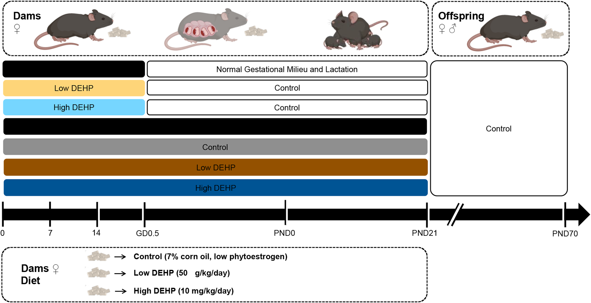
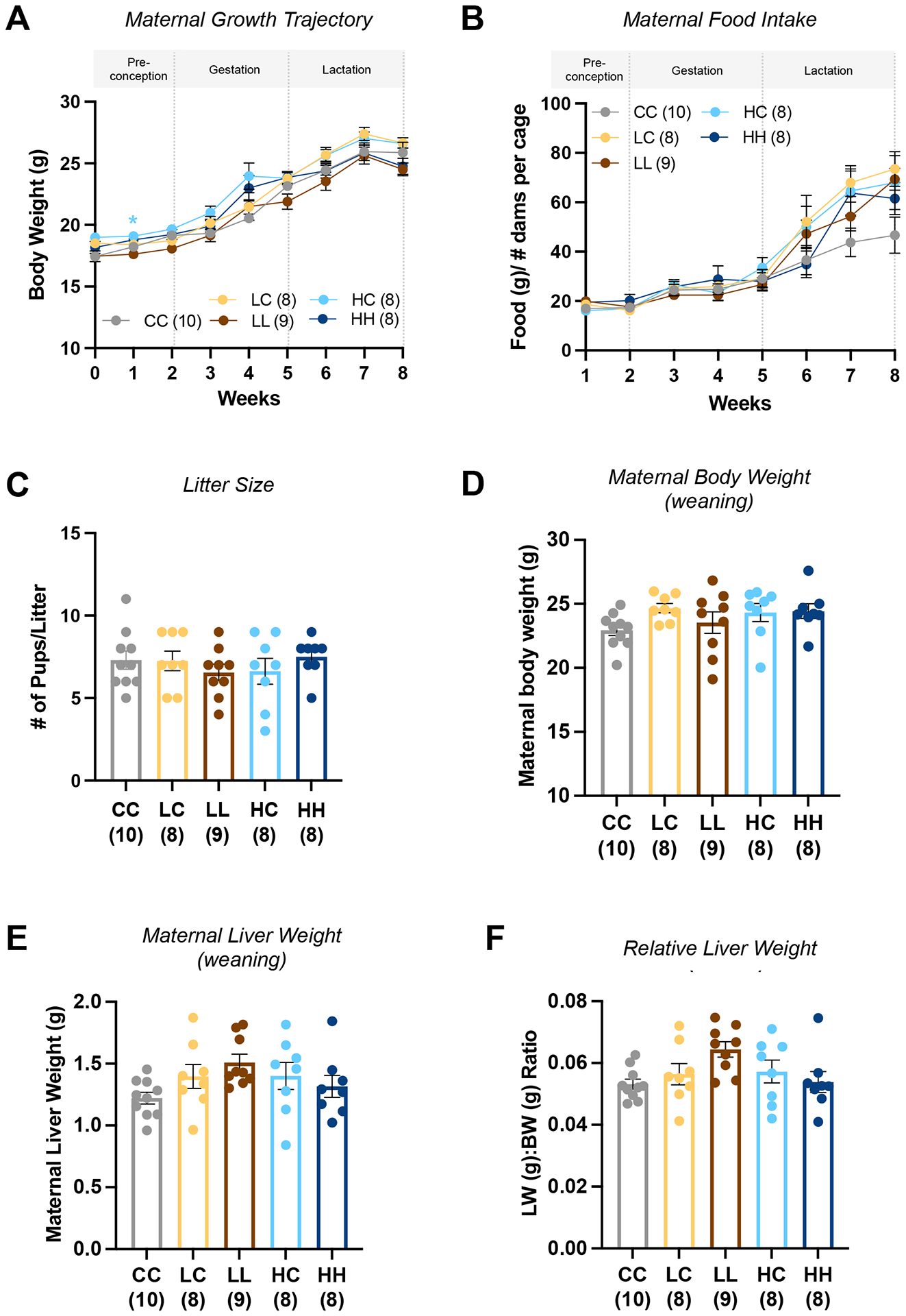
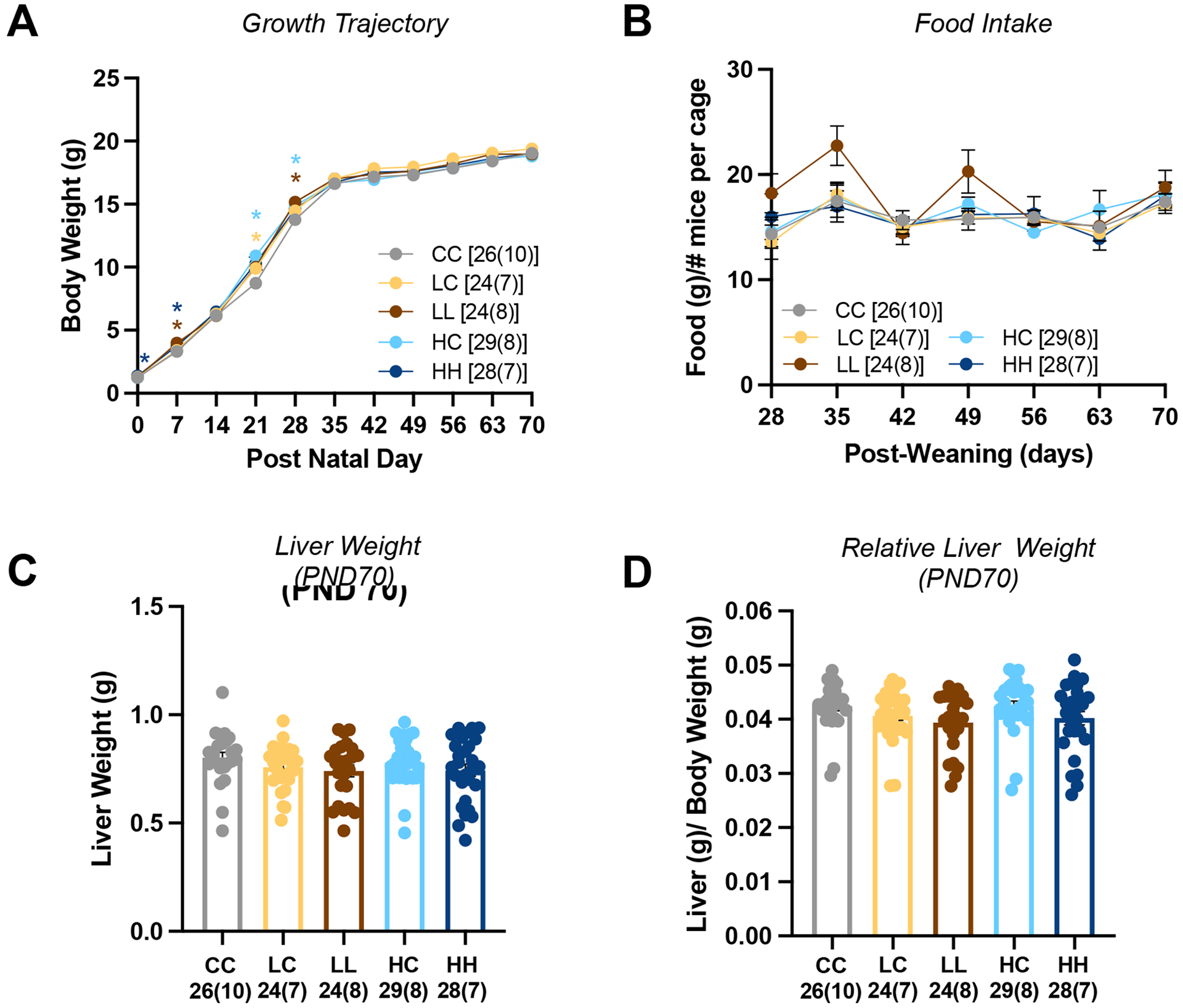
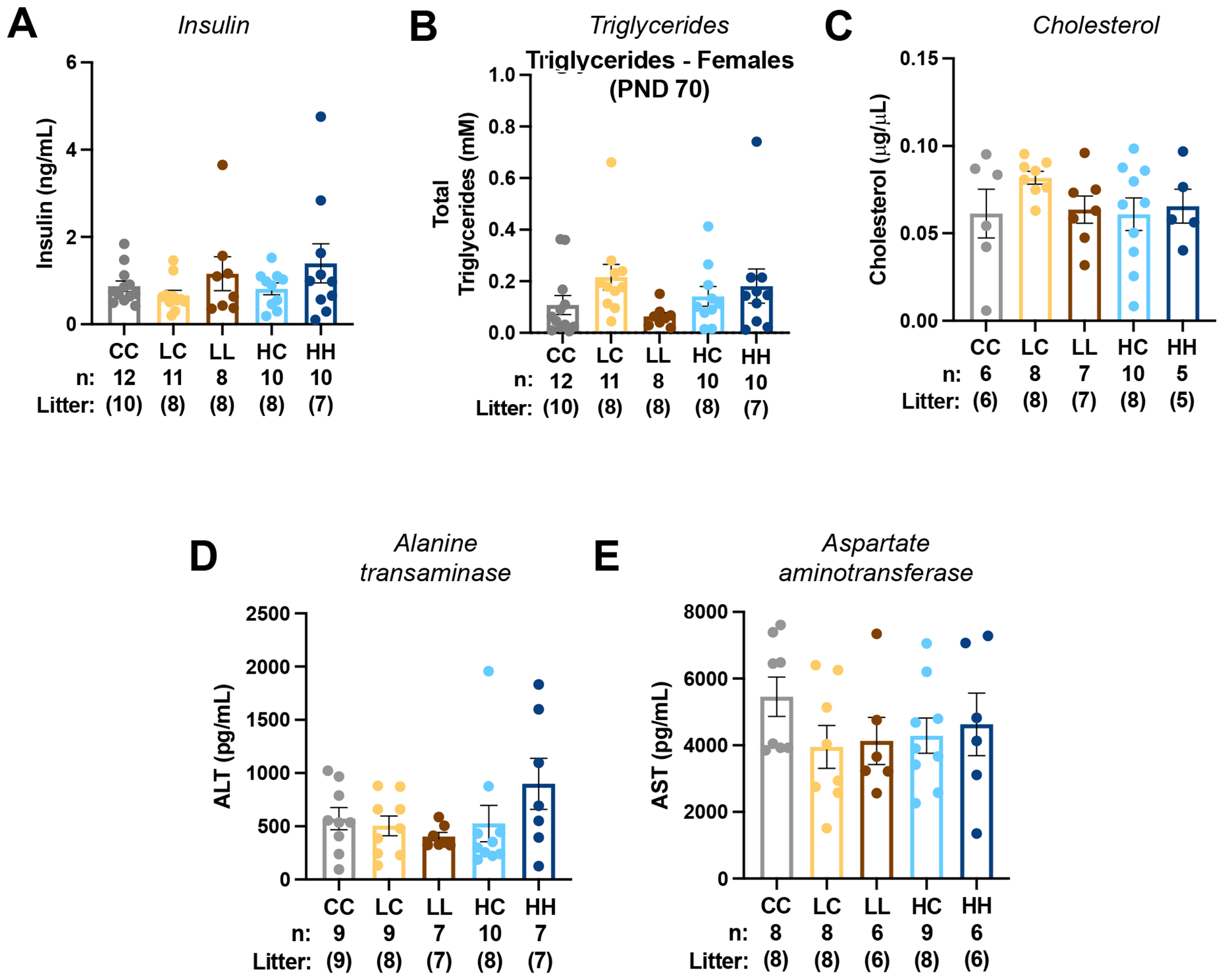
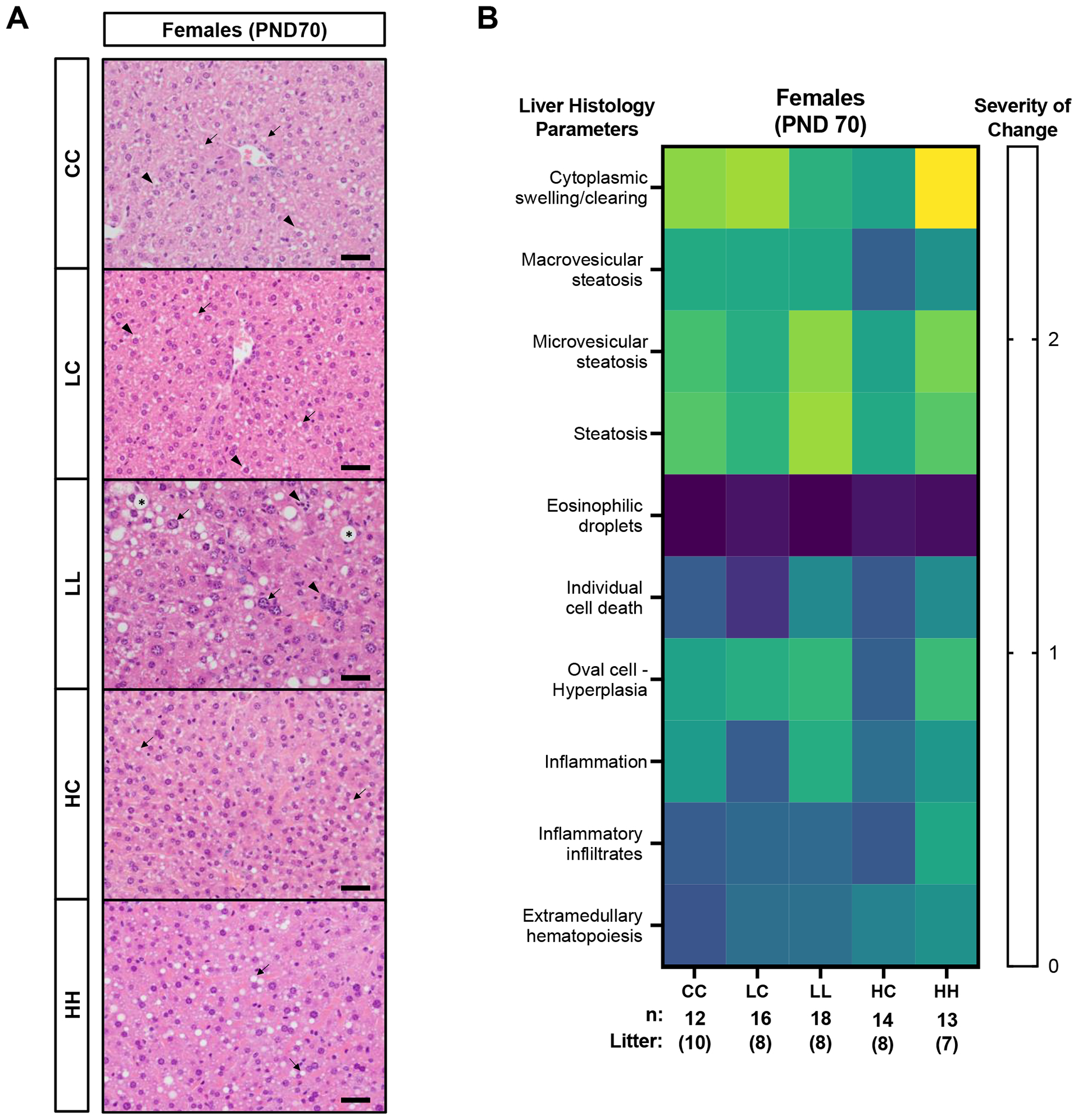
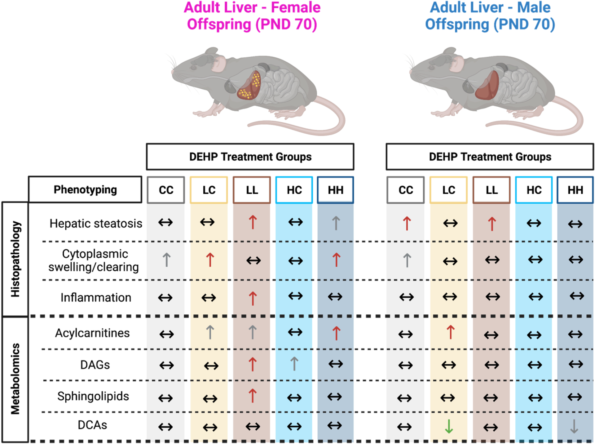
Environmental exposure to endocrine disrupting chemicals (EDCs) during critical periods of development is associated with an increased risk of metabolic diseases, including hepatic steatosis and obesity. Di-2-ethylhexyl-phthalate (DEHP) is an EDC strongly associated with these metabolic abnormalities. DEHP developmental windows of susceptibility are unknown yet have important public health implications. The purpose of this study was to identify these windows of susceptibility and determine whether developmental DEHP exposure alters hepatic metabolism later in life. Dams were exposed to control or feed containing human exposure relevant doses of DEHP (50 μg/kg BW/d) and high dose DEHP (10 mg/kg BW/d) from preconception until weaning or only exposed to DEHP during preconception. Post-weaning, all offspring were fed a control diet throughout adulthood. Using the Metabolon Untargeted Metabolomics platform, we identified 148 significant metabolites in female adult livers that were altered by preconception-gestation-lactation DEHP exposure. We found a significant increase in the levels of acylcarnitines, diacylglycerols, sphingolipids, glutathione, purines, and pyrimidines in DEHP-exposed female livers compared to controls. These changes in fatty acid oxidation and oxidative stress-related metabolites were correlated with hepatic changes including microvesicular steatosis, hepatocyte swelling, inflammation. In contrast to females, we observed fewer metabolic alterations in male offspring, which were uniquely found in preconception-only low dose DEHP exposure group. Although we found that preconception-gestational-lactation exposure causes the most liver pathology, we surprisingly found preconception exposure linked to an abnormal liver metabolome. We also found that two doses exhibited non-monotonic DEHP-induced changes in the liver. Collectively, these findings suggest that metabolic changes in the adult liver of offspring exposed periconceptionally to DHEP depends on the timing of exposure, dose, and sex.

Keywords: Developmental exposure; Di-2-ethylhexyl phthalate; Endocrine disrupting chemicals; Hepatic steatosis; Liver; Metabolomics.


Conflict of interest statement

Declaration of Competing Interest The authors declare that they have no known competing financial interests or personal relationships that could have appeared to influence the work reported in this paper.

Figures

Figure 1.
Figure 1.. Maternal DEHP exposure paradigm.
B6 female mice (designated dams) were assigned to a low phytoestrogen control diet with or without the addition of DEHP via oral administration (feed). Dams in the window of exposure 1 were exposed to control or DEHP-containing feed starting 2 weeks prior to conception. After a vaginal plug was identified (GD0.5), dams were placed on a control diet until weaning at PND 21. Dams in the window of exposure 2 were exposed to control or DEHP-containing feed starting 2 weeks prior to conception until PND21. Post-weaning, all the offspring were placed on a control feed diet until liver tissue collection at PND70. From PND0 to PND70, body weight and food intake was recorded. At PND70 liver tissues from male and female offspring were harvested for histopathology, serum biochemistry and global metabolomics analyses.
Figure 2.
Figure 2.. Dose-specific changes in growth for exposed dams.
(A) Maternal growth trajectory weekly from week 0 of exposure until weaning (week 8). (B) Maternal food consumption weekly, amount of food intake per week (g) normalized to number of dams per cage. (C) Litter size at delivery for each DEHP treatment group. (D) Maternal body weight and (E) liver weight at week 8 (weaning). (F) Maternal liver weight normalized by body weight at week 8 when maternal tissues were collected. Each data point corresponds to an individual dam. Number litters are indicated below each treatment group in parenthesis and correspond to animals from three different cohorts. Bars represent mean ± SEM. Treatment groups are represented by the exposure windows (preconception | gestation-lactation), CC: Control-Control, LC: Lower-Control, LL: Lower-Lower, HC: High-Control, HH: High-High. Statistical analysis: One-Way ANOVA Dunnett’s multiple comparison test and ANOVA repeated measures. *P < 0.05.
Figure 3.
Figure 3.. Female offspring growth and food consumption.
(A) Growth trajectory from PND0-PND70. (B) Food consumption was recorded on a weekly basis (post-weaning) and normalized to number of female mice per cage. (C) Liver weight at PND70. (D) Liver weight normalized to body weight at PND70. Each data point corresponds to an individual offspring. Number of pups and litters (in parentheses) is indicated below each treatment group. Number of offspring and number of litters denoted below each treatment group from three cohorts of exposures. The adult animal was used as the unit of measurement. Bars represent mean ± SEM. Treatment groups are represented by exposure windows (preconception | gestation-lactation). Statistical analysis: One-Way ANOVA Dunnett’s multiple comparison test and ANOVA repeated measures. *P < 0.05.
Figure 4.
Figure 4.. Serum biochemistry of female adult offspring exposed to DEHP through maternal diet.
Fasting serum from female offspring at PND70 was collected for analysis of (A) insulin, (B) total triglycerides and (C) total cholesterol. Liver injury markers of (D) ALT and (E) AST were also analyzed. Each data point corresponds to an individual offspring. Number of pups and litters are indicated below each treatment group. Number of offspring and number of litters denoted below each treatment group from three cohorts of exposures. The adult animal was used as the unit of measurement. Bars represent mean ± SEM. Treatment groups are represented by the exposure windows (preconception | gestation-lactation). Statistical analysis: One-Way ANOVA, Dunnett’s multiple comparison test.
Figure 5.
Figure 5.. Dose-specific effects on liver histopathology of adult female offspring exposed to DEHP via maternal diet.
(A) Representative examples of H&E staining of adult female offspring livers at PND70. (B) Heat map of liver histopathology lesions using a severity of change scale (0 = unremarkable, 1 = minimal, 2 = mild, 3 = moderate, and 4 = severe). N = 10–18 livers representative of 7–10 litters as showed in parenthesis. CC, LC: minimal to mild microvesicular fatty changes (small round vacuoles, arrows) combined with modest swelling and clearing (irregular cytoplasmic clearing arrowheads). HC: minimal to mild microvesicular fatty changes (small round vacuoles, arrows). HH: moderate microvesicular fatty changes (small round vacuoles, arrows). LL: marked micro and macrovesicular fatty changes (large round vacuoles, asterisks), multifocal mononuclear inflammatory cells (arrowheads), moderate hepatocellular karyomegaly (arrows). Number of offspring and number of litters denoted below each treatment group from three cohorts of exposures. The adult animal was used as the unit of measurement. Black scale bars represent 35 μm. Statistical analysis: ANOVA Kruskal-Wallis Test.
Figure 6.
Figure 6.. Lipotoxic effects of periconceptional DEHP exposure in liver of adult offspring (PND70).
Using histopathology analysis, we found that adult female livers exposed to low dose DEHP during preconception-gestation-lactation window (Low-Low) exhibited a significant increase in hepatic steatosis and mild inflammation. These findings were correlated with global metabolomics in which adult female livers showed significant increase in DAGs and sphingolipids and a moderate increase in carnitines. Adult female offspring exposed to high dose DEHP during preconception-gestation-lactation exhibited significant increase in cellular swelling, moderate increase in hepatic steatosis and significant increase in acylcarnitine metabolites. In females, DEHP groups exposed to DEHP during preconception only were less impacted by DEHP-induced lipotoxicity in the liver. In contrast to females, males exposed to low dose DEHP and control during preconception-gestation-lactation had significant increase in hepatic steatosis. On the other hand, males exposed to low dose DEHP during preconception only, showed a significant increase in carnitines and decrease DCAs. Sex differences were observed in our control groups. Control females had a trend towards increased cytoplasmic swelling while males had significant increase in steatosis and a trend towards increased cytoplasmic swelling which indicates oxidative stress. Sex hormones might be a potential mechanism behind these differences because females in reproductive age are more protected from hepatic steatosis and NAFLD due to the protected role of estrogens. DEHP treatment groups are represented by the window of exposure: CC: Control-Control, LC: Low-Control, LL: Low-Low, HC: High-Control, and HH: High-Control. Compared to controls, (↑) indicates significant increase, (↑) indicates moderate increase, (↓) indicates significant decrease, and (↔) indicates no significant changes observed. Figure created with BioRender.com

Similar articles

Cited by

References

    1. Kavlock RJ, Daston GP, DeRosa C, et al. Research needs for the risk assessment of health and environmental effects of endocrine disruptors: a report of the U.S. EPAsponsored workshop. Environ Health Perspect. 1996;104 Suppl 4(Suppl 4):715–740. doi:10.1289/ehp.96104s4715 - DOI - PMC - PubMed
    1. Endocrine-Disrupting Chemicals (EDCs). Published January 24, 2022. Accessed April 30, 2023. https://www.endocrine.org/patient-engagement/endocrine-library/edcs
    1. Metcalfe CD, Bayen S, Desrosiers M, Muñoz G, Sauvé S, Yargeau V. An introduction to the sources, fate, occurrence and effects of endocrine disrupting chemicals released into the environment. Environ Res. 2022;207:112658. doi:10.1016/j.envres.2021.112658 - DOI - PubMed
    1. Muncke J. Exposure to endocrine disrupting compounds via the food chain: Is packaging a relevant source? Sci Total Environ. 2009;407(16):4549–4559. doi:10.1016/j.scitotenv.2009.05.006 - DOI - PubMed
    1. Braun JM. Early-life exposure to EDCs: role in childhood obesity and neurodevelopment. Nat Rev Endocrinol. 2017;13(3):161–173. doi:10.1038/nrendo.2016.186 - DOI - PMC - PubMed

Substances

LinkOut - more resources