Review
. 2023 Oct 9;4(1):32.
doi: 10.1186/s43556-023-00144-0.

Spatial transcriptomics in development and disease

Affiliations
Review

Spatial transcriptomics in development and disease

Ran Zhou et al. Mol Biomed. .

Abstract

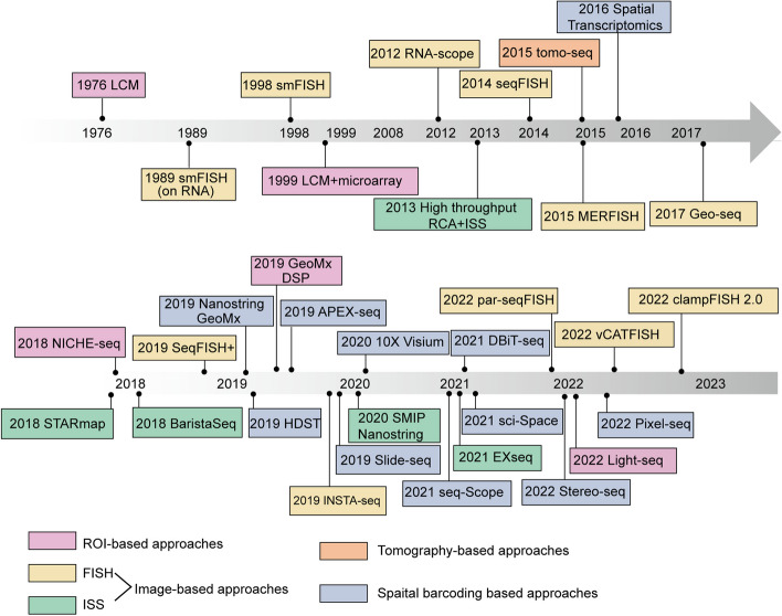
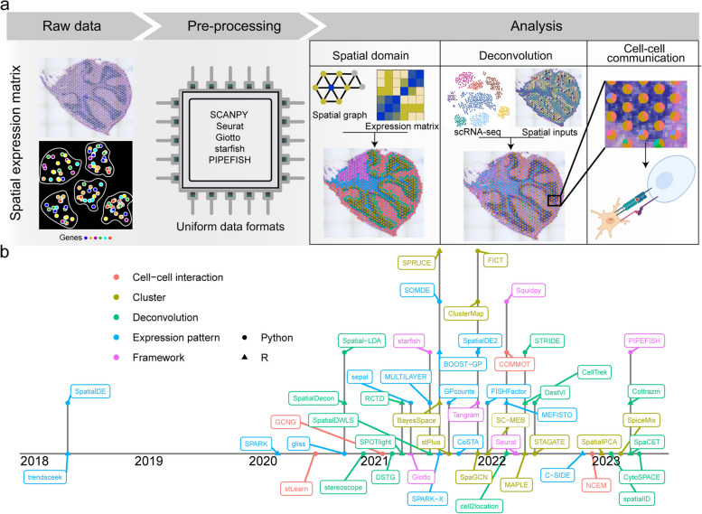
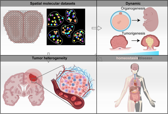
The proper functioning of diverse biological systems depends on the spatial organization of their cells, a critical factor for biological processes like shaping intricate tissue functions and precisely determining cell fate. Nonetheless, conventional bulk or single-cell RNA sequencing methods were incapable of simultaneously capturing both gene expression profiles and the spatial locations of cells. Hence, a multitude of spatially resolved technologies have emerged, offering a novel dimension for investigating regional gene expression, spatial domains, and interactions between cells. Spatial transcriptomics (ST) is a method that maps gene expression in tissue while preserving spatial information. It can reveal cellular heterogeneity, spatial organization and functional interactions in complex biological systems. ST can also complement and integrate with other omics methods to provide a more comprehensive and holistic view of biological systems at multiple levels of resolution. Since the advent of ST, new methods offering higher throughput and resolution have become available, holding significant potential to expedite fresh insights into comprehending biological complexity. Consequently, a rapid increase in associated research has occurred, using these technologies to unravel the spatial complexity during developmental processes or disease conditions. In this review, we summarize the recent advancement of ST in historical, technical, and application contexts. We compare different types of ST methods based on their principles and workflows, and present the bioinformatics tools for analyzing and integrating ST data with other modalities. We also highlight the applications of ST in various domains of biomedical research, especially development and diseases. Finally, we discuss the current limitations and challenges in the field, and propose the future directions of ST.


Conflict of interest statement

The authors declare no conflict of interest.

Figures

Fig. 1
Fig. 1
Timeline of the major published methods mentioned in this review. Every approach is categorized according to its foundational methodology, primarily segmented into the subsequent classifications: ROI-based methods (purple), image-based methods including FISH (yellow) and ISS (green), tomography-based methods (orange), and spatial barcoding-based methods (blue)
Fig. 2
Fig. 2
Overview of technologies for spatial transcriptomics. a Physical dissection or optical marking-based approaches involve the selection or marking of regions of interest. Following ROI marking, samples can be individually collected for RNA-seq or dissociated into single cells for scRNA-seq. b Image-based technologies achieve spatially resolved gene expression through decoding fluorescence signals. In situ sequencing and single molecule fluorescence in situ hybridization detect molecular abundance by directly reading transcript sequences within the tissue or target RNA barcodes, respectively. c Tomography-based methods, such as RNA tomo-seq, utilize a frozen section technique to linearly amplify cDNA from single tissue samples. Three identical biological samples are systematically frozen and sliced in three different directions, allowing for the completion of 2D transcriptional reconstruction through overlapping data. d Spatial barcoding-based methods generate spatial transcriptomes using reverse transcription primers with unique positional barcodes
Fig. 3
Fig. 3
Overview of spatial transcriptomics tasks and analysis tools. a The analysis framework standardizes diverse spatial molecular datasets into a consistent data format, followed by detecting spatial domains, deconvoluting spots, and analyzing cell–cell communication. b Spatial analysis tool timeline showcasing task categories through color coding, with dot shapes representing the language used by each tool
Fig. 4
Fig. 4
Spatial transcriptomics provides new insights into the molecular mechanisms underlying organ and embryonic development and human pathological tissues. a Stereo-seq maps spatiotemporal transcriptome dynamics in developing mouse embryos, detailing tissue-specific identities at different stages. b Using scRNA-seq and ST, the spatiotemporal landscape of the mouse stomach and intestine was established, unveiling distinct cell clusters and their interactions responsible for gastric compartmentalization. c ST provides a detailed transcriptional map of cell types within the developing heart at three stages, pinpointing cell-type-specific gene expression within distinct anatomical regions. d By utilizing coronal brain sections covering the entire anterior–posterior axis, ST generates a comprehensive molecular map of the mouse brain. e Combining various ST techniques using both mouse and human tissue samples unveiled widespread changes in the transcriptome and co-expression networks caused by amyloid plaques in (AD). f Employing ST into tissue repair uncovers molecular compartmentalization and transcriptome alterations in both steady state and mucosal healing. g ST elucidate the dynamic gene expression in the tissue during pathogen infection. h The complex cellular structure and heterogeneity of cutaneous lupus erythematosus has been characterized by integrating scRNA-seq and ST analysis in autoimmune diseases
Fig. 5
Fig. 5
Spatial transcriptomics techniques facilitate the study of tumor microenvironment heterogeneity and tumor heterogeneity. a ST examines the diversity within cancer-associated fibroblasts and immunosuppressive molecules within the microenvironment of breast cancer. b ST enables the identification and comprehensive exploration of unique tumor microenvironment regions, such as the tumor interface and tertiary lymph nodes. c ST analyzes the spatial distribution of PDAC-associated heterogeneity, identifying highly heterogeneous and transitional PDAC subpopulations. d ST applied to breast cancer biopsy identified that tumor cells harboring GATA3 mutations became more invasive, revealing the spatial heterogeneity of breast cancer
Fig. 6
Fig. 6
The future directions of spatial genomics. The ongoing advancement of spatial genomics techniques is driving research in areas such as tissue homeostasis, diseases, tumor and embryo development, and tumor heterogeneity. These advancements hold the potential to offer valuable insights into both biological understanding and clinical applications

References

    1. Palla G, Fischer DS, Regev A, Theis FJ. Spatial components of molecular tissue biology. Nat Biotechnol. 2022;40(3):308–318. doi: 10.1038/s41587-021-01182-1. - DOI - PubMed
    1. Fomitcheva-Khartchenko A, Kashyap A, Geiger T, Kaigala GV. Space in cancer biology: its role and implications. Trends Cancer. 2022;8(12):1019–1032. doi: 10.1016/j.trecan.2022.07.008. - DOI - PubMed
    1. Kulkarni A, Anderson AG, Merullo DP, Konopka G. Beyond bulk: a review of single cell transcriptomics methodologies and applications. Curr Opin Biotechnol. 2019;58:129–136. doi: 10.1016/j.copbio.2019.03.001. - DOI - PMC - PubMed
    1. John HA, Birnstiel ML, Jones KW. RNA-DNA hybrids at the cytological level. Nature. 1969;223(5206):582–587. doi: 10.1038/223582a0. - DOI - PubMed
    1. Moses L, Pachter L. Museum of spatial transcriptomics. Nat Methods. 2022;19(5):534–546. doi: 10.1038/s41592-022-01409-2. - DOI - PubMed

LinkOut - more resources