. 2023 Oct 5:6:0236.
doi: 10.34133/research.0236. eCollection 2023.

Desialylated Platelet Clearance in the Liver is a Novel Mechanism of Systemic Immunosuppression

Affiliations

Desialylated Platelet Clearance in the Liver is a Novel Mechanism of Systemic Immunosuppression

June Li et al. Research (Wash D C). .

Abstract

Platelets are small, versatile blood cells that are critical for hemostasis/thrombosis. Local platelet accumulation is a known contributor to proinflammation in various disease states. However, the anti-inflammatory/immunosuppressive potential of platelets has been poorly explored. Here, we uncovered, unexpectedly, desialylated platelets (dPLTs) down-regulated immune responses against both platelet-associated and -independent antigen challenges. Utilizing multispectral photoacoustic tomography, we tracked dPLT trafficking to gut vasculature and an exclusive Kupffer cell-mediated dPLT clearance in the liver, a process that we identified to be synergistically dependent on platelet glycoprotein Ibα and hepatic Ashwell-Morell receptor. Mechanistically, Kupffer cell clearance of dPLT potentiated a systemic immunosuppressive state with increased anti-inflammatory cytokines and circulating CD4+ regulatory T cells, abolishable by Kupffer cell depletion. Last, in a clinically relevant model of hemophilia A, presensitization with dPLT attenuated anti-factor VIII antibody production after factor VIII ( infusion. As platelet desialylation commonly occurs in daily-aged and activated platelets, these findings open new avenues toward understanding immune homeostasis and potentiate the therapeutic potential of dPLT and engineered dPLT transfusions in controlling autoimmune and alloimmune diseases.


Figures

Fig. 1.
Fig. 1.
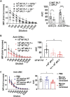
dPLTs induce immunosuppression. (A) Antibody titers in GPIbα−/− and β3−/− mice intravenously immunized with 108 WT platelets (WT PLT) or dPLTs (dPLT). (B) Antibody titers in GPIbα−/− mice cocurrently intravenously immunized with 108 WT platelets with increasing doses of dPLTs. (C) Anti-sRBC antibody titers in WT BALB/c mice preinjected with 2 × 10⁸ WT or dPLTs. Statistical analysis was done with one-way analysis of variance (ANOVA) with Tukey post hoc test. All data represented as means ± SD and quantified as area under the curve (AUC). *P < 0.05 and ****P < 0.001.
Fig. 2.
Fig. 2.
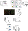
dPLTs target to the gut vasculature at early time points and are exclusively cleared in the liver. (A and B) CellTracker-labeled WT platelets or dPLTs were intravenously transfused into (A) WT or (B) splenectomized mice. At 15 min, 30 min, 2 h, and 16 h after injection, remaining labeled platelets in circulation was assessed by flow cytometry and calculated as percentage of baseline (percent in circulation at 1 min after injection). n = 10 per group. (C) Representative immunofluorescence images of ICG-labeled platelets. Arrows point to labeled platelets. (D) Representative images of MSOT scans of ICG-labeled platelets and unlabeled platelets in agar phantoms. Tracings represent mean optoacoustic intensities across the length of the agar phantom at nonspecific 900-nm and ICG-specific 810-nm excitation wavelengths. (E) Representative 3D reconstructed images of MSOT scans over 40 min of mice transfused with ICG-labeled dPLTs and platelets. Tracings represent mean optoacoustic intensities across 60 min of 2 regions in mice. Blue line, the liver and gut region; red line, spleen. (F) Representative dot plots and quantification of CellTracker+CD41+ platelets in indicated organs at early (<2 h) and late (>12 h) time points. Data represented as means ± SD. *P < 0.05, **P < 0.01, and ***P < 0.001. (G) Representative immunofluorescence of CellTracker+ platelets in various organs at 2 h after intravenous transfusion. Bar graph is quantification of % area of whole tissue section positive for CellTracker+ platelets as assessed by HALO software. White, CellTracker+ platelets; green, F4/80+ macrophages; blue, DAPI. Arrows point to localized CellTracker+ platelet.
Fig. 3.
Fig. 3.
Platelet hepatic targeting requires synergistic platelet GPIbα and hepatic AMR. (A) Percentage of CellTracker-labeled desialylated WT or GPIbα−/− platelets remaining in circulation at time of 15-min and 2-h time points following intravenous transfusion into WT of ASGR2−/− mice. Data shown as percentage of baseline (1 min after injection). Significant data points are compared to WT. n = 5 to 9. (B and C) Percentage of FarRed+ dPLTs as determined by flow cytometry within organs at 2 h after intravenous transfusion. n = 5 to 6 per group. (C) Percentage represented as (x / % FarRed+ dPLTs in the liver × 100). n = 6 to 8 per group (D and E) Representative immunofluorescence images of indicated organs harvested at 2 h after intravenous transfusion of FarRed+ dPLTs. Bar graph is % area of FarRed+ dPLT that is also positive for F4/80+. Green, F4/80+ macrophages; red, FarRed+ dPLTs; blue, DAPI. Arrows point to FarRed+ dPLTs. Statistical analysis was done by one-way ANOVA with Tukey post hoc, *P < 0.05, **P < 0.01, and ***P < 0.001.
Fig. 4.
Fig. 4.
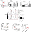
Kupffer cells are required for dPLT clearance and are functionally distinct from splenic macrophages. (A) Platelet counts of WT BALB/c mice intravenously transfused with NEU with or without Kupffer cell depletion. (B to D) In vitro coculture of (B) primary Kupffer and splenic macrophages, (C) RAW624.7, (D) differentiated human THP-1 with CellTracker+ WT (CTRL PLT), dPLT, or anti-αIIb-opsonized platelets. Macrophage-phagocytosed platelets are defied as (B) Kupffer F4/80+CellTracker+ platelets and (C and D) CellTracker+CD61 platelets. n = 6 to 7 per group. Statistical analysis was done by one-way ANOVA with Tukey post hoc test. (C) Representative immunofluorescence images comparing phagocytosis of anti-αIIb-opsonized platelets or dPLT by RAW624.7 cells. Green, CellTracker+ platelet; red, F4/80+ macrophages. Arrows point to orange internalized platelets localized intracellularly. (E) Percentage of FarRed+ dPLTs as determined by flow cytometry within harvested organs at 1 h after intravenous transfusion. Data shown are normalized to % FarRed+ platelets in circulation at a time of 1 min after injection. n = 5. (F) Representative density plot and graph gated on FarRed+ platelets of showing predominant association of anti-αIIb-opsonized FarRed+ platelets with monocyte-derived CD11b+F4/80 and dPLTs with CD11bloF4/80+ Kupffer. n = 5. *P < 0.05, **P < 0.01, and ***P < 0.001.
Fig. 5.
Fig. 5.
(A and B) In vitro analysis of cytokine changes by flow cytometry of primary Kupffer cells following coculture with dPLTs, LPS (10 μg/ml), or control (Kupffer cells alone). Data represented as fold change of % positive of F4/80+CD68+ cells per isotype control. n = 4 (duplicate). (C) Indicated concentration of LPS was added at 24 h after incubation with dPLT. n = 4 (duplicate). (D and E) Serum and liver TGF-β levels as measured by enzyme-linked immunosorbent assay day 1 after intravenous injection of 50 mU of NEU or anti-αIIb monoclonal antibody (0.2 μg/g). n = 6. Statistical analysis was done with one-way ANOVA with Tukey post hoc test. *P < 0.05, **P < 0.01, and ***P < 0.001.
Fig. 6.
Fig. 6.
dPLT increases circulating and splenic functional CD4+ Tregs. (A) Representative dot plot gated on CD4+ showing increased CD25+FOXP3+ Tregs in circulation on day 3 following intravenous transfusion with dPLTs. (A and B) Measurement of PBMC CD4+ Tregs on indicated days following intravenous transfusion of (A) 2 × 10⁸ dPLT or platelet (B) 50 mU of NEU or anti-αIIb antibody (0.02 μg/g). n = 6 per group. Statistical analysis done by with one-way ANOVA with Tukey post hoc test. (C) Graph and representative histogram of decreased CD4+ proliferation in vitro in presence of increasing dilution of splenic CD4+ Tregs isolated from desialylated or WT platelet transfused GPIbα−/− mice. (D) Anti-GPIbα titers from 3× WT immunized GPIbα−/− mice transfused with CD4+CD25+ splenic Treg sorted from either dPLT- or WT platelet-treated mice. (E) Measurement of PBMC CD4+ Tregs on indicated days following macrophage depletion by clodronate liposomes. Data represented as percentage change from day 0 (immediately prior to clodronate injection). n = 4. *P < 0.05 and ***P < 0.001.
Fig. 7.
Fig. 7.
dPLT infusion attenuates antibody generation in clinically relevant transfusion models. (A) Anti-sRBC antibody titers in WT BALB/c mice preinjected 3 days prior with 108 WT human dPLTs. Bar graphs show antibody titers quantified as AUC. n = 2 human donors and 3 mice per group. (B) Schematic diagram of the timeline of dPLT transfusion and FVIII immunization in naïve FVIIInull mice. (C) Flow cytometry analysis of dPLTs. Platelets without NEU treatment were used as a negative control. (D) Inhibitor titers of FVIIInull mice infused with desialylated 2bF8Tg platelets followed by rhFVIII immunization (50 U/kg per week, ×4). FVIIInull mice without 2bF8Tg dPLT infusion were used as a control in parallel. Statistically significant differences between the means of groups were determined by one-way ANOVA followed by Tukey’s multiple comparisons test. (E) Comparison of number animals that developed high inhibitory titers (>100 BU/ml) following rhFVIII immunization. Statistical significance was determined by Pearson’s test. **P < 0.01 and ****P < 0.0001. “n.s.” indicates no statistically significant difference between the 2 groups.

References

    1. Xu XR, Zhang D, Oswald BE, Carrim N, Wang X, Hou Y, Zhang Q, Lavalle C, McKeown T, Marshall AH, et al. Platelets are versatile cells: New discoveries in hemostasis, thrombosis, immune responses, tumor metastasis and beyond. Crit Rev Clin Lab Sci. 2016;53(6):409–430. - PubMed
    1. Jackson SP. Arterial thrombosis--insidious, unpredictable and deadly. Nat Med. 2011;17(11):1423–1436. - PubMed
    1. Ma X, Liang J, Zhu G, Bhoria P, Shoara AA, MacKeigan D, Khoury CJ, Slavkovic S, Lin L, Karakas D, et al. SARS-CoV-2 RBD and its variants can induce platelet activation and clearance: Implications for antibody therapy and vaccinations against COVID-19. Research. 2023;6:0124. - PMC - PubMed
    1. Smyth SS, McEver RP, Weyrich AS. Platelet functions beyond hemostasis. J Thromb Haemost. 2009;7(11):1759–1766. - PubMed
    1. Xu XR, Wang Y, Adili R, Ju L, Spring CM, Jin JW, Yang H, Neves MAD, Chen P, Yang Y, et al. Apolipoprotein A-IV binds alphaIIbbeta3 integrin and inhibits thrombosis. Nat Commun. 2018;9(1): 3608. - PMC - PubMed