. 2023 Sep 22:14:1243818.
doi: 10.3389/fmicb.2023.1243818. eCollection 2023.

The antibacterial activity of a photoactivatable diarylacetylene against Gram-positive bacteria

Affiliations

The antibacterial activity of a photoactivatable diarylacetylene against Gram-positive bacteria

Ryan Waite et al. Front Microbiol. .

Abstract

The emergence of antibiotic resistance is a growing threat to human health, and therefore, alternatives to existing compounds are urgently needed. In this context, a novel fluorescent photoactivatable diarylacetylene has been identified and characterised for its antibacterial activity, which preferentially eliminates Gram-positive over Gram-negative bacteria. Experiments confirmed that the Gram-negative lipopolysaccharide-rich outer surface is responsible for tolerance, as strains with reduced outer membrane integrity showed increased susceptibility. Additionally, bacteria deficient in oxidative damage repair pathways also displayed enhanced sensitivity, confirming that reactive oxygen species production is the mechanism of antibacterial activity. This new diarylacetylene shows promise as an antibacterial agent against Gram-positive bacteria that can be activated in situ, potentially for the treatment of skin infections.

Keywords: Gram-positive bacteria; antimicrobial resistance; lipopolysaccharides; photodynamic therapy; reactive oxygen species.


Conflict of interest statement

CTA, DC, CS, JH, ED, AW, and CAA were employed by the company LightOx Limited. CAA and AW own shares of LightOx Limited, the company licensed to pursue commercial applications of the novel chemicals described in this manuscript. The remaining authors declare that the research was conducted in the absence of any commercial or financial relationships that could be construed as a potential conflict of interest.

Figures

Figure 1
Figure 1
Screening lead compounds for antibacterial activity against Gram-positive and Gram-negative bacteria. Two-fold dilutions of six lead compounds (labelled 1–6) were applied in 6 μL volumes to the surface of a soft agar overlay inoculated with Bacillus subtilis, Staphylococcus epidermidis, Escherichia coli or Pseudomonas fluorescens. The LB agar plates were exposed to light at 365 nm for 5 min and then incubated for 24 h at 37°C prior to imaging. Controls without light activation or with application of DMSO are shown in Supplementary Figure S2.
Figure 2
Figure 2
Photophysical properties of Compound 2. (A) Synthesis and structure of Compound 2. (B) Normalised absorption spectra of Compound 2 in chloroform, DMSO and toluene. (C) Emission spectra of Compound 2 in chloroform, DMSO and toluene with excitation at the respective absorption peak maxima.
Figure 3
Figure 3
Visualisation of non-activated Compound 2 in bacterial cells. Mid-log phase bacteria were incubated with 2 μM of Compound 2 for 30 min before centrifugation and resuspension in 1x PBS. Resuspended cells were applied to a 1% agarose pad and imaged using confocal microscopy with cyan false colour imaging of the compound using a 405 nm laser with detection at 450–550 nm using the Airyscan function. The bar represents 1 μm.
Figure 4
Figure 4
Effect of Compound 2 on bacterial growth. Escherichia coli (A), B. subtilis (B) and S. epidermidis (C) were cultivated in LB broth at 37°C in 96-well plates in a plate reader with continuous shaking. Growth was monitored at OD600nm in samples exposed to light at 365 nm for 5 min (filled symbols) or without light treatment (open symbols). Samples contained 2 μM Compound 2 or 0.2% DMSO indicated by circles or squares, respectively.
Figure 5
Figure 5
Effect of Compound 2 on bacterial viability. Bacteria at mid-log phase of growth (30 μL) were transferred to a clear 24-well plate were mixed with 270 μL LB broth. Serial (10-fold) dilutions were performed and 10 μL of each dilution was applied to the surface of an LB agar plate in the presence of 2 μM Compound 2 or 0.2% DMSO. The plate was then activated by irradiation at 365 nm and samples diluted and spotted again. Both irradiated and non-irradiated plates were incubated for 24 h before enumeration of colonies to determine CFU/ml. Data are the mean and standard error of three independent experiments.
Figure 6
Figure 6
Effect of Compound 2 on bacterial membrane integrity. Bacteria were grown to mid-log phase in the presence or absence of 2 μM Compound 2 as described in the Material and Methods. The relative fluorescence units (RFU × 103) at an emission of 645 nm were normalised against controls containing appropriate control concentrations of DMSO. All samples were exposed to light at 365 nm after 20 min and incubation continued for another 60 min. An ethanol positive control for membrane damage using ethanol was conducted in parallel but is not included in the graph.
Figure 7
Figure 7
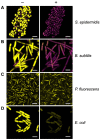
Real-time monitoring of bacterial membrane integrity. The BacLight assay of membrane integrity following photoactivation of Compound 2. S. epidermidis (A), B. subtilis (B), P. fluorescens (C) and E. coli (D) in mid-log phase of growth were stained with PI (magenta) and SYTO 9 (yellow) and imaged by confocal microscopy without light activation (−) or 10 min after photoactivation with the 405 nm laser (+). The laser was applied at 30% power for 1 min. The bar represents 3 μm.
Figure 8
Figure 8
Role of the E. coli outer membrane in protecting against Compound 2 toxicity. (A) The viability of E. coli O+, O and ΔrfaC cells was evaluated as in Figure 5 with exposure to 2 μM Compound 2 with or without light activation. (B) PI assay of E. coli strains to monitor loss of membrane integrity. (C) Real-time imaging of E. coli strains stained with PI (magenta) and SYTO 9 (yellow) by confocal microscopy. Images taken prior to light exposure (−) and 10 min after photoactivation (+) as indicated. The bar represents 3 μm.
Figure 9
Figure 9
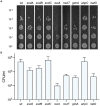
Susceptibility of E. coli strains deficient in oxidative damage and tolerance pathways. The viability of bacteria exposed to 2 μM Compound 2 after photoactivation is shown. Serial dilutions of bacteria were applied to LB agar plates and colonies counted to determine CFU/ml. A representative image of each strain is shown (A) and the CFU/ml (B) represents the mean and standard error of three independent experiments.

References

    1. Baba T., Ara T., Hasegawa M., Takai Y., Okumura Y., Baba M., et al. (2006). Construction of Escherichia coli K-12 in-frame, single-gene knockout mutants: the Keio collection. Mol. Syst. Biol. 2. doi: 10.1038/msb4100050 - DOI - PMC - PubMed
    1. Baptista M. S., Cadet J., Di Mascio P., Ghogare A. A., Greer A., Hamblin M. R., et al. (2017). Type I and type II photosensitized oxidation reactions: guidelines and mechanistic pathways. Photochem. Photobiol. 93, 912–919. doi: 10.1111/php.12716, PMID: - DOI - PMC - PubMed
    1. Benov L. C. L. Y., Day B., Fridovich I. (1995). Copper, zinc superoxide dismutase in Escherichia coli: periplasmic localization. Arch. Biochem. Biophys. 319, 508–511. doi: 10.1006/abbi.1995.1324, PMID: - DOI - PubMed
    1. Chisholm D. R., Hughes J. G., Blacker T. S., Humann R., Adams C., Callaghan D., et al. (2020). Cellular localisation of structurally diverse diphenylacetylene fluorophores. Org. Biomol. Chem. 18, 9231–9245. doi: 10.1039/D0OB01153C, PMID: - DOI - PubMed
    1. Chisholm D. R., Lamb R., Pallett T., Affleck V., Holden C., Marrison J., et al. (2019). Photoactivated cell-killing involving a low molecular weight, donor–acceptor diphenylacetylene. Chem. Sci. 10, 4673–4683. doi: 10.1039/C9SC00199A, PMID: - DOI - PMC - PubMed

LinkOut - more resources