Review
. 2023 Aug 24;3(5):oead083.
doi: 10.1093/ehjopen/oead083. eCollection 2023 Sep.

In-stent restenosis after percutaneous coronary intervention: emerging knowledge on biological pathways

Affiliations
Review

In-stent restenosis after percutaneous coronary intervention: emerging knowledge on biological pathways

Francesco Pelliccia et al. Eur Heart J Open. .

Abstract

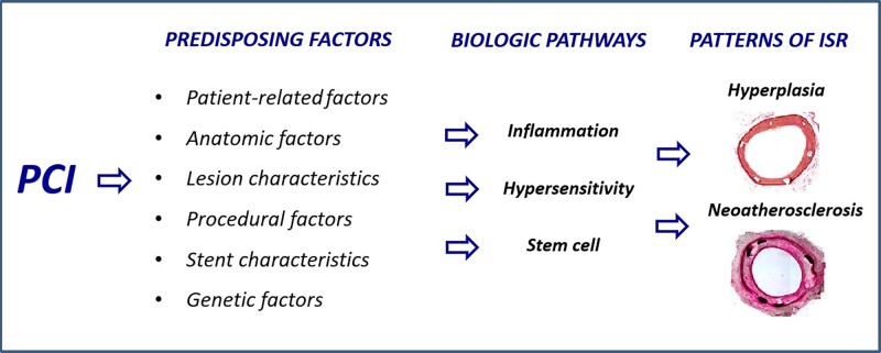
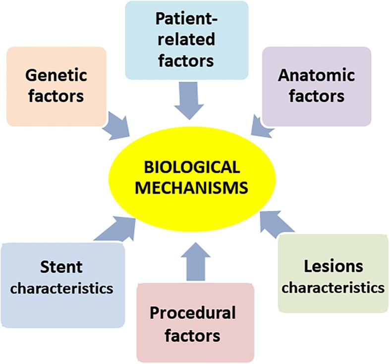
Percutaneous coronary intervention (PCI) has evolved significantly over the past four decades. Since its inception, in-stent restenosis (ISR)-the progressive reduction in vessel lumen diameter after PCI-has emerged as the main complication of the procedure. Although the incidence of ISR has reduced from 30% at 6 months with bare-metal stents to 7% at 4 years with drug-eluting stents (DESs), its occurrence is relevant in absolute terms because of the dimensions of the population treated with PCI. The aim of this review is to summarize the emerging understanding of the biological pathways that underlie ISR. In-stent restenosis is associated with several factors, including patient-related, genetic, anatomic, stent, lesion, and procedural characteristics. Regardless of associated factors, there are common pathophysiological pathways involving molecular phenomena triggered by the mechanical trauma caused by PCI. Such biological pathways are responses to the denudation of the intima during balloon angioplasty and involve inflammation, hypersensitivity reactions, and stem cell mobilization particularly of endothelial progenitor cells (EPCs). The results of these processes are either vessel wall healing or neointimal hyperplasia and/or neo-atherosclerosis. Unravelling the key molecular and signal pathways involved in ISR is crucial to identify appropriate therapeutic strategies aimed at abolishing the 'Achille's heel' of PCI. In this regard, we discuss novel approaches to prevent DES restenosis. Indeed, available evidence suggests that EPC-capturing stents promote rapid stent re-endothelization, which, in turn, has the potential to decrease the risk of stent thrombosis and allow the use of a shorter-duration dual antiplatelet therapy.

Keywords: Bare-metal stents; Drug-eluting stents; Endothelial progenitor cells; Hypersensitivity; In-stent restenosis; Inflammation; Percutaneous coronary intervention.


Conflict of interest statement

Conflict of interest: All authors have nothing to disclose.

Figures

Graphical Abstract
Graphical Abstract
Figure 1
Figure 1
The histology of neointimal hyperplasia. (A) van Gieson’s elastin–stained cross-section of the stented segments. Intima hyperplasia is limited in the segments treated with bevacizumab stent and more pronounced in the arterial segments treated with control stent (×40). (B) Representative photomicrographs with haematoxylin and eosin staining at the stented segments, showing the difference in the neointima hyperplasia between the two groups (×200). Reproduced with permission from Stefanadis et al.
Figure 2
Figure 2
Histological findings of neo-atherosclerosis. (A) Cross-sectional histology of a bare-metal stent implanted in the coronary artery for 7 years ante mortem (×20). (B) High-power image of the box in A (×100). A large necrotic core containing cholesterol crystals is identified within the neointima. The fibrous cap overlying the necrotic core is infiltrated by numerous foamy macrophages and is markedly thinned (arrowheads point to thinnest portion), which resembles vulnerable plaque encountered in native coronary arteries. The asterisks represent metal struts. (C) Cross-sectional histology of a paclitaxel-eluting stent implanted in the coronary artery for 4 years ante mortem (×40). (D) High-power image of the box in C (×200). A relatively small necrotic core containing cholesterol crystals is formed around metal struts (asterisk). The fibrous cap is infiltrated by numerous foamy macrophages and is markedly thinned (yellow arrowheads point to the thinnest portion). NC, necrotic core; PES, paclitaxel-eluting stent. Reproduced with permission from Park et al.
Figure 3
Figure 3
The central role of biological mechanisms over possible underlying factors in the occurrence of in-stent restenosis. Reproduced with permission from Maleknia et al.
Figure 4
Figure 4
The joint effects of cytokines and growth factors in the pathogenesis of in-stent restenosis. Representative optical coherence tomography findings from patients presenting with stent thrombosis: (i) persistent uncovered stent struts late after implantation; (ii) marked stent malapposition in the target vessel, this may have been present at the time of implantation or acquired due to late positive remodelling; (iii) neo-atherosclerotic plaque formation: diffuse low-signal intensity with higher backscatter in deeper neointimal layers may indicate underlying lipid-rich atherosclerotic tissue; (iv) severe stent underexpansion at the site of overlap of multiple stent layers. Reproduced with permission from Byrne et al.
Figure 5
Figure 5
The joint effects of cytokines and growth factors in the pathogenesis of in-stent restenosis. In the process of inflammation, the secretion of cytokines causes the invasion of inflammatory cells such as macrophages, monocytes, and T cells. Monocytes cause the proliferation of fibroblasts by secretion of platelet-derived growth factor. Also, platelet-derived growth factor causes more growth in monocytes. Macrophages with the secretion of inflammatory cytokines such as transforming growth factor-beta enhance the performance of platelet-derived growth factor, which increases vascular smooth muscle cells proliferation and their migration to the intima. On the other hand, the simultaneous secretion of tumour necrosis factor-alpha and interleukin 1 with basic fibroblast growth factor leads to stimulation of fibroblasts and endothelial cells. Endothelial cells through the secretion of vascular endothelial growth factor stimulate proliferation and migration of vascular smooth muscle cells to the intima, but in the presence of transforming growth factor-beta, their function is inhibited. On the other hand, in the presence of basic fibroblast growth factor, fibroblasts are affected by the interferon gamma, which increases the endothelial cell accumulation and provides conditions for inducing restenosis. Meanwhile, the release of TNF-α and interleukin 1 inhibits the biological function of the insulin-like growth factor, thus preventing the formation of the intima and reducing restenosis. bFGF, basic fibroblast growth factor; EC, endothelial cell; IL-1, interleukin 1; IFN-γ, interferon gamma; IGF, insulin-like growth factor; MQ, macrophage; PDGF, platelet-derived growth factor; TGF-β, transforming growth factor-beta; TNF-α, tumour necrosis factor-alpha; VEGF, vascular endothelial growth factor; VSMC, vascular smooth muscle cell. Reproduced with permission from Maleknia et al.
Figure 6
Figure 6
The role of allergic inflammation in adverse reactions after stent implantation. DES, drug-eluting stents; PCI, percutaneous coronary intervention. Reproduced with permission from Niccoli et al. Circulation 2018;138:1736–1748.
Figure 7
Figure 7
The paracrine activity of endothelial progenitor cells at the site of endothelial damage. When the endothelial layer is damaged, circulating endothelial progenitor cells are stimulated to act through paracrine mechanisms leading to the secretion of various cytokines and pro-angiogenic growth factors, such as vascular endothelial growth factor, stromal-derived factor-1, and nitirc oxide. The paracrine signalling mediated by endothelial progenitor cells results in the production of an angiogenic microenvironment that stimulates the nearby endothelium to proliferate. EPCs, endothelial progenitor cells; SDF-1, stromal-derived factor-1; VEGF, vascular endothelial growth factor. Reproduced with permission from Pelliccia et al.

References

    1. Piccolo R, Giustino G, Mehran R, Windecker S. Stable coronary artery disease: revascularisation and invasive strategies. Lancet 2015;386:702–713. - PubMed
    1. Madhavan MV, Kirtane AJ, Redfors B, Généreux P, Ben-Yehuda O, Palmerini T, Benedetto U, Biondi-Zoccai G, Smits PC, von Birgelen C, Mehran R, McAndrew T, Serruys PW, Leon MB, Pocock SJ, Stone GW. Stent-related adverse events >1 year after percutaneous coronary intervention. J Am Coll Cardiol 2020;75:590–604. - PubMed
    1. Giustino G, Baber U, Sartori S, Mehran R, Mastoris I, Kini AS, Sharma SK, Pocock SJ, Dangas GD. Duration of dual antiplatelet therapy after drug-eluting stent implantation: a systematic review and meta-analysis of randomized controlled trials. J Am Coll Cardiol 2015;65:1298–1310. - PubMed
    1. Virmani R, Farb A. Pathology of in-stent restenosis. Curr Opin Lipidol 1999;10:499–506. - PubMed
    1. Otsuka F, Byrne RA, Yahagi K, Mori H, Ladich E, Fowler DR, Kutys R, Xhepa E, Kastrati A, Virmani R, Joner M. Neoatherosclerosis: overview of histopathologic findings and implications for intravascular imaging assessment. Eur Heart J 2015;36:2147–2159. - PubMed