. 2023 Aug 23;9(4):101304.
doi: 10.1016/j.jvscit.2023.101304. eCollection 2023 Dec.

Successful Wallstent exclusion of iliofemoral venous aneurysms-a new treatment paradigm

Affiliations

Successful Wallstent exclusion of iliofemoral venous aneurysms-a new treatment paradigm

Arjun Jayaraj. J Vasc Surg Cases Innov Tech. .

Abstract

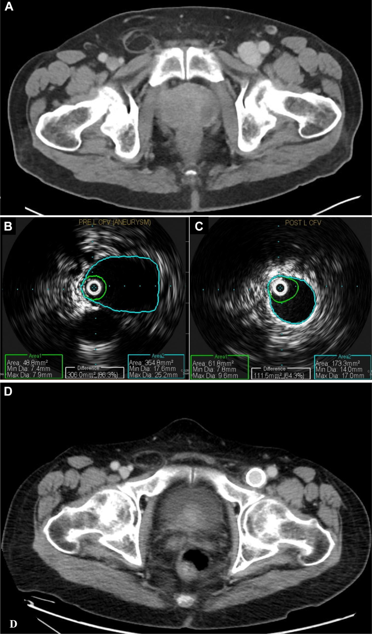
Treatment of venous aneurysms involving the iliac and femoral veins has generally been an open surgical approach, with a few case reports noting use of an endovascular approach. We report three cases: (1) a patient with an iliocaval occlusion involving an occluded TrapEase filter who presented with a large left external iliac vein aneurysm; (2) a patient with a left common femoral vein aneurysm; and (3) a patient with left profunda femoris vein aneurysms with associated pulmonary embolism. All three patients were successfully managed with the use of appropriately sized bare metal woven stents (Wallstents; Boston Scientific). Their clinical presentation, technical considerations, and outcomes are reviewed.

Keywords: Endovascular stenting; Iliac vein aneurysm; Venous aneurysms; Wallstent; Woven venous stent.


Conflict of interest statement

None.

Figures

Fig 1
Fig 1
Computed tomography images depicting left external iliac vein aneurysm (red ovals) in patient 1 on coronal (A) and axial (B) views. Intraprocedural intravascular ultrasound (IVUS) images demonstrating left external iliac aneurysm in patient 1 before stenting (C) and after two-layer stent exclusion (D). Note the continued filling of the aneurysm sac outside the stent after exclusion (hypoechoic area outside stent). This is expected, given the nature of the stent, and successful thrombosis was noted on duplex ultrasound (DUS) at 4 weeks of follow-up. E, Stent configuration in patient 1. A composite Wallstent Z stent configuration was used because the procedure involved recanalization of the bilateral common iliac–distal inferior vena cava (IVC), in addition to exclusion of the left external iliac venous aneurysm. A two-layer Wallstent was used to successfully exclude the aneurysm over its extent on the left side. Technical details are provided in the text. Computed tomography venography (CTV) images showing patent bilateral femoroiliocaval stents and patent IVC filter (red circle) on coronal view (F) and demonstrating successful stent exclusion at 14 months after the procedure (orange arrows) on axial view (G). A, Artery.
Fig 2
Fig 2
A, Axial computed tomography venography (CTV) image depicting the left common femoral vein aneurysm in patient 2 before intervention. Intraprocedural IVUS images demonstrating the left common femoral venous aneurysm before stenting (B) and after successful stent exclusion (C). D, Axial CTV image depicting the left common femoral vein aneurysm in patient 2 after successful stent exclusion and remodeling at 4 months after the procedure.
Fig 3
Fig 3
Axial computed tomography venography (CTV) images depicting left profunda femoris vein aneurysms in patient 3: superior aneurysm (A) and inferior aneurysm (B). CTV demonstrating left profunda femoris vein aneurysms in patient 3: before stent exclusion (C) and after stent exclusion (D). Note the continued filling of the aneurysm sacs after exclusion. This is expected, with thrombosis occurring subsequently, given the venous flow properties. D, Coronal CTV image 8 months after intervention depicting left profunda femoris vein aneurysms in patient 3 that have since undergone successful stent exclusion. Arrows depict thrombosed sac: orange arrows, superior aneurysm; and green arrow, inferior aneurysm.

References

    1. Calligaro K.D., Ahmad S., Dandora R., et al. Venous aneurysms: surgical indications and review of the literature. Surgery. 1995;117:1–6. - PubMed
    1. Teter K.A., Maldonado T.M., Adelman M.A. A systematic review of venous aneurysms by anatomic location. J Vasc Surg Venous Lymphat Disord. 2018;6:408–413. - PubMed
    1. Gabriel S., Eisenberg N., Kim D., Jaberi A., Roche-Nagle G. Primary venous aneurysms: a 20-year retrospective analysis. Vascular. 2020;28:577–582. - PubMed
    1. Zarrintan S., Tadayon N., Kalantar-Motamedi S.M.R. Iliac vein aneurysms: a comprehensive review. J Cardiovasc Thorac Res. 2019;11:1–7. - PMC - PubMed
    1. Fanshawe A.E., Hamilton H.E.C., Constantinou J. External iliac vein aneurysm: a case report and review of the literature. J Surg Case Rep. 2018;2018:rjy115. - PMC - PubMed

LinkOut - more resources