[Preprint]. 2025 Jan 26:2023.09.26.559449.
doi: 10.1101/2023.09.26.559449.

Cell autonomous polarization by the planar cell polarity signaling pathway

Affiliations

Cell autonomous polarization by the planar cell polarity signaling pathway

Alexis T Weiner et al. bioRxiv. .

Abstract

Planar Cell Polarity (PCP) signaling polarizes epithelial cells in a plane orthogonal to their apical-basal axis. A core PCP signaling module segregates two distinct molecular subcomplexes to opposite sides of cells and coordinates the direction of polarization between neighboring cells. Homodimers of the atypical cadherin Flamingo are thought to scaffold these subcomplexes and are required for intercellular polarity signaling. Feedback is required for polarization, but whether feedback requires intercellular and/or intracellular pathways is unknown, and traditional genetic tools have limited utility in dissecting these mechanisms. Using novel tools, we show that cells lacking Flamingo, or bearing a homodimerization-deficient Flamingo, do polarize, indicating that functional PCP subcomplexes form and segregate cell-autonomously. We identify feedback pathways and propose a competitive binding-based asymmetry amplifying mechanism that each operate cell-autonomously. The intrinsic logic of PCP signaling is therefore more similar to that in single cell polarizing systems than was previously recognized.


Conflict of interest statement

Conflict of interest The authors declare that the research was conducted in the absence of any commercial or financial relationships that could be construed as a potential conflict of interest.

Figures
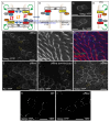
Figure 1.
Figure 1.
Offline cells polarize. A,B. Schematic of PCP complex composition and organization. Asymmetric Fmi bridges linking Fz-Dsh-containing sub-complexes to Vang-Pk-containing sub-complexes (A) sort themselves to yield predominantly one sub-complex on one side of the cell and the other sub-complex on the opposite side of the cell (B). Green and blue arrows represent hypothesized positive feedback pathways and red and orange arrows represent hypothesized negative feedback pathways. Blue and orange pathways require intercellular communication whereas green and red might work cell-autonomously. C. Vang::YFP and Fz::YFP clones in a wild-type background. D-D”. Vang::YFP clones in offline (fminull) wings reveal asymmetric segregation as occurs in wildtype wings. The direction of polarization deviates from the proximal-distal axis (arrows) and corresponds to the direction of prehairs (D,D’). Prehairs arise on the side of the cell opposite to the Vang::YFP crescents as occurs in wildtype (D”; circles). E. Fz::YFP clones in an offline wing. F. Vang::YFP clones in an offline+fmiΔcad wing. G. Vang::YFP clone near the anterior crossvein in the region of the swirl. The location corresponds to the yellow box in Supplemental Figure 1B. H, I. Vang::YFP clones visualized with TIRF microscopy in wild-type Fmi or offline wings Scale bars = 5 μm.
Figure 2.
Figure 2.
Velcroed Fz recruits Dsh and Velcroed Dsh recruits Fz in wildtype and offline backgrounds. A. Schematic of the Fz Velcro system. Clonal N-Cad::V5-Fz expression is expected to recruit N-Cad::V5-Fz selectively to shared cell junctions (C,D). No endogenous Fz is present. B. Schematic of the Dsh Velcro system. Clonal expression of N-Cad::HA-3DX in wings uniformly expressing Dsh-myc at low levels is expected to recruit Dsh-myc to shared cell junctions. C. Schematic of clonal expression (yellow dots outline hypothetical clone) recruiting Velcroed components (red) to shared cell junctions. At the periphery of the clone, recruitment is expected to be to some but not all cell junctions D. N-Cad::V5-Fz stained with anti-V5, showing localization to shared cell junctions. E-E”. N-Cad::V5-Fz clones in fznul1 background (Velcroed Fz) stained for V5, Dsh (E’) and merged (E”). Note that the Dsh antibody is not sensitive enough to detect endogenous Dsh, so it only detects Dsh colocalizing with N-Cad::V5-Fz where it is recruited to sufficient levels for detection. Arrows point to examples of colocalization in the zoomed in region shown. F-F”. N-Cad::HA-3DX clone in a Dsh-myc expressing wing (Velcroed Dsh) stained for HA to detect N-Cad::HA-3DX (F), Fz (F’) and merged F”. Note that the Fz antibody is not sensitive enough to detect endogenous Fz, so it only detects Fz colocalizing with N-Cad::HA-3DX where it is recruited to sufficient levels for detection. G-G”. N-Cad::V5-Fz clones in fznul1, fminul1 background (Velcroed Fz, offline) stained for V5 (G), Dsh (G’) and merged (G”). H-H”. N-Cad::V5-Fz clones in fznul1, fminul1, arm-fmiΔcad background (Velcroed Fz, offline with fmiΔcad) stained for V5 (H), Dsh (H’) and merged (H”). Scale bars = 5 μm.
Figure 3.
Figure 3.
Velcroed Fz excludes Pk in wildtype and offline backgrounds. A. Schematic of Velcroed Fz excluding the Vang-Pk subcomplex. B. Potential outcomes of the exclusion experiment showing localization of Pk: no exclusion (orange arrow), exclusion and accumulation in cytoplasm (green arrow), and exclusion and capture of Pk at cell boundaries lacking N-Cad::V5-Fz (yellow arrow; clone “boundary”). To quantify Pk capture at clone boundaries, intensity line scans were done at clone boundaries (white arrowhead) and compared to intensity linescans of nearby external cell boundaries (gray arrowhead). C-C”. N-Cad::V5-Fz clones in fznul1 background (Velcroed Fz) stained for V5 (C), Pk (C’) and merge (C”). Pk is excluded from internal clone boundaries where N-Cad::V5-Fz localizes (red triangle) and cytoplasmic level of Pk is increased. Pk is captured at clone boundaries (quantified in F; because Pk from the cell outside the clone also may accumulate at that cell interface, a contribution due to exclusion from the clone cell was scored as a level higher than at external cell junctions). D-D”. N-Cad::V5-Fz clones in fznull, fminul1 background (Velcroed Fz, offline) stained for V5 (D), Pk (D’) and merge (D”). Pk is excluded from internal clone boundaries where N-Cad::V5-Fz localizes and cytoplasmic level is increased. Pk is excluded from internal clone boundaries where N-Cad::V5-Fz localizes and cytoplasmic level is increased. Pk is not captured at clone boundaries (quantified in F). E-E”. N-Cad::V5-Fz clones in fznul1, fminul1, arm-fmiΔcad background (Velcroed Fz, offline with fmiΔcad) stained for V5 (E), Pk (E’) and merge (E”). Pk is excluded from internal clone boundaries where N-Cad::V5-Fz localizes and cytoplasmic level is increased. Pk is excluded from internal clone boundaries where N-Cad::V5-Fz localizes and cytoplasmic level is increased. Pk is captured at clone boundaries (quantified in F). F. Quantification of cell boundary intensity linescans as described in B for the results in C-E (see Materials and Methods). Significance was tested with a one way ANOVA to compare mean variance. ** denotes p < .01 and * denotes p < .05. Scale bars = 5 μm.
Figure 4.
Figure 4.
Negative feedback requires Dsh-dependent clustering. A-A”. N-Cad::V5-Fz clones in a dshG63D, fzR52 mutant background, stained for V5 (N-Cad::V5-Fz; A), Dsh (A’) and Pk (A”). DshG63D is recruited to Velcroed Fz, but in the presence of a clustering deficient Dsh, fails to exclude Pk. Furthermore, Pk is recruited to higher levels than in the fz mutant background. For an essential control, see Supp Fig. 4. B-B”. N-Cad::V5-Fz clones in a dshG63D, fzR52 mutant background, stained for V5 (N-Cad::V5-Fz; B), Vang (B’) and Pk (B”). Vang is recruited to Velcroed Fz, perhaps mimicking the population of distal Vang that is observed in wild type cells. C-C”. N-Cad::V5-Fz clones in a dshG63D, fzR52, vangstbm mutant background, stained for V5 (N-Cad::V5-Fz; C), Vang (C’) and Pk (C”). In the absence of Vang, Velcroed Fz with a clustering deficient Dsh no longer recruits Pk. Scale bars = 5 μm.

Similar articles

References

    1. Butler M.T., and Wallingford J.B. (2017). Planar cell polarity in development and disease. Nature reviews. Molecular cell biology 18, 375–388. 10.1038/nrm.2017.11. - DOI - PMC - PubMed
    1. Hale R., and Strutt D. (2015). Conservation of Planar Polarity Pathway Function Across the Animal Kingdom. Annu Rev Genet 49, 529–551. 10.1146/annurev-genet-112414-055224. - DOI - PubMed
    1. Simons M., and Mlodzik M. (2008). Planar Cell Polarity Signaling: From Fly Development to Human Disease. Annu Rev Genet 42, 517–540. - PMC - PubMed
    1. Chae J., Kim M.J., Goo J.H., Collier S., Gubb D., Charlton J., Adler P.N., and Park W.J. (1999). The Drosophila tissue polarity gene starry night encodes a member of the protocadherin family. Development 126, 5421–5429. - PubMed
    1. Usui T., Shima Y., Shimada Y., Hirano S., Burgess R.W., Schwarz T.L., Takeichi M., and Uemura T. (1999). Flamingo, a seven-pass transmembrane cadherin, regulates planar cell polarity under the control of Frizzled. Cell 98, 585–595. - PubMed

Publication types