[Preprint]. 2023 Oct 4:2023.09.22.558628.
doi: 10.1101/2023.09.22.558628.

A comprehensive study of SARS-CoV-2 main protease (Mpro) inhibitor-resistant mutants selected in a VSV-based system

Affiliations

A comprehensive study of SARS-CoV-2 main protease (Mpro) inhibitor-resistant mutants selected in a VSV-based system

Francesco Costacurta et al. bioRxiv. .

Update in

  • A comprehensive study of SARS-CoV-2 main protease (Mpro) inhibitor-resistant mutants selected in a VSV-based system.
    Costacurta F, Dodaro A, Bante D, Schöppe H, Peng JY, Sprenger B, He X, Moghadasi SA, Egger LM, Fleischmann J, Pavan M, Bassani D, Menin S, Rauch S, Krismer L, Sauerwein A, Heberle A, Rabensteiner T, Ho J, Harris RS, Stefan E, Schneider R, Dunzendorfer-Matt T, Naschberger A, Wang D, Kaserer T, Moro S, von Laer D, Heilmann E. Costacurta F, et al. PLoS Pathog. 2024 Sep 11;20(9):e1012522. doi: 10.1371/journal.ppat.1012522. eCollection 2024 Sep. PLoS Pathog. 2024. PMID: 39259728 Free PMC article.

Abstract

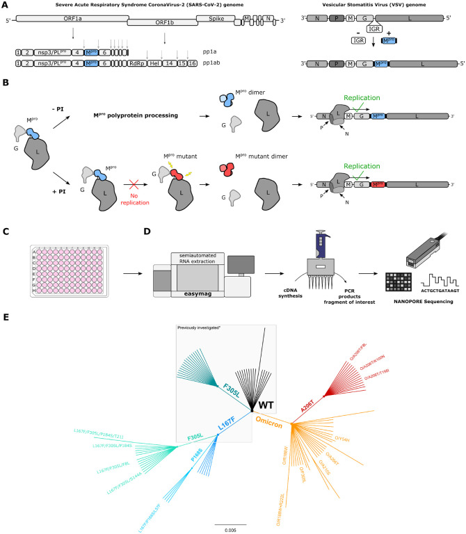
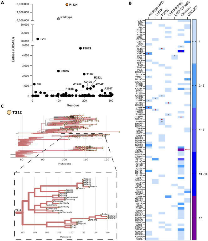
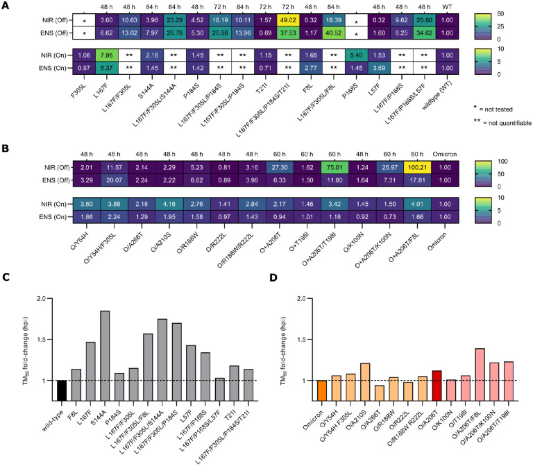
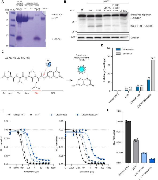
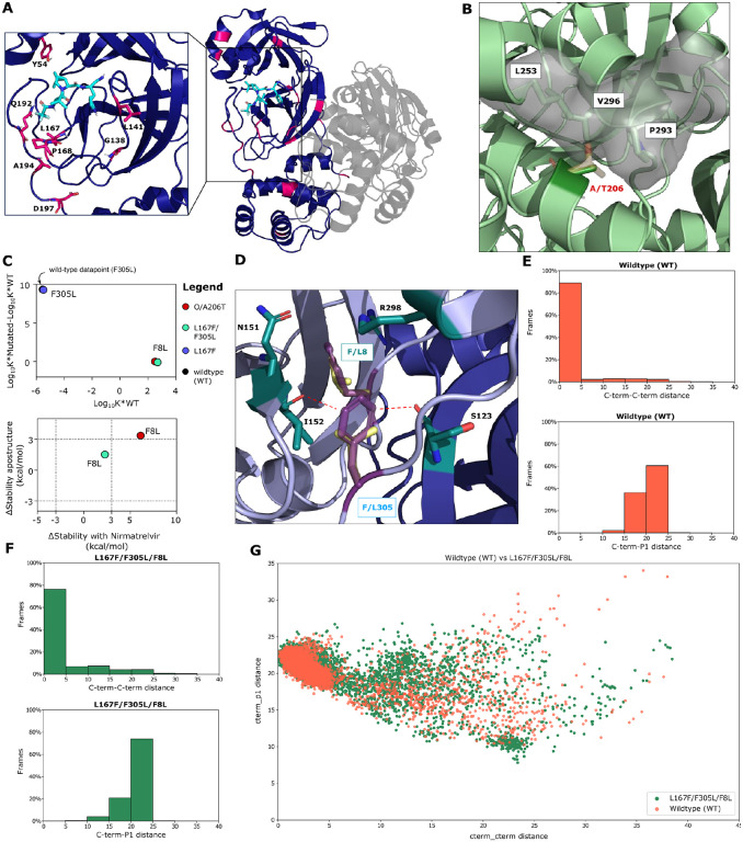
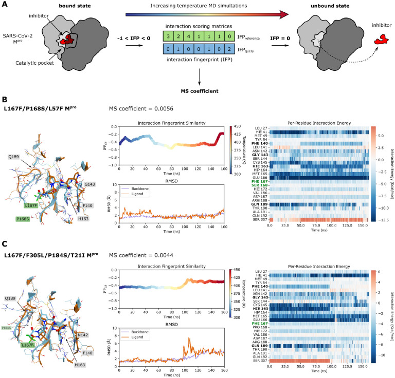
Nirmatrelvir was the first protease inhibitor (PI) specifically developed against the SARS-CoV-2 main protease (3CLpro/Mpro) and licensed for clinical use. As SARS-CoV-2 continues to spread, variants resistant to nirmatrelvir and other currently available treatments are likely to arise. This study aimed to identify and characterize mutations that confer resistance to nirmatrelvir. To safely generate Mpro resistance mutations, we passaged a previously developed, chimeric vesicular stomatitis virus (VSV-Mpro) with increasing, yet suboptimal concentrations of nirmatrelvir. Using Wuhan-1 and Omicron Mpro variants, we selected a large set of mutants. Some mutations are frequently present in GISAID, suggesting their relevance in SARS-CoV-2. The resistance phenotype of a subset of mutations was characterized against clinically available PIs (nirmatrelvir and ensitrelvir) with cell-based and biochemical assays. Moreover, we showed the putative molecular mechanism of resistance based on in silico molecular modelling. These findings have implications on the development of future generation Mpro inhibitors, will help to understand SARS-CoV-2 protease-inhibitor-resistance mechanisms and show the relevance of specific mutations in the clinic, thereby informing treatment decisions.

Keywords: Antimicrobial resistance; Omicron; SARS-CoV-2; ensitrelvir; nirmatrelvir; non-gain-of-function research; protease inhibitor resistance mutations; selection pressure experiments.


Conflict of interest statement

D.v.L. is founder of ViraTherapeutics GmbH. D.v.L serves as a scientific advisor to Boehringer Ingelheim and Pharma KG. E.H. and D.v.L have received an Austrian Science Fund (FWF) grant in the special call “SARS urgent funding”. E.H. is a registered consultant at Guidepoint. D. Bante holds stocks of Pfizer Inc. and Oxford Nanopore Technologies plc. S.A.M and R.S.H. are inventors on the patent application “Live cell assay for protease inhibition”, application number WO/2022/094463. All other authors declare they have no competing interests.

Figures

Fig. 1.
Fig. 1.. VSV-G-Mpro-L construct: molecular mechanism, sequencing workflow, and mutant lineage phylogeny.
(A) Schematic representation of SARS-CoV-2 genome, polyproteins pp1a and pp1ab and the VSV-based mutation selection tool. The InterGenic Region (IGR) between proteins G and L of WT VSV was replaced with the SARS-CoV-2 Mpro Wuhan-1 sequence and its cognate autocleavage sites. Mpro genome positions in SARS-CoV-2 and in VSV-Mpro are highlighted in light blue. (B) The virus is fully dependent on Mpro for replication. Upon translation of G-Mpro-L, two outcomes are possible: 1. without an inhibitor, Mpro is free to process the polyprotein, and the transcription and replication complexes can assemble; 2. with an inhibitor, Mpro is inhibited, the polyprotein is not processed, and the virus is thus not able to replicate, unless it acquires a mutation rendering the Mpro less susceptible to the inhibitor. Then, the virus can replicate despite the inhibitor. (C) BHK21 cells in a 96-well plate are infected with VSV-Mpro. Here, the two outcomes described in panel b can occur. (D) Nanopore sequencing workflow. (E) Unrooted phylogenetic tree showing the relationship between original/parental viruses and mutants. Mpro variants belonging to the same parental virus are coloured accordingly: WT (black), F305L (sea green), L167F (blue), L167F/F305L (light sea green), L167F/P168S (light blue), Omicron (orange), Omicron/A206T (red). For clarity, only the names of the mutants investigated in this work are displayed. *Previously generated/investigated set of mutants in our first study (34).
Fig. 2.
Fig. 2.. Frequency of Mpro mutations in the GISAID database.
(A) The total number of nsp5/Mpro substitution entries for each mutation found during the selection experiments. WT and Omicron-Mpro sequences are displayed in grey and orange, respectively. The dotted line at 500 represent the cut-off value. (B) Heat map representing the number of a specific substitution’s independent occurrences from different samples of VSV-Mpro selection experiments. The red arrow indicates the substitution A206T. Red dots indicate residues within the catalytic site and grey dots indicate residues near the catalytic site. (C) Phylogenetic subtree of nsp5/Mpro-T21I mutant generated with the Ultrafast Sample placement on Existing tRee (UShER) tool and magnified view of a subtree area, showing transmission of this variant possibly from a single founder event (9th of April 2023). Only sequences deposited after the Omicron emergence were used.
Fig. 3.
Fig. 3.. Resistance data and viral replication kinetics of WT, Omicron and mutant main proteases.
Rectangular fields with one and two stars represent constructs/variants that have not been tested or for which IC50 values were not quantifiable, respectively. Hours after infection / hours post infection (hpi). (A) Heat maps of IC50 fold-changes of Mpro-On (bottom) and Mpro–Off (top) dose responses for nirmatrelvir and ensitrelvir. (B) Heat maps of IC50 fold-changes for Omicron-Mpro-On (bottom) and Omicron-Mpro–Off (top) dose responses for nirmatrelvir and ensitrelvir. (C) Bar plot of TM50 fold-changes vs the WT protease. The WT Mpro is coloured in black. (D) Bar plot of TM50 fold-changes vs Omicron (orange) and Omicron/A206T (bordeaux) proteases. Variants are coloured in light orange and light bordeaux according to the parental protease from which they originated.
Fig. 4.
Fig. 4.. Biochemical assay dose responses of nirmatrelvir and ensitrelvir against selected mutant recombinant proteases.
(A) Representative Coomassie-staining for the Mpro-L57F variant after FPLC purification and His-tag removal with HRV-3CP followed by a negative Immobilized Metal Affinity Chromatography (IMAC). The dotted line indicated that the marker lane photograph was cut and placed near the production of the recombinant Mpro lanes. Lane 1: Tag removal reaction of Mpro with HRV-3CP. Combined band of Mpro and HRV 3CP at 35 kDa and free 6xHis-tag (GP-6H) at around 10 kDa. Lane 2: Flow-through (FT) of negative IMAC, yields the Mpro-L57F without His tag. Lane 3: Wash step, indicating a slight loss of protein. Lane 4: The elution step leads to the release of the bound HRV-3CP protease (His tagged) and cleaved His tag of Mpro. Lane 5: 1.5 μg Mpro-L57F without His Tag after desalting. (B) Western blot analysis of the cleavage of the “long” cutting reporter after the addition of the different Mpro variants. The RLuc-F[2] band at approximately 26 kDa indicates successful reporter cleavage. One representative western blot of n = 3 independent experiments is shown. (C) Schematic representation of the fluorescence cleavage-based Mpro activity assay. (D) Fold-changes of IC50 values of nirmatrelvir (blue) and ensitrelvir (light grey) against WT, L57F, L167F/P168S and L167F/P168S/L57F proteases. (E) Dose response experiment with nirmatrelvir (left) and ensitrelvir (right) with WT, L57F, L167F/P168S and L167F/P168S/L57F (± SEM; n = 2). (F) WT and mutant proteases relative activity at 4 hours (± SD; n = 4). The WT signal in the absence of inhibitor was normalized to 1 and mutants’ relative activities were normalized accordingly using WT as reference scale.
Fig. 5.
Fig. 5.. Structural analysis of mutants.
(A) Overview of investigated mutants (pink) mapped onto the Mpro homodimer (dark violet and grey) bound to nirmatrelvir (light blue sticks, PDB 8DZ2) (48). Side chains of residues within the drug binding site are shown as sticks to highlight their distance to nirmatrelvir. (B) The WT alanine side chain of residue 206 (dark green sticks) is tightly packed against the hydrophobic side chains of residues L253, P293, and V296 (green sticks, PDB entry 7ALI) (51). The threonine side chain (yellow sticks) is polar and would clash with these residues as indicated by the light grey surface. (C) Dimerization affinity plot of F8L and F305L mutants (top) and Δ_Stability values (bottom) of apo and nirmatrelvir bound structures for the F8L mutants within different variant backgrounds. The additional dotted lines at x = ± 3 and y = ± 3 in the stability plot are cut-off values that we considered as a meaningful difference in protease stability (a positive value indicates a decrease in protease stability and a negative value indicates an increase in protein stability). (D) The F8 and F305 aromatic side chains (dark violet) form a pi-pi T-stack and F8 in addition interacts with N151, I152, and R298 (green sticks, PDB entry 7ALI (51)). F8 is located within the homodimer interface, with chains A and B colored light violet and blue, respectively. The conjugated p-orbitals of the F305 side chain are located nearby of the I152 and S123 backbone oxygen atoms, potentially leading to electronic repulsion. In the F305L mutant, the distance of the leucine side chain (yellow sticks) to these oxygen atoms is considerably increased. (E) Bar plots showing the percentage of frames against C-term/C-term distance or C-term/P1 distance for WT (F) Bar plots showing the percentage of frames against C-term/C-term distance or C-term/P1 distance for L167F/F305L/F8L mutant. (G) Scatter/cluster plot of WT (orange) and the L167F/F305L/F8L (dark green) mutant C-term-C-term and C-term/P1 distances.
Fig. 6.
Fig. 6.. Thermal Titration Molecular Dynamics (TTMD) experiments of L167F/P168S/L57F and L167F/F305L/P184S/T21I.
(A) Schematic representation of the TTMD simulation workflow. Interaction FingerPrints (IFP) of reference (bound state) and query (protease-inhibitor complex at each temperature condition over time) are continuously sampled and compared during the simulation. The closer from negative to zero the IFPquery is, the more the protease-inhibitor complex differs from the bound state. (B) TTMD simulation data of the nirmatrelvir-L167F/P168S/L57F-Mpro complex. Left: overlay of Mpro structure at the beginning (turquoise) and at the end of the simulation (orange); middle top: a rainbow plot including the IFPCS (left y-axis) is plotted against time in nanoseconds (x-axis). Additionally, the temperature in Kelvin is indicated by colours from blue to red on the right y-axis; middle bottom: the root-mean-square-deviation (left y-axis) is plotted against time in nanoseconds (x-axis); right: a heat map of interaction energies between ligand and surrounding residues. (C) TTMD simulation data of the nirmatrelvir-L167F/F305L/P184S/T21I-Mpro complex. Left: overlay of Mpro structure at the beginning (turquoise) and at the end of the simulation (orange); middle top: a rainbow plot including the IFPCS (left y-axis) is plotted against time in nanoseconds (x-axis). Additionally, the temperature in Kelvin is indicated by colours from blue to red on the right y-axis; middle bottom: the root-mean-square-deviation (left y-axis) is plotted against time in nanoseconds (x-axis); right: a heat map of interaction energies between ligand and surrounding residues. Residues that are mentioned in the results/discussion are highlighted in black, and mutated residues are highlighted in dark green.

References

    1. Mohammed I., Nauman A., Paul P., Ganesan S., Chen K. H., Jalil S. M. S., Jaouni S. H., Kawas H., Khan W. A., Vattoth A. L., Al-Hashimi Y. A., Fares A., Zeghlache R., Zakaria D., The efficacy and effectiveness of the COVID-19 vaccines in reducing infection, severity, hospitalization, and mortality: a systematic review. Hum. Vaccines Immunother. 18 (2022), doi:10.1080/21645515.2022.2027160. - DOI - PMC - PubMed
    1. Davies N. G., Abbott S., Barnard R. C., Jarvis C. I., Kucharski A. J., Munday J. D., Pearson C. A. B., Russell T. W., Tully D. C., Washburne A. D., Wenseleers T., Gimma A., Waites W., Wong K. L. M., van Zandvoort K., Silverman J. D., Diaz-Ordaz K., Keogh R., Eggo R. M., Funk S., Jit M., Atkins K. E., Edmunds W. J., Estimated transmissibility and impact of SARS-CoV-2 lineage B.1.1.7 in England. Science (80-. ). 372 (2021), doi:10.1126/science.abg3055. - DOI - PMC - PubMed
    1. Kustin T., Harel N., Finkel U., Perchik S., Harari S., Tahor M., Caspi I., Levy R., Leshchinsky M., Ken Dror S., Bergerzon G., Gadban H., Gadban F., Eliassian E., Shimron O., Saleh L., Ben-Zvi H., Keren Taraday E., Amichay D., Ben-Dor A., Sagas D., Strauss M., Shemer Avni Y., Huppert A., Kepten E., Balicer R. D., Netzer D., Ben-Shachar S., Stern A., Evidence for increased breakthrough rates of SARS-CoV-2 variants of concern in BNT162b2-mRNA-vaccinated individuals. Nat. Med. 27, 1379–1384 (2021). - PMC - PubMed
    1. Rössler A., Riepler L., Bante D., von Laer D., Kimpel J., Correspondence SARS-CoV-2 Omicron Variant Neutralization in Serum from Vaccinated and Convalescent Persons, 1–3 (2021). - PMC - PubMed
    1. Carabelli A. M., Peacock T. P., Thorne L. G., Harvey W. T., Hughes J., de Silva T. I., Peacock S. J., Barclay W. S., de Silva T. I., Towers G. J., Robertson D. L., SARS-CoV-2 variant biology: immune escape, transmission and fitness. Nat. Rev. Microbiol. 21, 162–177 (2023). - PMC - PubMed

Publication types