[Preprint]. 2023 Sep 29:2023.09.27.559733.
doi: 10.1101/2023.09.27.559733.

Replenishing Age-Related Decline of IRAK-M Expression in Retinal Pigment Epithelium Attenuates Outer Retinal Degeneration

Affiliations

Replenishing Age-Related Decline of IRAK-M Expression in Retinal Pigment Epithelium Attenuates Outer Retinal Degeneration

Jian Liu et al. bioRxiv. .

Update in

Abstract

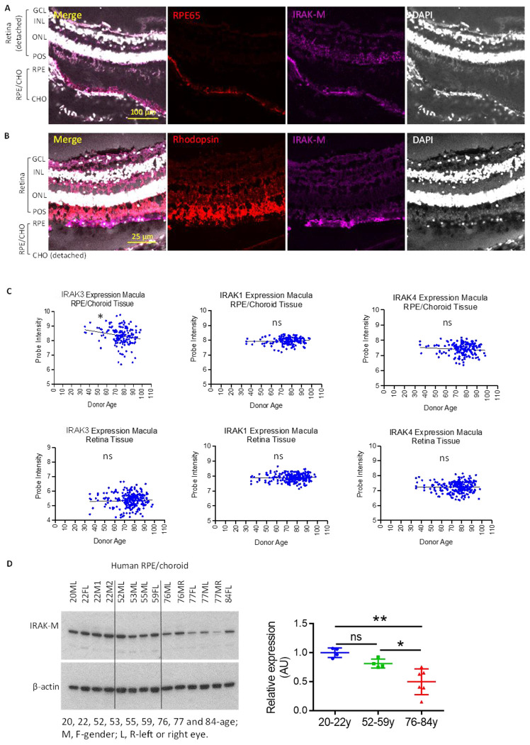
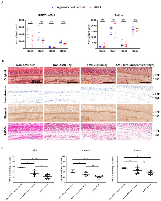
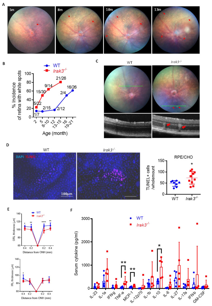
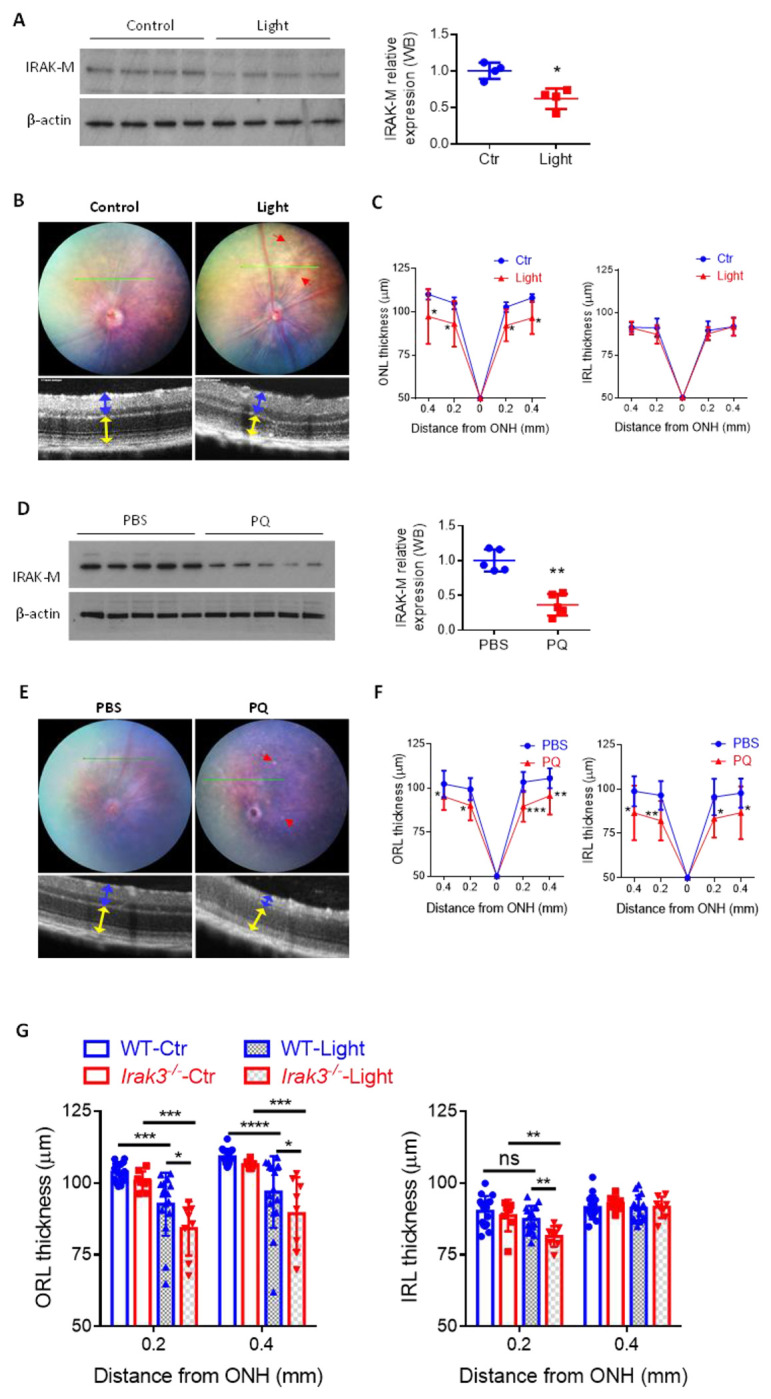
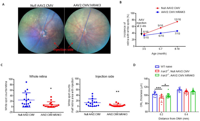
Unchecked, chronic inflammation is a constitutive component of age-related diseases, including age-related macular degeneration (AMD). Here we identified interleukin-1 receptor-associated kinase (IRAK)-M as a key immunoregulator in retinal pigment epithelium (RPE) that declines with age. Rare genetic variants of IRAK-M increased the likelihood of AMD. IRAK-M expression in RPE declined with age or oxidative stress and was further reduced in AMD. IRAK-M-deficient mice exhibited increased incidence of outer retinal degeneration at earlier ages, which was further exacerbated by oxidative stressors. The absence of IRAK-M disrupted RPE cell homeostasis, including compromised mitochondrial function, cellular senescence, and aberrant cytokine production. IRAK-M overexpression protected RPE cells against oxidative or immune stressors. Subretinal delivery of AAV-expressing IRAK-M rescued light-induced outer retinal degeneration in wild-type mice and attenuated age-related spontaneous retinal degeneration in IRAK-M-deficient mice. Our data support that replenishment of IRAK-M expression may redress dysregulated pro-inflammatory processes in AMD, thereby treating degeneration.


Conflict of interest statement

Competing interests ADD, JL and YKC are named inventors on an International Patent Application No: PCT/EP2022/082518. ADD is consultant for Hubble Tx, Affibody, 4 DMT, Novartis, Roche, UCB, Amilera, Janssen, and ActivBio. RG is consultant for Roche, Genentech, Apellis, Novartis, and Bayer.

Figures

Fig. 1.
Fig. 1.. IRAK-M is expressed in RPE and its expression level is reduced with age and in AMD.
(A&B) Confocal images of human retinal sections from a 20-year-old donor (without recorded ocular disease) demonstrate IRAK-M immunopositivity at the RPE layer (anti-RPE65 stain). DAPI and anti-Rhodopsin were used to stain nuclei and POS, respectively. (C) Affymetrix chip-based transcriptome analyses show an age-related reduction in the expression level of IRAK3 mRNA in macular RPE/choroid tissues, but not in the retina. Neither IRAK1 nor IRAK4 mRNA level is changed with age in RPE/choroid or retina. (D) Western blot and densitometry quantification show reduced levels of IRAK-M protein expression in aged human RPE/choroidal lysates. The IRAK-M levels were normalized to β-actin (n=4-6). *P < 0.05; **P < 0.01; ns, nonsignificant. Comparison by simple linear regression (C) or one-way ANOVA (D).
Fig. 2.
Fig. 2.. IRAK-M expression level in RPE is reduced in AMD.
(A) PORT-normalized gene counts from RNA-Seq data (GSE99248) show decreased IRAK3 mRNA expression in RPE/Choroid/Sclera of AMD donors versus age-matched normal controls. IRAK1, IRAK2 and IRAK4 mRNA levels in RPE/Choroid/Sclera have no difference. Nor mRNA levels of any IRAKs in retina show difference (n=7-8). (B) Magnification of boxed regions of representative IHC images (Fig. S5) of human retinal sections from two non-AMD (59-year and 97-year old, respectively), a mild AMD (76-year old), and an unidentified stage AMD donors (85-year old) were color-deconvoluted using ImageJ to separate IRAK-M staining (red), pigment (brown) and nuclei (blue). Note a nonspecific staining of thickened BM in AMD. (C) Quantification of mean staining intensity of macular area shows more severely reduced IRAK-M expression in both aged and AMD RPE, while the reduced expression in choroid is only significant with old age. There are no changes in retina with ageing or in AMD (n=2 for young control, n=5 for old control and n=11 for AMD). *P < 0.05; ***P < 0.001; ****P < 0.0001; ns, nonsignificant. Comparison by two-way ANOVA (A) or one-way ANOVA (C).
Fig. 3.
Fig. 3.. Irak3−/− mice spontaneously display early retinal abnormalities.
(A) Representative fundal images show age-related appearance of white spots (red line arrow) in Irak3−/− mouse retinas. (B) Time course of incidence of flecked retina (number of spots > 3) shows increased incidence of retinal spots in Irak3−/− mice compared to WT controls. Each value is a ratio of number of flecked retina to total number of retina at each time point. (C) Representative fundal and OCT images demonstrate that the white spots (red line arrow) are associated with outer retinal abnormalities (red arrow) in 5m-old Irak3−/− mice. (D) TUNEL staining on RPE/choroidal flatmounts reveals elevated number of apoptotic cells in Irak3−/− mice versus WT controls (5m-old) (n=8-10). (E) Quantification of OCT images indicates significant outer retinal thinning in Irak3−/− mice aged 12-13m. The change in inner retinal thickness is negligible (n=6-12). (F) Multiplex cytokine array demonstrates an overall higher levels of serum cytokines in Irak3−/− compared to WT mice (12-13m-old), where the increases of TNF-α, MCP-1 and IL-10 serum concentrations are statistically significant (n=5-6). *P < 0.05; **P < 0.01; ****P < 0.0001. Comparison by unpaired two-tailed Student’s t-test (D) or two-way ANOVA (E and F).
Fig. 4.
Fig. 4.. Wild-type mice exhibit reduced RPE-IRAK-M expression level by oxidative stress and Irak3−/− mice are more vulnerable to light-induced retinal degeneration.
Retinal oxidative stresses were induced in 8-week-old C57BL/6J mice by either fundus-light induction (100kLux for 20min, A-C) or intravitreal administration of paraquat (PQ, 2μl at 1.5mM, D-F). (A&D) Western blot analyses of IRAK-M expression in RPE lysate on day 7 post oxidative damage (n=4 or 5). (B&E) Representative fundoscopy and OCT images obtained on day 14 demonstrate appearance of retinal lesions (red line arrows), and reduced thickness of outer retina (yellow double-arrow lines) in light model (n=8, C), or both outer and inner retina (blue double-arrow lines) in PQ model (n=9-11, F). (G) Eight-week-old WT and Irak3−/− mice were subjected to retina oxidative insults by light induction. OCT quantification of retinal thickness (average of temporal and nasal measurements) demonstrates exaggerated retinal thinning in Irak3−/− mice compared to WT controls on 14 days post light induction, which is more pronounced in outer retinal layers (n=8-16). *P < 0.05; **P < 0.01; ***P < 0.001; ****P < 0.0001. Comparison by unpaired two-tailed Student’s t-test (A and D) or two-way ANOVA (C, F and G).
Fig. 5.
Fig. 5.. Overexpression of IRAK-M in RPE cells supports metabolic activities and inhibits cell death against stressors.
(A&B) Metabolic flux analyses demonstrate that increasing endogenous IRAK-M expression in human iPSC-RPE cells via CRISPR/Cas9 activation plasmid maintains both mitochondrial respiration (OCR, A) and glycolytic capacity (ECAR, B), upon 24h treatment with 30 μM H2O2 or 1 μg/ml LPS (n=3-7). (C) Stably transfected cell lines selected from mouse B6-RPE07 cells were established to persistently express human IRAK-M. Time course of LDH release over 5 days since confluence of monolayers shows sustained cell viability by human IRAK-M transfection (n=4-8). (D) Human IRAK-M expression inhibits PQ (125 μM) or LPS (40 ng/ml)-induced cytotoxicity post 72h of treatment in stably transfected B6-RPE07 cells (n=2-4). (E&F) Primary mouse Irak3−/− RPE cells were subjected to transient transfection for human IRAK-M expression using pUNO1 plasmid and 48h later, the cells were treated with 60 μM H2O2 for another 24h. OCR analysis (E) shows protected mitochondrial maximal respiration by human IRAK-M against oxidative stress treatment. ECAR analysis (F) does not show any changes in glycolysis activity by H2O2 treatment or IRAK-M transfection (n=3). *P < 0.05; **P < 0.01; ****P < 0.0001; ns, nonsignificant. Comparison by two-way ANOVA.
Fig. 6.
Fig. 6.. Subretinal delivery of AAV.hIRAK3 protects retina against light damage in wild-type mice.
(A) Two weeks post subretinal injection of AAV2.CMV.hIRAK3 or AAV2.CMV (high dose 2×10⁹ versus low dose 4×10⁸ gc/eye), RPE/choroid and retina were analyzed for IRAK3 transgene expression using qRT-PCR, normalized by RPS29 mRNA (n=5). (B) Retinal cryosections were examined for high dose AAV-mediated IRAK-M expression using an antibody recognizing both human and mouse IRAK-M (Ab1), or an antibody specific to human IRAK-M (Ab2). Representative confocal images were shown. (C-E) Two weeks after subretinal injection with the high dose of AAV2.CMV.hIRAK3 or null vector, each mouse was subjected to light-induced retinal degeneration in one eye and left thereafter for a further two weeks, followed by assessment of retinal pathology and therapeutic response. (C) Representative fundoscopy/OCT images and quantification show light-induced retinal lesions and averaged outer retinal thickness (n=10-11). (D) Representative confocal images of TUNEL staining on retinal sections and quantification of 3 sections from each eye (n=3-6). (E) Confocal images of MitoView Green staining for mitochondrial content and MFI measurement in 3 different fields from two sections of each eye (n=3-6). *P < 0.05; **P < 0.01; ***P < 0.001; ****P < 0.0001. Comparison by two-way ANOVA.
Fig. 7.
Fig. 7.. Subretinal delivery of AAV.hIRAK3 prevents from age-related spontaneous retinal degeneration in Irak3−/− mice.
2×10⁹ gc of AAV2.CMV.hIRAK3 was injected subretinally in one eye of each Irak3−/− mouse (2-4m old), with null vector inj ected to the contralateral eye. Mice were then monitored by fundoscopy and OCT for 6 months thereafter. (A) Representative fundal images show retinal spots in 8m-old Irak3−/− mice with AAV administration at the age of 2m. Blue lines separate the retina into two sides based on the injection site. (B) Time course of incidence of flecked retina shows IRAK3 gene therapy decelerated the appearance of retinal spots in ageing Irak3−/− mice (n=15 or 16). (C) Number of retinal spots in whole retina or at the injection side, was blind-counted for comparison between AAV2.CMV.hIRAK3 and null vector groups (8-10m-old, n=15 or 16). (D) OCT quantification shows a reduction in outer retinal thickness close to the centre region (0.2 mm distant from optic nerve head) in 8-10m-old Irak3−/− mice compared to age-matched WT littermates, which is revoked by AAV.hIRAK3 gene delivery (n=12-16). *P < 0.05; **P < 0.01; ***P < 0.001. Comparison by unpaired two-tailed Student’s t-test (C) or two-way ANOVA (D).

References

    1. Moretti J., Blander J. M., Cell-autonomous stress responses in innate immunity. J Leukoc Biol 101, 77–86 (2017). - PMC - PubMed
    1. Franceschi C., Garagnani P., Parini P., Giuliani C., Santoro A., Inflammaging: a new immune-metabolic viewpoint for age-related diseases. Nat Rev Endocrinol 14, 576–590 (2018). - PubMed
    1. Tong Y., Wang S., Not All Stressors Are Equal: Mechanism of Stressors on RPE Cell Degeneration. Front Cell Dev Biol 8, 591067 (2020). - PMC - PubMed
    1. Copland D. A., Theodoropoulou S., Liu J., Dick A. D., A Perspective of AMD Through the Eyes of Immunology. Invest Ophthalmol Vis Sci 59, AMD83–AMD92 (2018). - PubMed
    1. Handa J. T. et al., A systems biology approach towards understanding and treating non-neovascular age-related macular degeneration. Nat Commun 10, 3347 (2019). - PMC - PubMed

Publication types