[Preprint]. 2023 Sep 26:2023.09.25.559341.
doi: 10.1101/2023.09.25.559341.

Nuclear lamin A-associated proteins are required for centromere assembly

Affiliations

Nuclear lamin A-associated proteins are required for centromere assembly

Adriana Landeros et al. bioRxiv. .

Abstract

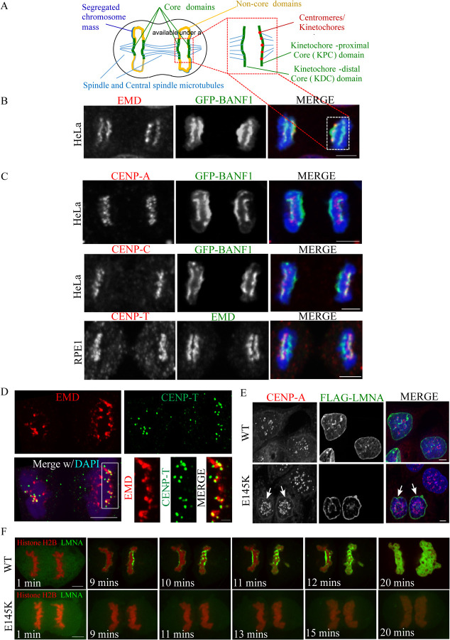
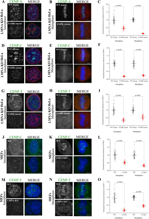
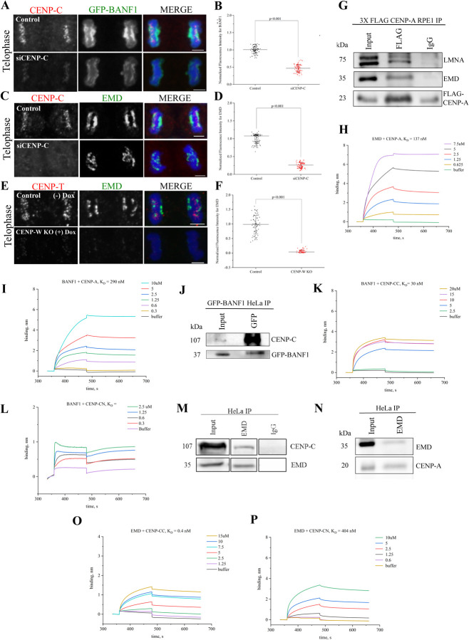
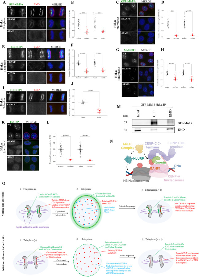
Many Lamin A-associated proteins (LAAP's) that are key constituents of the nuclear envelope (NE), assemble at the "core" domains of chromosomes during NE reformation and mitotic exit. However, the identity and function of the chromosomal core domains remain ill-defined. Here, we show that a distinct section of the core domain overlaps with the centromeres/kinetochores of chromosomes during mitotic telophase. The core domain can thus be demarcated into a kinetochore proximal core (KPC) on one side of the segregated chromosomes and the kinetochore distal core (KDC) on the opposite side, close to the central spindle. We next tested if centromere assembly is connected to NE re-formation. We find that centromere assembly is markedly perturbed after inhibiting the function of LMNA and the core-localized LAAPs, BANF1 and Emerin. We also find that the LAAPs exhibit multiple biochemical interactions with the centromere and inner kinetochore proteins. Consistent with this, normal mitotic progression and chromosome segregation was severely impeded after inhibiting LAAP function. Intriguingly, the inhibition of centromere function also interferes with the assembly of LAAP components at the core domain, suggesting a mutual dependence of LAAP and centromeres for their assembly at the core domains. Finally, we find that the localization of key proteins involved in the centromeric loading of CENP-A, including the Mis18 complex and HJURP were markedly affected in LAAP-inhibited cells. Our evidence points to a model where LAAP assembly at the core domain serves a key function in loading new copies of centromeric proteins during or immediately after mitotic exit.

Keywords: BANF1; CENP-A; Centromere; Emerin; LaminA; Mitosis.


Conflict of interest statement

Conflict of Interest The authors declare no competing financial interests.

Figures

Figure 1.
Figure 1.. Lamin A-associated proteins (LAAPs) co-assemble with centromeric components at the kinetochore-proximal core (KPC) during telophase.
(A) A schematic representation of the chromosomal core domains formed during mitotic telophase. The core domains assembled on the segregated chromosome mass are indicated in green, the non-core domains in yellow, the spindle and central spindle microtubules in blue and the centromeres/kinetochores in red. (B) Visualization of the core domain via the immunostaining of LAAPs, BANF1 (GFP, green) and EMD (red), with the chromosomes counterstaining using DAPI (blue). The selected regions from the schematic on top and the mitotic cell at the bottom indicate the two core domains formed in telophase. (C) Immunostaining of CENP-A combined with GFP-BANF1 in HeLa cells (top panel); CENP-C with GFP-BANF1 in HeLa cells (middle panel) and CENP-T with EMD in RPE1 cells (bottom panel). Scale bar, 5 μm. (D) HeLa cells in telophase immunostained using anti-EMD and anti-CENP-T antibodies with the chromosomes counterstained using DAPI, were subjected to Structured-Illumination (SIM) super-resolution imaging. Bar, 5 μm. Insets from C are shown at the bottom right. Bar, 1 μm. (E) HeLa cells expressing FLAG-tagged WT or E145K mutants of LMNA were immunostained for the FLAG-tag and the centromeric protein, CENP-A, with the nuclei counterstained using DAPI. (F) Live imaging of HeLa cells stably expressing Histone H2B-RFP transfected with either GFP-tagged WT (upper panel) or E145K LMNA (lower panel) constructs. Bar, 5 μm.
Figure 2.
Figure 2.. LMNA is required for proper centromere assembly in interphase and mitotic cells.
(A-B) LMNA KO interphase (A) or mitotic (B) HeLa cells rescued with either WT or the E145K mutant of LMNA were immunostained for CENP-A (green) and the expressed FLAG-tagged LMNA (red) with the chromosomes counterstained using DAPI (blue). (C) Quantification of CENP-A immunofluorescence in the two conditions depicted in A and B. (D-E). Same as the conditions in A and B but in this case, CENP-C was detected instead of CENP-A. (F) Quantification of CENP-C immunofluorescence from D and E. (G-H). Same as the conditions in A and B, but in this case, CENP-T was detected instead of CENP-A. (I) Quantification of CENP-T immunofluorescence from G and H. (J-K) WT or LMNA (also labelled as LA) KO interphase (J) or mitotic (K) Mouse Embryonic Fibroblasts (MEFs) were immunostained for CENP-C (green) with the chromosomes counterstained using DAPI. (L) Quantification of CENP-C immunofluorescence in the two conditions depicted in J and K. (M-N). Same as the conditions in J and K, but in this case, CENP-T was detected instead of CENP-C. (O) Quantification of CENP-C immunofluorescence in the two conditions depicted in M and N. Scale bars, 5 μm.
Figure 3.
Figure 3.. Biochemical interaction between LAAPs and the centromeric proteins facilitate their codependent assembly at the KPC.
(A) HeLa cells in Mitotic telophase were immunostained for CENP-C (red) and GFP-BANF1 (green) in control cells or those treated with siRNA against CENP-C (siCENP-C) with the chromosomes counterstained using DAPI (blue). (B) Quantification of BANF1 immunofluorescence in WT or siCENP-C cells as depicted in A. (C) Similar to the conditions as in A, but cells were immunostained for EMD (green) instead of GFP-BANF1. (D) Quantification of EMD immunofluorescence in control or siCENP-C cells as depicted in C. (E) Telophase cells immunostained for CENP-T (red) and EMD (green) in WT and inducible CENP-W KO HeLa cells. (F) Quantification of EMD immunofluorescence in control or CENP-W KO cells as depicted in E. (G) RPE1 cells over-expressing FLAG-tagged CENP-A were immunoprecipitated using FLAG antibody and probed for LMNA, EMD, and FLAG. IgG was used as negative control. (H) Biolayer interferometry (BLI) sensograms obtained using EMD as the ligand with various concentrations (0.625–7.5 μM) of CENP-A as the analyte. Buffer only sample was used to set the baseline. The graph depicts the binding and dissociation phases along with equilibrium dissociation constant (KD) that was determined. (I) BLI sensograms obtained using BANF1 as the ligand with various concentrations (0.3–10 μM) of CENP-A as the analyte. (J) GFP-BANF1 HeLa cell lysates were immunoprecipitated with anti-GFP antibody and probed for CENP-C. (K) BLI sensogram of BANF1 with various concentrations (2.5–20 μM) of the C-terminal of CENP-C. (L) BLI sensogram of BANF1 with various concentrations (0.3–2.5 μM) of the N-terminal region of CENP-C. (M-N) Immunoprecipitation of HeLa cell lysates showing EMD pulling down both CENP-C (M) as well as CENP-A (N). (O) BLI sensogram of EMD with various concentrations (1.25–15 μM) of the C-terminal region of CENP-C. (P) BLI sensogram of EMD with various concentrations (0.6–10 μM) of the N-terminal region of CENP-C. Scale bars, 5 μm.
Figure 4.
Figure 4.. The loading of CENP-A adaptor Mis18 and the CENP-A chaperone, HJURP, to the centromeres is severely affected in LAAP-inhibited cells.
(A) Control, siBANF1- or siEMD-treated mitotic telophase HeLa cells constitutively expressing the GFP-Mis18α subunit of the Mis18 complex were immunostained for GFP-Mis18α (green) and EMD (red) with chromosomes counterstained using DAPI (blue). (B) Quantification of GFP-Mis18α immunofluorescence in the conditions as depicted in A. (C) same conditions as in A but with interphase cells. (D) Quantification of GFP-Mis18α immunofluorescence in the conditions as depicted in C. (E) Control, siBANF1 or siEMD mitotic telophase HeLa cells were immunostained for the Mis18 complex subunit, Mis18-BP1 (green), and EMD (red) with chromosomes counterstained using DAPI. (F) Quantification of Mis18-BP1 immunofluorescence in the conditions as depicted in E. (G) same conditions as in E but with interphase cells. (H) Quantification of Mis18-BP1 immunofluorescence in the conditions as depicted in G. (I) WT and LMNA KO HeLa cells in mitotic telophase were immunostained for Mis18-BP1 (green) and EMD (red). (J) Quantification of Mis18-BP1 immunofluorescence in the conditions as depicted in I. (K) Control, siBANF1, and siEMD HeLa cells in interphase were immunostained for HJURP (green) with the chromosomes counterstained using DAPI. (L) Quantification of HJURP immunofluorescence in the conditions as depicted in K. Scale bars, 5 μm. (M) Immunoprecipitation of GFP-Mis18α HeLa cell lysates using anti-GFP and anti-EMD antibodies with the blot being reversibly probed for both these proteins. (N) A Molecular Model for LAAP-centromere co-assembly. The centromeric DNA and the CCAN is instrumental in the recruitment of LAAPs during telophase. Once the core is assembled, both these structures (CCAN and the core) seems to contribute to the loading of the Mis18 complex later in telophase. Mis18 along with HJURP can now recruit new copies of CENP-A in late telophase and G1. (O) A Schematic model of core function in normal cell division (top) and cells in which LAAPs are perturbed (bottom). During normal core assembly, LMNA and the LAAPs (green) assemble at the two core domains during telophase, contributing to the recruitment of CENP-A chaperones (red). The existing centromeric and CCAN components contribute to the assembly of LAAPs at the core domains. Following cytokinesis and entry into the next G1, LMNA and the LAAPs relocate from the cores to the nuclear envelope while new CENP-A as well as new CCANs components (yellow) gets loaded onto the centromeres so that there is adequate amounts of CENP-A and CCAN components retained for the succeeding round of mitosis. However, when LMNA or LAAPs are perturbed, these proteins obviously do not assemble at the core domains during telophase. This results in a substantial reduction of CENP-A chaperone recruitment in telophase, which in turn interferes with new CENP-A and CCAN recruitment to centromeres. When cells progress through multiple rounds of cell division cycle, without LMNA or LAAPs assembly at the core, there is a continuous loss of CENP-A and CCANs proteins from the centromeres, leading to the manifestation of severe kinetochore assembly defects and rampant chromosome mis-segregation.

References

    1. Takeuchi K., and Fukagawa T. (2012). Molecular architecture of vertebrate kinetochores. Experimental cell research 318, 1367–1374. - PubMed
    1. Hori T., and Fukagawa T. (2012). Establishment of the vertebrate kinetochores. Chromosome Res 20, 547–561. - PubMed
    1. Zasadzińska E., and Foltz D.R. (2017). Orchestrating the Specific Assembly of Centromeric Nucleosomes. Prog Mol Subcell Biol 56, 165–192. - PMC - PubMed
    1. Ray-Gallet D., and Almouzni G. (2021). The Histone H3 Family and Its Deposition Pathways. Adv Exp Med Biol 1283, 17–42. - PubMed
    1. Cheeseman I.M., and Desai A. (2008). Molecular architecture of the kinetochore-microtubule interface. Nat Rev Mol Cell Biol 9, 33–46. - PubMed

Publication types