[Preprint]. 2023 Sep 25:2023.09.23.559091.
doi: 10.1101/2023.09.23.559091.

Improved CAR-T cell activity associated with increased mitochondrial function primed by galactose

Affiliations

Improved CAR-T cell activity associated with increased mitochondrial function primed by galactose

Golda Gross et al. bioRxiv. .

Update in

Abstract

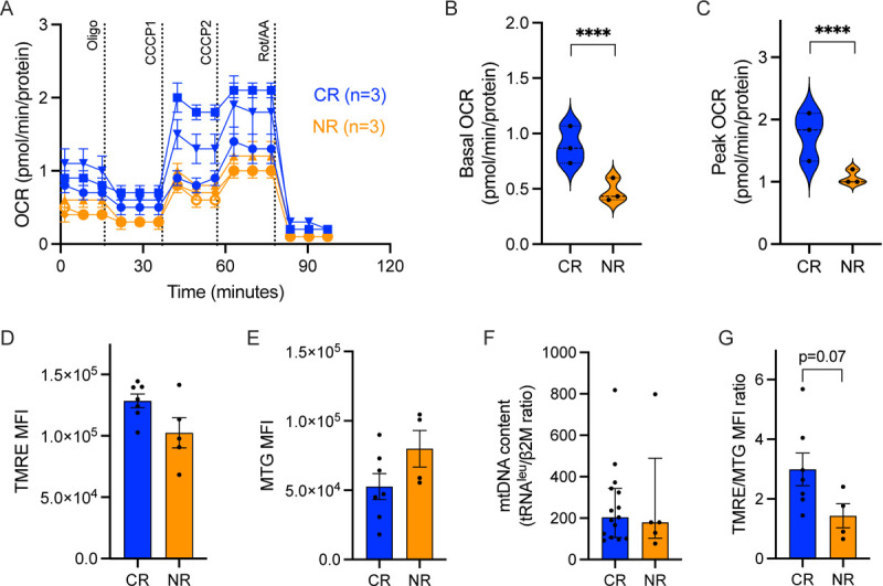
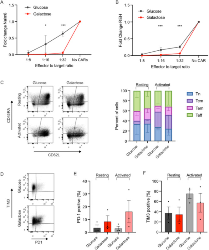
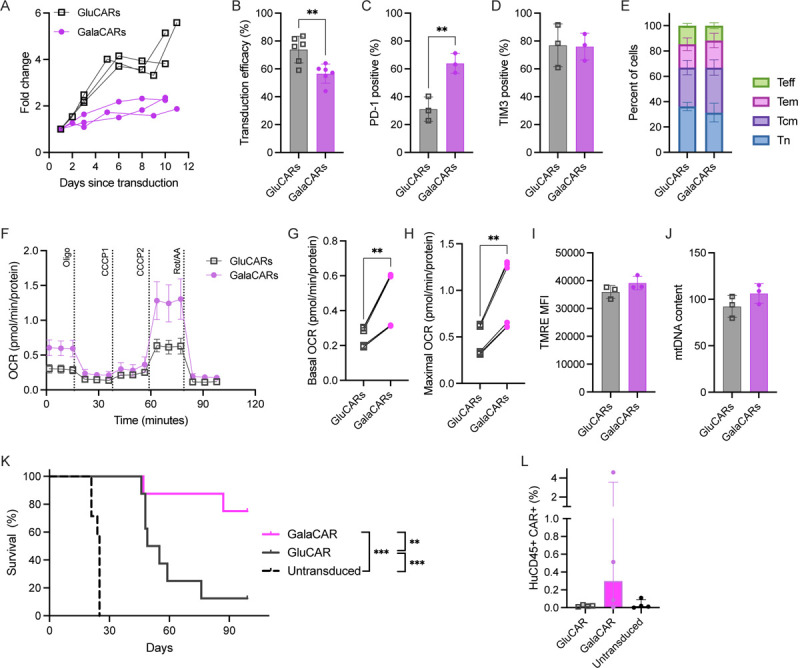
CD19 CAR-T cells have led to durable remissions in patients with refractory B-cell malignancies; nevertheless, most patients eventually relapse in the long term. Many interventions aimed at improving current products have been reported, with a subset of them focusing on a direct or indirect link to the metabolic state of the CAR-T cells. We assessed clinical products from an ongoing clinical trial utilizing CD19-28z CAR-T cells from patients with acute lymphoblastic leukemia. CAR-T clinical products leading to a complete response had significantly higher mitochondrial function (by oxygen consumption rate) irrespective of mitochondrial content. Next, we replaced the carbon source of the media from glucose to galactose to impact cellular metabolism. Galactose-containing media increased mitochondrial activity in CAR-T cells, and improved in vitro efficacy, without any consistent phenotypic change in memory profile. Finally, CAR-T cells produced in galactose-based glucose-free media resulted in increased mitochondrial activity. Using an in vivo model of Nalm6 injected mice, galactose-primed CAR-T cells significantly improved leukemia-free survival compared to standard glucose-cultured CAR-T cells. Our results prove the significance of mitochondrial metabolism on CAR-T cell efficacy and suggest a translational pathway to improve clinical products.

Keywords: CAR-T; chimeric antigen receptor; galactose; immunotherapy; metabolism; mitochondria.


Conflict of interest statement

Competing Interests statement: The authors report no financial conflict of interests. This work was funded by the Dotan center for hematologic malignancies grant (EJ) and NIH grant 5R01CA259635 (TY).

Figures

Fig. 1:
Fig. 1:. Mitochondrial parameters of clinical CAR-T cell products of six pediatric ALL patients.
(A) Oxygen Consumption Rate (OCR) measurements were obtained over time (minutes) using the Seahorse XFe96 analyzer and normalized to the amount of protein per well. Using the mitochondrial stress assay, we tested basal mitochondrial respiration, mitochondrial proton leak by adding the ATP synthase inhibitor Oligomycin A (1.5μM), followed by the mitochondrial uncoupler cyanide 3-chlorophenylhydrazone (CCCP) twice, (4μM total) for measuring maximal respiration, and Rotenone (0.5μM) together with Antimycin A (0.5μM) for non-mitochondrial respiration. (B) Basal mitochondrial OCR and (C) maximal respiration. (D) Mitochondrial activity measured by mean fluorescent index (MFI) of TMRE staining by flow cytometry. (E) Mitochondrial mass measured by MFI of MitoTracker Green (MTG) staining by flow cytometry; (F) Mitochondrial content measured by mtDNA copy number, by qPCR of tRNAleu and β2-microglobulin (β2M), and (G) Ratio of the MFI of TMRE to the MFI of MTG per sample. Complete response patients, CR (blue, N=3), non-responding patients, NR (orange, n=3). P value ****<0.0001.
Fig. 2:
Fig. 2:. CD19 CAR-T cell metabolism and activity in glucose and galactose.
(A) ATP content was assessed by luminescence assay in Oligomycin A-treated (1uM) resting (left) or activated (right) CAR-T cells vs untreated cells in glucose (grey) or galactose (red). (B) Ghost red staining indicating cell death in glucose and galactose cultured CAR-T cells with or without Oligomycin A, read by flow cytometry. (C-E) Resting and Nalm6-activated CAR-T cells were stained with TMRE or MTG followed by flow cytometry. Mean fluorescence index (MFI) of MTG (D), TMRE (E) and the per-sample ratio of TMRE/MTG MFI are shown in all conditions. P value *<0.05, **<0.01.
Fig. 3:
Fig. 3:. Functional properties of CAR-T cells are impacted by galactose.
(A-B) Nalm6 (A) and REH (B) leukemia cells were co-cultured with CAR-T cells for a week under effector to target ratios of 1:8, 1:16 and 1:32, in either glucose or galactose. Fold change of Nalm6 cells (A) or REH cells (B) compared to their growth in the same media without CAR-T cells is shown. Target cells were identified by flow cytometry as CD10+. (C-F) Glucose or galactose-cultured CAR-T cells, either resting or activated with CD19+ targets, were subjected to flow cytometry analysis for memory subsets (C), PD-1 (D-E) and TIM3 (D,F) expression. Memory subsets were based on CD45RA and CD62L expression, and reported as naïve (Tn, blue), central memory (Tcm, violet), effector memory (Tem, pink) and effector (Teff, green). P value *< 0.05 ***<0.001.
Fig. 4:
Fig. 4:. CD19 CAR-T cells were produced in glucose (GluCARs, grey) or galactose (GalaCARs, magenta).
(A) Growth of the T-cells in culture during production starting at transduction. (B-E) GluCARs and GalaCARs were assessed by flow cytometry for (B) transduction efficacy; (C) PD-1 expression; (D) TIM3 expression; and (E) memory profile [naïve (Tn, blue), central memory (Tcm, violet), effector memory (Tem, pink) and effector (Teff, green)]. (F-J) Mitochondrial parameters of GluCARs and GalaCARs were assessed by Seahorse XF96. Oxygen consumption rate (OCR) measured in cells from a single donor is shown in (F). Basal OCR (G) and maximal respiration (H) on multiple products from 2 donors are shown. (I) Mean fluorescent index of TMRE is shown in GluCARs (grey) and GalaCARs (magenta). (J) mtDNA content by RT-qPCR for mitochondrial gene tRNAleu and nuclear gene β2-microglobulin. (K) Survival curves for NSG mice injected with Nalm6 cells that were treated with untransduced T-cells, GluCARs or GalaCARs (n=8 per group, 2 independent experiments). (L) Peripheral blood was drawn from treated mice on day 14 after CAR-T cell or mock T-cell treatment and analyzed for human CD45 and CAR expression. Percent of cells in blood is shown. Experiments done on 3–6 independent donors. P value **<0.01. ***<0.001

References

    1. Kishton RJ, Sukumar M, Restifo NP. Metabolic Regulation of T Cell Longevity and Function in Tumor Immunotherapy. Cell Metab. 2017;26(1):94–109. doi:10.1016/j.cmet.2017.06.016 - DOI - PMC - PubMed
    1. Chang CH, Curtis JD, Maggi LB, et al. Posttranscriptional control of T cell effector function by aerobic glycolysis. Cell. 2013;153(6):1239. doi:10.1016/j.cell.2013.05.016 - DOI - PMC - PubMed
    1. Kondo T, Ando M, Nagai N, et al. The NOTCH–FOXM1 axis plays a key role in mitochondrial biogenesis in the induction of human stem cell memory–like CAR-T cells. Cancer Res. 2020;80(3):471–483. doi:10.1158/0008-5472.CAN-19-1196 - DOI - PubMed
    1. Vardhana SA, Hwee MA, Berisa M, et al. Impaired mitochondrial oxidative phosphorylation limits the self-renewal of T cells exposed to persistent antigen. Nat Immunol. 2020;21(9):1022–1033. doi:10.1038/s41590-020-0725-2 - DOI - PMC - PubMed
    1. Labanieh L, Mackall CL. CAR immune cells: design principles, resistance and the next generation. Nature. 2023;614(7949):635–648. doi:10.1038/s41586-023-05707-3 - DOI - PubMed

Publication types