[Preprint]. 2024 Jul 1:2023.04.19.537477.
doi: 10.1101/2023.04.19.537477.

Evidence for a role of human blood-borne factors in mediating age-associated changes in molecular circadian rhythms

Affiliations

Evidence for a role of human blood-borne factors in mediating age-associated changes in molecular circadian rhythms

Jessica E Schwarz et al. bioRxiv. .

Update in

Abstract

Aging is associated with a number of physiologic changes including perturbed circadian rhythms; however, mechanisms by which rhythms are altered remain unknown. To test the idea that circulating factors mediate age-dependent changes in peripheral rhythms, we compared the ability of human serum from young and old individuals to synchronize circadian rhythms in culture. We collected blood from apparently healthy young (age 25-30) and old (age 70-76) individuals at 14:00 and used the serum to synchronize cultured fibroblasts. We found that young and old sera are equally competent at initiating robust ~24h oscillations of a luciferase reporter driven by clock gene promoter. However, cyclic gene expression is affected, such that young and old sera promote cycling of different sets of genes. Genes that lose rhythmicity with old serum entrainment are associated with oxidative phosphorylation and Alzheimer's Disease as identified by STRING and IPA analyses. Conversely, the expression of cycling genes associated with cholesterol biosynthesis increased in the cells entrained with old serum. Genes involved in the cell cycle and transcription/translation remain rhythmic in both conditions. We did not observe a global difference in the distribution of phase between groups, but found that peak expression of several clock-controlled genes (PER3, NR1D1, NR1D2, CRY1, CRY2, and TEF) lagged in the cells synchronized ex vivo with old serum. Taken together, these findings demonstrate that age-dependent blood-borne factors affect circadian rhythms in peripheral cells and have the potential to impact health and disease via maintaining or disrupting rhythms respectively.


Figures
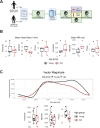
Fig 1.
Fig 1.. Healthy elderly individuals tend to have lower heart rate, increased heart rate variability, and phase advanced activity patterns relative to young subjects.
Experimental protocol for enrolment and monitoring of human subjects (A, made with BioRender.com). As assessed by Zyphyr BioPatch, electrocardiogram (EKG) measurements suggest that MESOR of average heart rate decreases with age (p=0.056) and that MESOR of heart rate variability increases with age (p=0.056). N=5 per group. MESOR and amplitude were tested using two-sided Wilcoxon rank sum exact test, while phase was tested by Kuiper’s two-sample test (B). Average activity counts across three axes (vector magnitude), as recorded by the actigraph device plotted throughout the day (top) and analyzed for circadian rhythm (bottom) (C). While the amplitudes of activity did not differ between the age groups, the older individuals trended towards an early phase (p~0.055, Kupier’s two-sample test) compared to young individuals. N=7 for young and N=8 for old. Lines in the top panel of C are smoothed means (fit with penalized cubic regression splines) for data from each age group. Dots in the bottom panel of C are subject-level cosinor parameter estimates derived from cosinor fits to the actigaraphy data. Boxplot midlines correspond to median values, while the lower and upper hinges correspond to the first and their quartiles, respectively. Boxplot whiskers extend to the smallest/largest points within 1.5 * IQR (Inter Quartile Range) of the lower/upper hinge.
Fig 2.
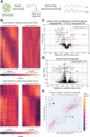
Fig 2.. The circadian transcriptome is differentially affected by entrainment with sera from young and old human subjects.
A visual representation of the serum starvation-serum addition protocol to synchronize the BMAL1-luciferase BJ-5TA fibroblasts (A, made with BioRender.com). When comparing the circadian transcriptome entrained by either young or old sera (n=4 sera per group), 1519 genes lost rhythmicity with age (B, top) while only 637 genes gained rhythmicity with age (B, bottom). Weighted BIC criterion with a threshold of 0.75 was used to assess rhythmicity (B). The number of genes rhythmic in young and old (according to weighted BIC > 0.75 criterion) that show a detectable change in MESOR (q < 0.05 criterion for MESOR difference) is 568, with MESOR increasing in 163 and decreasing in 405 genes. Out of these, 40 genes with increased MESOR (red) and 98 genes with decreased MESOR (blue) also satisfy the condition |log₂ FC| > 0.25 (C). We were only able to detect change in amplitude for a small number of genes, 39 genes had decreased amplitude in old and 2 had increased amplitude using CircaCompare. Only 30 genes with decreased amplitude also satisfy the condition |log₂ FC| > 0.1 (blue) (D). For phase, using a test provided by CircaCompare, under q < 0.05 cutoff for age-related phase differences in genes rhythmic in young and old (with BIC>0.75 cycling criteria), we detected 20 genes with advanced phase, and 34 with delayed phase (E).
Fig 3.
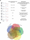
Fig 3.. Each type of circadian change is associated with different KEGG pathways by STRING analysis, but a similar set of transcription factors identified by LISA
Entrainment of the fibroblasts in culture with old serum significantly altered the circadian transcriptome despite use of the same cells in both young and old conditions, suggesting the aged serum itself affects the regulation of specific pathways in the cell. Significant KEGG Pathways (FDR <1×10⁻⁴ unless specified) are indicated next to each category of genes. Age related pathways such as Alzheimer’s Disease/oxidative phosphorylation are associated with a loss of rhythms in the old condition. Cell cycle and DNA replication pathways remain rhythmic in the old serum condition, but cycle with a decreased MESOR. Rhythmic genes were determined using CircaCompare. Rhythmicity was determined using the weighted BIC > 0.75 criterion while p-values for difference in MESOR and phase were determined using CircaCompare (A). LinC similarity analysis (LISA), based on known transcription factor binding in fibroblasts and RNA expression, suggests that 59 total transcription factors (q<0.05) show significant changes in activity in conjunction with the following cycling phenotypes: decreased in MESOR, increased in MESOR, phase delay, gain of rhythmicity, and loss of rhythmicity as were defined above (B).
Fig 4.
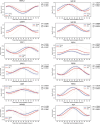
Fig 4.. Synchronizing with old serum phase delays the expression profile of several core clock genes
Traces of molecular clock mRNA transcripts, the average curve (bold) of results of individual sera (faded). P-values for the difference in MESOR (P_M), amplitude (P_A), and phase (P_P) are shown for the comparison of young and old conditions. Several clock genes are significantly phase delayed (CRY1, CRY2, NR1D1, NR1D2, PER3, TEF) in response to synchronization with old serum. While BMAL1 is not phase delayed, the MESOR significantly increases with age. N=4 subjects per timepoint for both young and old groups.

References

    1. Monk T. H., Buysse D. J., Reynolds C. F. & Kupfer D. J. Inducing jet lag in older people: Adjusting to a 6-hour phase advance in routine. Experimental Gerontology 28, 119–133 (1993). - PubMed
    1. Monk T. H., Buysse D. J., Carrier J. & Kupfer D. J. Inducing jet-lag in older people: Directional asymmetry. Journal of Sleep Research 9, 101–116 (2000). - PubMed
    1. Hood S. & Amir S. The aging clock: circadian rhythms and later life. Journal of Clinical Investigation 127, 437–446 (2017). - PMC - PubMed
    1. Farajnia S. et al. Evidence for Neuronal Desynchrony in the Aged Suprachiasmatic Nucleus Clock. J. Neurosci. 32, 5891–5899 (2012). - PMC - PubMed
    1. Nakamura T. J. et al. Age-Related Decline in Circadian Output. J Neurosci 31, 10201–10205 (2011). - PMC - PubMed

Publication types