[Preprint]. 2023 Sep 25:2023.09.25.559083.
doi: 10.1101/2023.09.25.559083.

CELLULAR RNA INTERACTS WITH MAVS TO PROMOTE ANTIVIRAL SIGNALING

Affiliations

CELLULAR RNA INTERACTS WITH MAVS TO PROMOTE ANTIVIRAL SIGNALING

Nandan S Gokhale et al. bioRxiv. .

Update in

  • Cellular RNA interacts with MAVS to promote antiviral signaling.
    Gokhale NS, Sam RK, Somfleth K, Thompson MG, Marciniak DM, Smith JR, Genoyer E, Eggenberger J, Chu LH, Park M, Dvorkin S, Oberst A, Horner SM, Ong SE, Gale M Jr, Savan R. Gokhale NS, et al. Science. 2024 Dec 20;386(6728):eadl0429. doi: 10.1126/science.adl0429. Epub 2024 Dec 20. Science. 2024. PMID: 39700280

Abstract

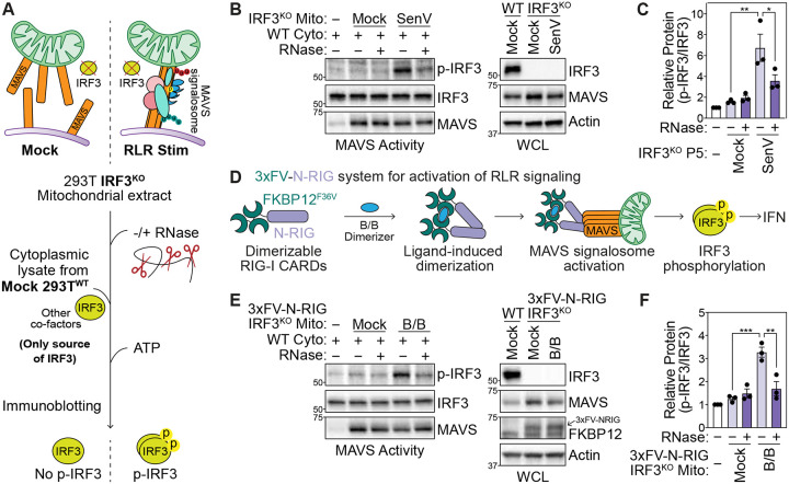
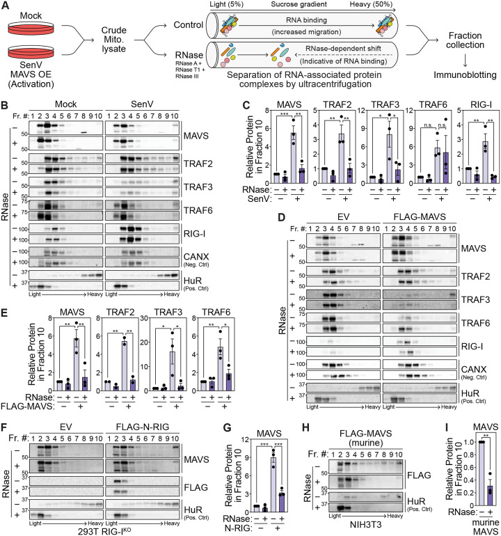
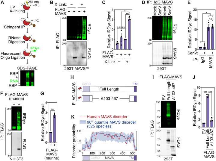
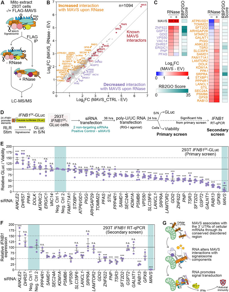
Immune signaling needs to be well-regulated to promote clearance of pathogens, while preventing aberrant inflammation. Interferons (IFNs) and antiviral genes are activated by the detection of viral RNA by RIG-I-like receptors (RLRs). Signal transduction downstream of RLRs proceeds through a multi-protein complex organized around the central adaptor protein MAVS. Recent work has shown that protein complex function can be modulated by RNA molecules providing allosteric regulation or acting as molecular guides or scaffolds. Thus, we hypothesized that RNA plays a role in organizing MAVS signaling platforms. Here, we show that MAVS, through its central intrinsically disordered domain, directly interacts with the 3' untranslated regions of cellular mRNAs. Importantly, elimination of RNA by RNase treatment disrupts the MAVS signalosome, including newly identified regulators of RLR signaling, and inhibits phosphorylation of the transcription factor IRF3. This supports the hypothesis that RNA molecules scaffold proteins in the MAVS signalosome to induce IFNs. Together, this work uncovers a function for cellular RNA in promoting signaling through MAVS and highlights a generalizable principle of RNA regulatory control of cytoplasmic immune signaling complexes.


Conflict of interest statement

COMPETING INTERESTS M.G. is a founder and shareholder in Kineta, Inc., and of HDT Bio.

Figures

Fig. 1:
Fig. 1:. Cellular RNA promotes IRF3 phosphorylation at the MAVS signalosome.
(A) MAVS activity assay for in vitro phosphorylation of IRF3. Crude mitochondrial (Mito) extracts from 293T IRF3KO cells are incubated with cytoplasmic extracts (Cyto) from unstimulated 293TWT cells in the presence of ATP. IRF3 phosphorylation is analyzed by immunoblot. (B) MAVS activity assay to analyze in vitro IRF3 phosphorylation by mitochondrial extracts −/+ RNase treatment from mock or SenV-infected (100 HAU/mL, 16 hpi) 293T IRF3KO cells. WCL = whole cell lysates. (C) Quantification of p-IRF3 (S386) relative to IRF3 from experiments in (B). (D) Outline of 3xFV-N-RIG system that activates the MAVS signalosome in the absence of viral RNA. Tandem FKBP12(F36V) dimerization domains are fused to the N-terminal CARDs of RIG-I. Treatment with the small molecule B/B multimerizes 3xFV-N-RIG to activate downstream signaling. (E) MAVS activity assay to analyze in vitro IRF3 phosphorylation by mitochondrial extracts −/+ RNase treatment from mock or B/B-treated (10 nM, 3 hpt) 293T IRF3KO cells stably expressing 3xFV-N-RIG. (F) Quantification of p-IRF3 relative to IRF3 (S386) from experiments in (E). Data in (B) and (E) are representative of 3 biological replicates. Values in (C) and (F) are mean ± SEM of 3 biological replicates. * p ≤ 0.05, ** p ≤ 0.01, *** p ≤ 0.001 by one-way ANOVA with Tukey’s multiple comparison test.
Fig. 2:
Fig. 2:. The MAVS signalosome is associated with cellular RNA.
(A) RNase-dependent shift assay to identify RNA-associated proteins. Mitochondrial lysates treated with RNase inhibitor (− RNase) or an RNase cocktail (+ RNase; RNase A, I, T1, III) are separated on a sucrose gradient by ultracentrifugation. RNA-associated proteins have reduced migration to heavy fractions in the presence of RNase. (B) Immunoblot analysis of fractions collected after sucrose gradient ultracentrifugation of mitochondrial lysates −/+ RNase treatment from mock- and SenV-infected (100 HAU/mL, 14 hpi) 293T cells. (C) Quantification of the indicated protein in fraction 10 relative to that in fraction 3 from experiments in (B). (D) Immunoblot analysis of fractions collected after sucrose gradient ultracentrifugation of mitochondrial lysates −/+ RNase treatment from 293T cells transfected with empty vector (EV) and FLAG-tagged MAVS (24 hpt). (E) Quantification of the indicated protein in fraction 10 relative to that in fraction 3 from experiments in (D). (F) Immunoblot analysis of fractions collected after sucrose gradient ultracentrifugation of mitochondrial lysates −/+ RNase treatment from 293T RIG-IKO cells transfected with empty vector and FLAG-tagged N-RIG. (G) Quantification of MAVS in fraction 10 relative to that in fraction 3 from experiments in (F). (H) Immunoblot analysis of fractions collected after sucrose gradient ultracentrifugation of mitochondrial lysates −/+ RNase treatment from murine NIH3T3 cells transfected with FLAG-tagged murine MAVS. (I) Quantification of FLAG-tagged murine MAVS in fraction 10 relative to that in fraction 3 from experiments in (H). Data in (B), (D), (F), and (H) are representative of 3 biological replicates. Values in (C), (E), (G), and (I) are mean ± SEM of 3 biological replicates. * p ≤ 0.05, ** p ≤ 0.01, *** p ≤ 0.001 by one-way ANOVA with Tukey’s multiple comparison test ((C), (E), and (G)) or unpaired t test (I). n.s. = not significant.
Fig. 3:
Fig. 3:. MAVS interacts with RNA through its central intrinsically disordered region.
(A) Infrared-dye crosslinking and immunoprecipitation (irCLIP) strategy to visualize RNA-protein complexes. The protein of interest is stringently immunoprecipitated and UV-crosslinked RNA is digested to fragments with RNase A. Following ligation of an IRDye-800 conjugated oligonucleotide, complexes are resolved by SDS-PAGE. RNA-protein complexes are detected by IRDye-800 fluorescence and immunoprecipitation is validated by immunoblot analysis. (B) irCLIP −/+ crosslinking of FLAG-tagged MAVS expressed in 293T MAVSKO cells (24 hpt). EV = empty vector. (C) Quantification of IRDye signal in experiments in (B). (D) irCLIP of endogenous MAVS from mock- and SenV-infected (100 HAU/mL, 16 hpi) 293T cells. (E) Quantification of IRDye signal in experiments in (D). (F) irCLIP of FLAG-tagged murine MAVS expressed in murine NIH3T3 cells (24 hpt). (G) Quantification of IRDye signal in experiments in (F). (H) Schematic of FLAG-tagged full length MAVS and MAVSΔ103–467 used in (I). (I) irCLIP of the indicated FLAG-tagged MAVS constructs expressed in 293T MAVSKO cells (24 hpt). (J) Quantification of IRDye signal in experiments in (I). (K) Prediction of disorder in human MAVS (red) and in 325 mammalian species (blue) using IUPred3. Data in (B), Data in (D), (F), and (I) are representative of 3 biological replicates. Values in (C), (E), and (G), and (J) are mean ± SEM of 3 biological replicates. * p ≤ 0.05, ** p ≤ 0.01, *** p ≤ 0.001 by one-way ANOVA with Tukey’s multiple comparison test. n.s. = not significant.
Fig. 4:
Fig. 4:. MAVS interacts with the 3′ UTRs of mRNAs.
(A) Targeted APOBEC1-mediated editing approach used to profile MAVS-interacting RNAs. C-to-T edits in sequenced cDNA enhanced with APOBEC1-MAVSFL overexpression relative to non-specific (APOBEC1 alone; CTRL) and mitochondria-localized (APOBEC1-MAVSC-term) background controls identifies MAVS-associated RNAs. (B) Summary of C-to-T editing sites and edited transcripts identified when comparing the indicated APOBEC1-MAVS constructs with background controls across biological triplicates. (C) Percent C-to-T editing (purple) at called sites in PMAIP1 and IFIT2 3′UTRs (mean of 3 replicates). Read counts from one representative experiment are shown in yellow. (D) Left: CLIP-RT-qPCR analysis of normalized enrichment relative to input of PMAIP1 and IFIT2 mRNA by immunoprecipitation of the indicated FLAG-tagged MAVS constructs in transfected 293T MAVSKO cells (24 hpt). Values are mean ± SEM of 4 biological replicates. * p ≤ 0.05, ** p ≤ 0.01, by unpaired t test. Right: Representative immunoblot of immunoprecipitated fractions. (E) Bottom: CLIP-RT-qPCR analysis of normalized enrichment relative to input of PMAIP1 and IFIT2 mRNA by immunoprecipitation of endogenous MAVS from mock- and SenV-infected (100 HAU/mL, 16 hpt) 293T cells. IgG was used as background control. Values are mean ± SEM of 3 biological replicates. * p ≤ 0.05 by one-way ANOVA with Tukey’s multiple comparison test. Top: Representative immunoblot of immunoprecipitated fractions.
Fig. 5:
Fig. 5:. RNA alters functional MAVS protein-protein interactions
(A) Immunoprecipitation and mass spectrometry strategy to identify RNA-dependent MAVS interactors. (B) Scatterplot of 1094 proteins significantly enriched with MAVS (Log2FC ≥ 2, p ≤ 0.05, found in < 30% of CRAPome datasets) −/+ RNase over empty vector control across 5 biological replicates. Dashed lines delineate |Log2FC| = 0.5 from the diagonal. Known MAVS-interacting proteins are in red, those with decreased MAVS-interaction upon RNase treatment are in purple, and those with increased MAVS-interaction upon RNase treatment are in ochre. (C) Heatmap of the enrichment of RNA-dependent MAVS-interactors over empty vector control −/+ RNase (blue-red gradient), and the RBP2GO score, a measure of empirically determined propensity to interact with RNA, for each protein (teal). (D) Schematic of targeted siRNA screen for RNA-dependent MAVS interactors. (E) Relative Gaussia luciferase (GLuc) activity normalized to viability following poly-U/UC RNA transfection (50 ng, 24 hpt) in 293T IFNB12X-GLuc reporter cells upon depletion of RNA-dependent MAVS interactors by siRNA treatment (36 hours). Controls are highlighted in teal. (F) Normalized IFNB1 mRNA expression relative to HPRT1 following poly-U/UC RNA transfection (50 ng, 24 hpt) in 293T cells upon depletion of RNA-dependent MAVS interactors by siRNA treatment (36 hours), determined by RTqPCR. Controls are highlighted in teal. (G) Proposed model for RNA regulation of MAVS signalosome function. Values are the mean ± SEM of 5 (E) or 3 (F) biological replicates. * p ≤ 0.05, ** p ≤ 0.01, *** p ≤ 0.001 by one-way ANOVA with Tukey’s multiple comparison test. n.s. = not significant.

References

    1. Rehwinkel J., Gack M. U., RIG-I-like receptors: their regulation and roles in RNA sensing. Nat Rev Immunol. 20, 537–551 (2020). - PMC - PubMed
    1. Ivashkiv L. B., Donlin L. T., Regulation of type I interferon responses. Nat. Rev. Immunol. 14, 36–49 (2014). - PMC - PubMed
    1. Kato H., Takeuchi O., Sato S., Yoneyama M., Yamamoto M., Matsui K., Uematsu S., Jung A., Kawai T., Ishii K. J., Yamaguchi O., Otsu K., Tsujimura T., Koh C.-S., Reis e Sousa C., Matsuura Y., Fujita T., Akira S., Differential roles of MDA5 and RIG-I helicases in the recognition of RNA viruses. Nature. 441, 101–105 (2006). - PubMed
    1. Horner S. M., Liu H. M., Park H. S., Briley J., Gale M., Mitochondrial-associated endoplasmic reticulum membranes (MAM) form innate immune synapses and are targeted by hepatitis C virus. Proc Natl Acad Sci U S A. 108, 14590–14595 (2011). - PMC - PubMed
    1. Dixit E., Boulant S., Zhang Y., Lee A. S. Y., Odendall C., Shum B., Hacohen N., Chen Z. J., Whelan S. P., Fransen M., Nibert M. L., Superti-Furga G., Kagan J. C., Peroxisomes are signaling platforms for antiviral innate immunity. Cell. 141, 668–681 (2010). - PMC - PubMed

Publication types