Review
. 2023 Sep 6;15(9):e44801.
doi: 10.7759/cureus.44801. eCollection 2023 Sep.

The Spectrum of Shoulder Pathologies on Magnetic Resonance Imaging: A Pictorial Review

Affiliations
Review

The Spectrum of Shoulder Pathologies on Magnetic Resonance Imaging: A Pictorial Review

Anshul Sood et al. Cureus. .

Abstract

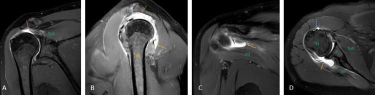
Patients present to the orthopedic outpatient department with complaints of shoulder pain on movement or restriction of movement in the shoulder joint and are referred for magnetic resonance imaging (MRI) of the shoulder joint. Almost all the patients have similar complaints but may have a wide range of pathology affecting the joint and causing pain. Rotator cuff tears or tendinopathy are the most common causes of shoulder pain. Ultrasound (USG) and MRI are the most commonly used imaging modalities for assessing rotator cuff pathologies. There is a wide range of pathologies affecting the shoulder joint, other than rotator cuff tendinopathies or tears, for which USG is less sensitive and specific in detecting accurate pathology. MRI is the choice of imaging for shoulder joint pathologies. We present a pictorial review discussing and depicting MRI features of a wide list of pathologies of the shoulder joint complex that should be kept in mind when the patient presents with shoulder pain.

Keywords: mri; pathology; pictorial; radiology; shoulder.


Conflict of interest statement

The authors have declared that no competing interests exist.

Figures

Figure 1
Figure 1. A 39-year-old male with hemophilic arthropathy. Axial fat-suppressed proton density (A), sagittal fat-suppressed proton density (B), and coronal susceptibility-weighted imaging (C) show erosive destruction of the head of humerus {H} with loss of contour and erosions of the visualized part including the greater and lesser tuberosity and glenoid margins (arrows) with hypo intensity of visualized subscapularis {Sub}, supraspinatus {S} and infraspinatus {Inf} on fat-suppressed proton density and blooming on susceptibility-weighted imaging.
Image credits: Anshul Sood
Figure 2
Figure 2. A 56-year-old female with tuberculous arthropathy. Contrast-enhanced T1 weighted axial (A), sagittal (B), and coronal (C) images show enhancing hyperintensities in the acromion {A} and distal clavicular {C} joint with decreased joint spaces and heterogeneously enhancing collection in the subacromial and subcoracoid bursa (arrows).
Image credits: Anshul Sood
Figure 3
Figure 3. A seven-year-old male with sickle cell disease with marrow reconversion and myositis. Coronal T1 weighted imaging (A) shows a hypointense signal in the proximal shaft and neck of the humerus (orange arrows) with mild expansion of the involved areas. Coronal fat-suppressed proton density (B) shows hyperintense signals in the proximal shaft and neck of the humerus with mild expansion of the involved areas (orange arrows) suggesting bone marrow reconversion. Also, hyperintense signal is seen in the short head of the triceps suggesting myositis (blue arrow).
H = humerus Image credits: Anshul Sood
Figure 4
Figure 4. A 43-year-old female with lung cancer metastasis. Coronal T2 weighted imaging (A), T1 weighted imaging (B), and contrast-enhanced T1 weighted imaging (C) show peripherally enhancing collection (orange arrows) and destruction of neck and head of humerus {H}, and glenoid labrum (green arrow) with increased joint space.
Image credits: Anshul Sood
Figure 5
Figure 5. A 45-year-old male with rheumatoid arthritis of the acromioclavicular joint. Coronal fat-suppressed proton density sequence (A), and short tau inversion recovery sequence shows peri-articular erosion at the acromion {A - green color} and distal clavicular {C} joint with marrow edema and hyperintense signal in peri-articular soft tissues (arrow).
H = humerus Image credits: Anshul Sood
Figure 6
Figure 6. A 55-year-old male with soft tissue myxoma. Coronal short tau inversion recovery sequence (A), fat-suppressed proton density sequence (B), and contrast-enhanced T1 weighted imaging (C) show well-defined heterogeneously enhancing lobulated soft tissue mass lesion in the posterior aspect of the axilla (arrows) involving the triceps muscle {T} and abutting the subscapularis muscle {Sub}.
H = humerus Image credits: Anshul Sood
Figure 7
Figure 7. A 27-year-old male with avascular necrosis of shoulder joint. Coronal fat-suppressed proton density sequence (A) and coronal T2 weighted imaging (B) show geographical lesions with irregular margins with heterogenous signal intensity (arrow) involving the neck of the humerus {H} with erosion and destruction of the head of the humerus.
Image credits: Anshul Sood
Figure 8
Figure 8. A 17-year-old male with Perthes. Axial (A) and coronal (B) fat-suppressed proton density images show detachment of the anteroinferior labrum with medially stripped but intact periosteum.
H = humerus Image credits: Anshul Sood
Figure 9
Figure 9. A 20-year-old male with posterior gleno-labral articular disruption (GLAD). Coronal fat-suppressed proton density (A) and (B) images show discontinuity in the glenoid labrum with hyperintensity in the defect (arrows).
H = humerus Image credits: Anshul Sood
Figure 10
Figure 10. A 40-year-old male with a bony Hill-Sachs lesion. Axial fat-suppressed proton density (A) shows loss of normal circular shape of the postero-lateral surface of the head of humerus (arrow); and coronal fat-suppressed proton density (B) shows fraying of the anteroinferior glenoid labrum (arrow).
H = humerus Image credits: Anshul Sood
Figure 11
Figure 11. A 55-year-old male with partial articular surface supraspinatus tendon avulsion (PASTA). Coronal fat-suppressed proton density sequence (A) and coronal T1 weighted imaging (B) show the hyperintense signal at the greater tuberosity of the humerus {H} at the insertion of the supraspinatus muscle {S} with surface irregularity (arrow).
Image credits: Anshul Sood
Figure 12
Figure 12. A 35-year-old male with an avulsion fracture of the supraspinatus tendon. Coronal T1 weighted imaging (A) and fat-suppressed proton density imaging (B) show avulsion fracture of the supraspinatus {S} tendon at its insertion at greater tuberosity of the humerus {H} which shows marrow edema (arrow).
Image credits: Anshul Sood
Figure 13
Figure 13. A 45-year-old female with a malunited fracture of the proximal humerus (neck). Coronal T1 weighted image (A), T2 weighted image (B), and fat-suppressed proton density image (C) show a malunited fracture of the proximal humerus (neck).
Image credits: Anshul Sood
Figure 14
Figure 14. A 39-year-old female with infraspinatus myositis. Axial fat-suppressed proton density sequence (A) and sagittal fat-suppressed proton density sequence (B) show hyperintense signal in the infraspinatus muscle {Inf} at the greater tubercle.
H = humerus Image credits: Anshul Sood
Figure 15
Figure 15. A 27-year-old male with a sprain of supraspinatus in an operated case of arthroscopic repair of supraspinatus. Coronal (A) and sagittal (B) fat-suppressed proton density images show a hyperintense signal at the insertion of the supraspinatus tendon (orange arrows) with implanted screw (blue arrow) noted in the anterosuperior part of the humerus {H}.
Image credits: Anshul Sood
Figure 16
Figure 16. A 46-year-old male with supraspinatus tendinopathy due to external impingement. Coronal (A) and sagittal (B) fat-suppressed proton density sequence shows acromion impingement over the supraspinatus tendon with hyperintensity near its humeral attachment (orange arrow). Degenerative changes at the acromioclavicular joint in the form of a decrease in joint space and increased signal intensity (yellow arrow).
H = humerus Image credits: Anshul Sood
Figure 17
Figure 17. A 51-year-old male with a complete tear of the supraspinatus tendon, a partial tear of the infraspinatus tendon, and the bicipital tendinopathy. Coronal (A) and (C), sagittal (B), and axial (D) fat-suppressed proton density sequence show hyperintensity along the fibers of supraspinatus near its attachment to the humeral head with complete disruption of fibers suggesting complete tear (orange arrow); hyperintensity along the fibers of infraspinatus near its attachment to the humeral head with partial disruption of fibers suggesting partial tear (yellow arrow). Hyperintensity along the tendon of the long head of the biceps in the bicipital groove suggesting bicipital tendinopathy (blue arrow).
H = humerus; S = scapula; Sub = subscapularis; Sup = supraspinatus; Inf = infraspinatus Image credits: Anshul Sood

References

    1. Role of magnetic resonance imaging in the evaluation of rotator cuff tears. Koganti DV, Lamghare P, Parripati VK, Khandelwal R, Reddy AD. Cureus. 2022;14:0. - PMC - PubMed
    1. Prevalence and incidence of shoulder pain in the general population; a systematic review. Luime JJ, Koes BW, Hendriksen IJ, Burdorf A, Verhagen AP, Miedema HS, Verhaar JA. Scand J Rheumatol. 2004;33:73–81. - PubMed
    1. Shoulder disorders in general practice: incidence, patient characteristics, and management. van der Windt DA, Koes BW, de Jong BA, Bouter LM. Ann Rheum Dis. 1995;54:959–964. - PMC - PubMed
    1. Magnetic resonance imaging of the shoulder. Ashir A, Lombardi A, Jerban S, Ma Y, Du J, Chang EY. Pol J Radiol. 2020;17:420–439. - PMC - PubMed
    1. Dixon A. Haemophilic arthropathy. [ Jul; 2023 ]. 2022. https://radiopaedia.org/articles/haemophilic-arthropathy https://radiopaedia.org/articles/haemophilic-arthropathy

LinkOut - more resources