. 2023 Sep 15;9(9):e20201.
doi: 10.1016/j.heliyon.2023.e20201. eCollection 2023 Sep.

Engineered elastin-like polypeptide improves the efficiency of adipose-derived stem cell-mediated cutaneous wound healing in type II diabetes mellitus

Affiliations

Engineered elastin-like polypeptide improves the efficiency of adipose-derived stem cell-mediated cutaneous wound healing in type II diabetes mellitus

Seung-Hwa Woo et al. Heliyon. .

Abstract

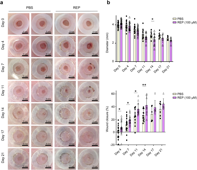
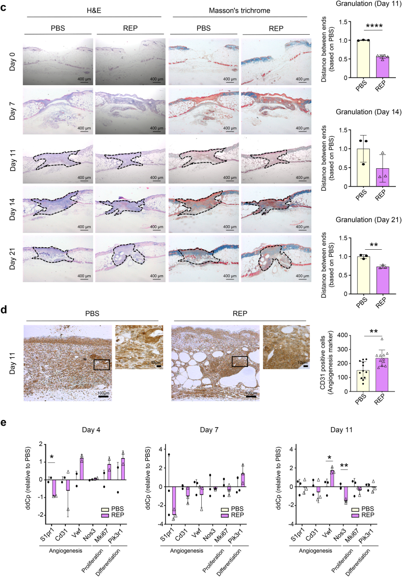
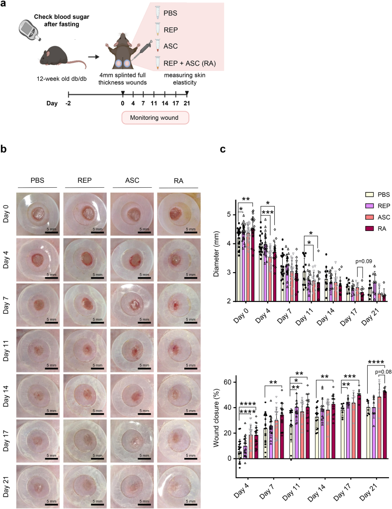
Impaired cutaneous wound healing is a major complication in patients with diabetes mellitus (DM), leading to increased amputation and mortality rates in affected patients. Adipose-derived stem cells (ASCs) are widely used seed cells for promoted tissue regeneration to improve wound closure under diabetic conditions. However, ASCs-based therapies remain limited due to difficulties in maintaining cell quality during transplantation. To overcome this problem, extracellular matrix mimetic biomaterials have been developed for use in biomedical engineering field, including tissue engineering and regenerative medicine. Herein, a biosynthesized arginine-glycine-aspartate amino acid residues (RGD motif, known as a cell adhesion motif)-containing elastin-like polypeptides (REPs) improved the efficacy of ASCs in enhancing wound closure and skin elasticity in diabetic wounds by promoting the expression of angiogenic growth factors. Therefore, REPs can be used as potential supplements to stem cell-based therapeutic approach to accelerate diabetic wound repair.

Keywords: Adipose-derived stem cells; Engineered elastin-like polypeptide; Skin elasticity; Type II diabetes mellitus; Wound healing.


Conflict of interest statement

The authors declare the following financial interests/personal relationships which may be considered as potential competing interests: Won Bae Jeon is the founder and CEO of Excellamol Inc., and has employment and financial relationships with Excellamol Inc., including patent inventions related with REPs. All other authors have no financial conflicts of interests.

Figures

Fig. 1
Fig. 1
REPs accelerate skin wound closure in db/db mice. (a) Schematic of the experimental procedure. Twelve-week-old db/db mice were randomly divided into four groups after measuring their blood sugar levels. Two excisional wounds were generated in the dorsal skin with a 4-mm biopsy punch. The wounds in each group were treated with PBS or REPs (50, 100, and 150 μM). The wound healing process was monitored for 11 days. (b) Representative images of the wound healing process. Scale bar = 5 mm. (c) Changes in the average wound diameter and percent wound closure. Each data point corresponds to a single wound (the number of wound ≥20 in each group). (d) H&E staining (top) and Masson's trichrome staining (bottom) of the wound after day 11. Scale bar = 200 μm. The granulation tissue border is marked with a dashed line. Changes in the granulation size (right). Each data point corresponds to a single wound (the number of wound = 4–5 in each group). The data are presented as the mean ± SD. For statistical analyses in (cd): *p < 0.05 according to multiple t-test (c) and **p < 0.01 according to unpaired t-test (d) compared to the PBS group.
Fig. 2
Fig. 2
REPs regulate angiogenic factor expression in db/db mice. (a) Twelve-week-old db/db mice were randomly divided into two groups after measuring their blood glucose levels. Two excisional wounds were generated in the dorsal skin with a 4-mm biopsy punch. The wounds in each group were treated with PBS or 100 μM REP. The wound healing process was monitored for 21 days. Scale bar = 5 mm. (b) Changes in the average wound diameter and percent wound closure. Each data point corresponds to a single wound (the number of wound ≥6 in each group). (c) H&E staining (left) and Masson's trichrome staining (middle) of the skin at wound site during healing. Scale bar = 400 μm. The granulation tissue border is marked with a dashed line. Changes in the granulation size (right). Each point corresponds to a single wound (the number of wound = 3 in each group). (d) Immunostaining for CD31 in the control group and the REP group. Scale bar = 10 μm (higher magnification images) or 100 μm (lower magnification). The number of CD31-positive cells (in brown color) was measured with ImageJ software (IHC tool). The amount of stained cells in each view field is represented by each data point (n = 11–12). (e) mRNA expression of wound healing markers, including genes that are associated with angiogenesis, proliferation, and differentiation. The data shown are mean ± SD. n = 3 (biological replicate). For statistical analyses in (be): *p < 0.05, **p < 0.01 according to multiple t-test (b, e) and **p < 0.01, ****p < 0.0001 according to unpaired t-test (c) compared to the PBS group.
Fig. 2
Fig. 2
REPs regulate angiogenic factor expression in db/db mice. (a) Twelve-week-old db/db mice were randomly divided into two groups after measuring their blood glucose levels. Two excisional wounds were generated in the dorsal skin with a 4-mm biopsy punch. The wounds in each group were treated with PBS or 100 μM REP. The wound healing process was monitored for 21 days. Scale bar = 5 mm. (b) Changes in the average wound diameter and percent wound closure. Each data point corresponds to a single wound (the number of wound ≥6 in each group). (c) H&E staining (left) and Masson's trichrome staining (middle) of the skin at wound site during healing. Scale bar = 400 μm. The granulation tissue border is marked with a dashed line. Changes in the granulation size (right). Each point corresponds to a single wound (the number of wound = 3 in each group). (d) Immunostaining for CD31 in the control group and the REP group. Scale bar = 10 μm (higher magnification images) or 100 μm (lower magnification). The number of CD31-positive cells (in brown color) was measured with ImageJ software (IHC tool). The amount of stained cells in each view field is represented by each data point (n = 11–12). (e) mRNA expression of wound healing markers, including genes that are associated with angiogenesis, proliferation, and differentiation. The data shown are mean ± SD. n = 3 (biological replicate). For statistical analyses in (be): *p < 0.05, **p < 0.01 according to multiple t-test (b, e) and **p < 0.01, ****p < 0.0001 according to unpaired t-test (c) compared to the PBS group.
Fig. 3
Fig. 3
REPs improve the efficiency of adipose-derived stem cell-mediated wound healing with increasing skin elasticity. (a) Schematic of the experimental procedure. Ten-week-old db/db mice were randomly selected into four groups after measuring their blood glucose levels. Two full-thickness wounds were generated in the dorsal skin of mice with a 4-mm biopsy punch. The wounds in each group were treated with PBS (control group), 100 μM REPs (REP group), ASCs (ACS group), or 100 μM REP + ASC (RA group). The wound healing process was monitored for 21 days. (b) Representative images of the wound healing process. Scale bar = 5 mm. (c) Changes in the average wound diameter and percent wound closure. Each point corresponds to a single wound (the number of wound ≥8 in each group). (d) Representative images of H&E and Masson's trichrome staining of the wound during healing (left). Scale bar = 200 μm. The granulation tissue border is marked with a dashed line. Changes in the granulation size (right). Each point corresponds to a single wound (the number of wound = 2–6 in each group). (e) Immunostaining for CD31 in the control, REP, ASC, and RA group. Scale bar = 10 μm (higher magnification) or 100 μm (lower magnification). (f) Quantification of CD31-positive cells in each group. The amount of positively stained cells in each view field is represented by each data point (n = 25–30). (g) Skin elasticity level on day 21 after wounding. Each point corresponds to a single wound (the number of wound = 16 in each group). The data shown are mean ± SD. For statistical analyses in (cg): *p < 0.05, **p < 0.01, ***p < 0.001, and ****p < 0.0001 according to multiple t-test (c) and according to unpaired t-test (dg).
Fig. 3
Fig. 3
REPs improve the efficiency of adipose-derived stem cell-mediated wound healing with increasing skin elasticity. (a) Schematic of the experimental procedure. Ten-week-old db/db mice were randomly selected into four groups after measuring their blood glucose levels. Two full-thickness wounds were generated in the dorsal skin of mice with a 4-mm biopsy punch. The wounds in each group were treated with PBS (control group), 100 μM REPs (REP group), ASCs (ACS group), or 100 μM REP + ASC (RA group). The wound healing process was monitored for 21 days. (b) Representative images of the wound healing process. Scale bar = 5 mm. (c) Changes in the average wound diameter and percent wound closure. Each point corresponds to a single wound (the number of wound ≥8 in each group). (d) Representative images of H&E and Masson's trichrome staining of the wound during healing (left). Scale bar = 200 μm. The granulation tissue border is marked with a dashed line. Changes in the granulation size (right). Each point corresponds to a single wound (the number of wound = 2–6 in each group). (e) Immunostaining for CD31 in the control, REP, ASC, and RA group. Scale bar = 10 μm (higher magnification) or 100 μm (lower magnification). (f) Quantification of CD31-positive cells in each group. The amount of positively stained cells in each view field is represented by each data point (n = 25–30). (g) Skin elasticity level on day 21 after wounding. Each point corresponds to a single wound (the number of wound = 16 in each group). The data shown are mean ± SD. For statistical analyses in (cg): *p < 0.05, **p < 0.01, ***p < 0.001, and ****p < 0.0001 according to multiple t-test (c) and according to unpaired t-test (dg).

Similar articles

Cited by

References

    1. Yoon H.S., Baik S.H., Oh C.H. Quantitative measurement of desquamation and skin elasticity in diabetic patients. Skin Res. Technol. 2002;8(4):250–254. - PubMed
    1. Greenhalgh D.G. Wound healing and diabetes mellitus. Clin. Plast. Surg. 2003;30(1):37–45. - PubMed
    1. Seirafi H., et al. Biophysical characteristics of skin in diabetes: a controlled study. J. Eur. Acad. Dermatol. Venereol. 2009;23(2):146–149. - PubMed
    1. Baumann L B.E., Weiss A.S., Bates D., Humphrey S., Silberberg M., Daniels R. Clinical relevance of elastin in the structure and function of skin. Aesthet Surg J Open Forum. 2021 May 14;3(3):ojab019. - PMC - PubMed
    1. Van Putte L D.S.S., Moortgat P. The effects of advanced glycation end products (AGEs) on dermal wound healing and scar formation: a systematic review. Scars Burn Heal. 2016 Dec 5;2 - PMC - PubMed

LinkOut - more resources