Review
. 2023 Sep 7;9(9):e19929.
doi: 10.1016/j.heliyon.2023.e19929. eCollection 2023 Sep.

Nanomaterials-based biosensor and their applications: A review

Affiliations
Review

Nanomaterials-based biosensor and their applications: A review

Sumit Malik et al. Heliyon. .

Abstract

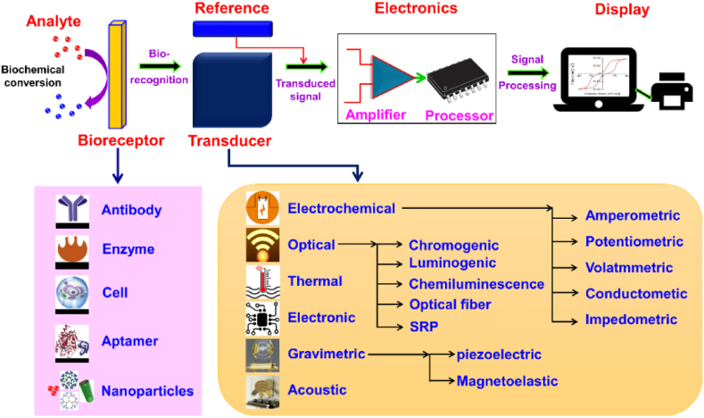
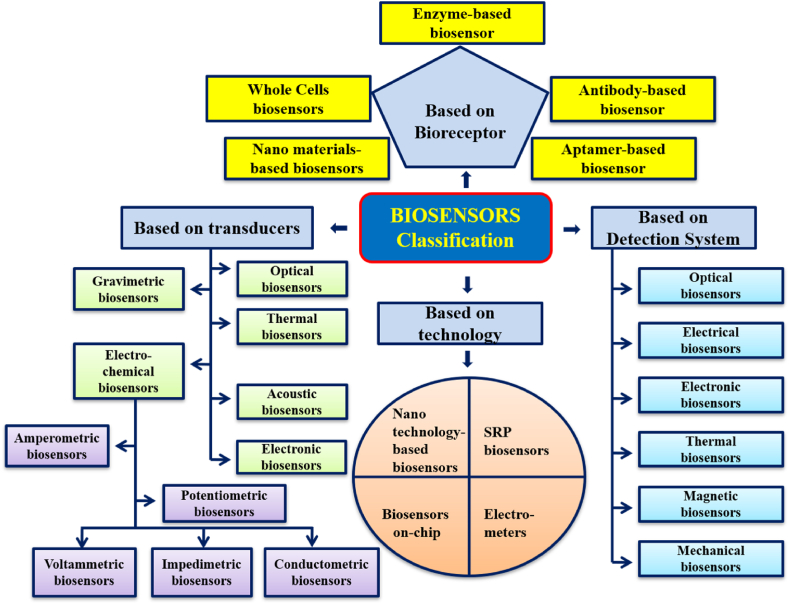
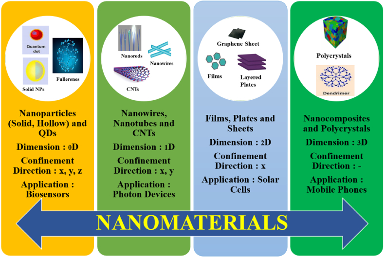
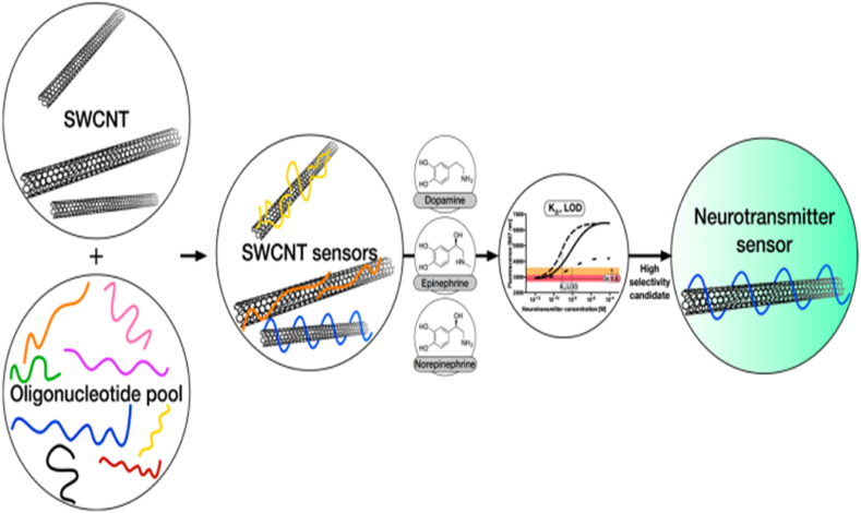
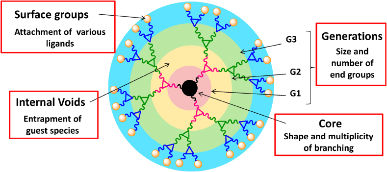
A sensor can be called ideal or perfect if it is enriched with certain characteristics viz., superior detections range, high sensitivity, selectivity, resolution, reproducibility, repeatability, and response time with good flow. Recently, biosensors made of nanoparticles (NPs) have gained very high popularity due to their excellent applications in nearly all the fields of science and technology. The use of NPs in the biosensor is usually done to fill the gap between the converter and the bioreceptor, which is at the nanoscale. Simultaneously the uses of NPs and electrochemical techniques have led to the emergence of biosensors with high sensitivity and decomposition power. This review summarizes the development of biosensors made of NPssuch as noble metal NPs and metal oxide NPs, nanowires (NWs), nanorods (NRs), carbon nanotubes (CNTs), quantum dots (QDs), and dendrimers and their recent advancement in biosensing technology with the expansion of nanotechnology.

Keywords: Carbon nanotubes; Dendrimers; Nanorods; Nanowires; Quantum dots.


Conflict of interest statement

The authors declare that they have no known competing financial interests or personal relationships that could have appeared to influence the work reported in this paper.

Figures

Fig. 1
Fig. 1
Schematic diagram of biosensor consisting of bioreceptor, transducer, and amplifier. Reproduced with permission from Ref. [3], with the permission of the Creative Commons Attribution 4.0 International License (http://creativecommons.org/licenses/by/4.0/). Copyright 2021, MDPI.
Fig. 2
Fig. 2
The different milestones achieved in the field of biosensors. Reproduced with permission from Ref. [12], with the permission of the Creative Commons Attribution 4.0 International License (http://creativecommons.org/licenses/by/4.0/). Copyright 2022, MDPI.
Fig. 3
Fig. 3
Classification of biosensors based on detection system, transducer, technology and bio-receptors.
Fig. 4
Fig. 4
Different types of NMs with different dimensions utilized in designing biosensors.
Fig. 5
Fig. 5
Top-down and bottom-up methodologies for preparing nanomaterials. Reproduced with permission from Ref. [18], with the permission of the Creative Commons Attribution 4.0 International License (http://creativecommons.org/licenses/by/4.0/). Copyright 2022, MDPI.
Fig. 6
Fig. 6
The four different shapes of ZnO nanostructures with their characteristics. Reproduced with permission from Ref. [22], with the permission of the Creative Commons Attribution 4.0 International License (http://creativecommons.org/licenses/by/4.0/). Copyright 2019, MDPI.
Fig. 7
Fig. 7
ZnO based biosensor for pesticide detection. Reproduced with permission from Ref. [27], with the permission of the Creative Commons Attribution 4.0 International License (http://creativecommons.org/licenses/by/4.0/). Copyright 2022, MDPI.
Fig. 8
Fig. 8
Schematic for the paper based colorimetric sensor using mesoporous copper oxide CuO hollow sphere for the detection of hydrogen peroxide. Reproduced with permission from Ref. [43], with the permission of the Creative Commons Attribution 4.0 International License (http://creativecommons.org/licenses/by/4.0/). Copyright 2021, MDPI.
Fig. 9
Fig. 9
Fe2O3/GO/GCE based electrochemical sensor for the detection of dopamine and uric acid. Reproduced with permission from Ref. [58], with the permission of the Creative Commons Attribution 4.0 International License (http://creativecommons.org/licenses/by/4.0/). Copyright 2019, MDPI.
Fig. 10
Fig. 10
Graphene quantum dots (GQDs)-based immunosensor for Y. enterocolitica detection. Reproduced with permission from Ref. [94], with the permission of the Creative Commons Attribution 4.0 International License (http://creativecommons.org/licenses/by/4.0/). Copyright 2019, MDPI.
Fig. 11
Fig. 11
The schematic illustration for Si-NW-sensors to detect biomolecules. Reproduced with permission from Ref. [114], with the permission of the Creative Commons Attribution 4.0 International License (http://creativecommons.org/licenses/by/4.0/). Copyright 2021, MDPI.
Fig. 12
Fig. 12
ZnO nanorods coated optical fiber sensor for volatile organic compounds (VOC) biomarker detection. Reproduced with permission from Ref. [122], with the permission of the Creative Commons Attribution 4.0 International License (http://creativecommons.org/licenses/by/4.0/). Copyright 2022, MDPI.
Fig. 13
Fig. 13
Fluorescent carbon nanotube-based neurotransmitter sensors. Reproduced with permission from Ref. [147], with the permission of the Creative Commons Attribution 4.0 International License (http://creativecommons.org/licenses/by/4.0/). Copyright 2017, MDPI.
Fig. 14
Fig. 14
Different elements of Dendrimers. Reproduced with permission from Ref. [148], with the permission of the Creative Commons Attribution 4.0 International License (http://creativecommons.org/licenses/by/4.0/). Copyright 2015, MDPI.

References

    1. Theavenot D.R., Toth K., Durst R.A., Wilson G.S. Electrochemical biosensors: recommended definitions and classification. Biosens. Bioelectron. 2001;16:121–131. - PubMed
    1. Heineman W.R., Jensen W.B., Clark Leland C., Jr. Biosens. Bioelectron. 2006;8(21):1403–1404. 1918–2005.
    1. Naresh V., Lee N. A review on biosensors and recent development of nanostructured materials-enabled biosensors. Sensors. 2021;21(4):1109. doi: 10.3390/s21041109. - DOI - PMC - PubMed
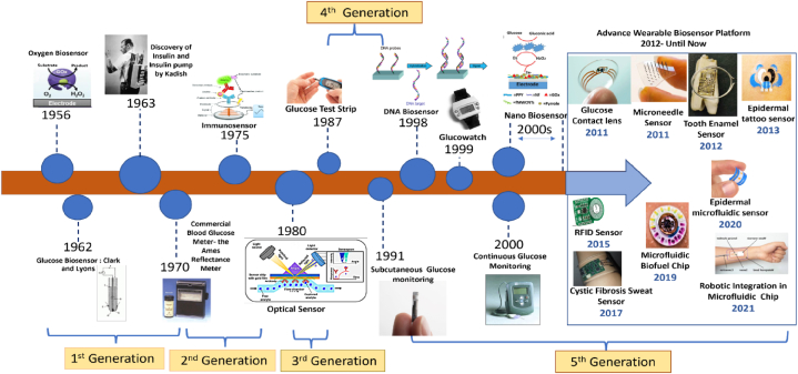
    1. Clark L.C., Lyons C. Electrode systems for continuous monitoring in cardiovascular surgery. Ann. N. Y. Acad. Sci. 1962;102:29–45. - PubMed
    1. Updike S.J., Hicks G.P. The enzyme electrode. Nature. 1967;214:986–988. - PubMed

LinkOut - more resources