. 2023 Aug 20;9(9):e19341.
doi: 10.1016/j.heliyon.2023.e19341. eCollection 2023 Sep.

In silico and in vitro inhibition of host-based viral entry targets and cytokine storm in COVID-19 by ginsenoside compound K

Affiliations

In silico and in vitro inhibition of host-based viral entry targets and cytokine storm in COVID-19 by ginsenoside compound K

Vinothini Boopathi et al. Heliyon. .

Abstract

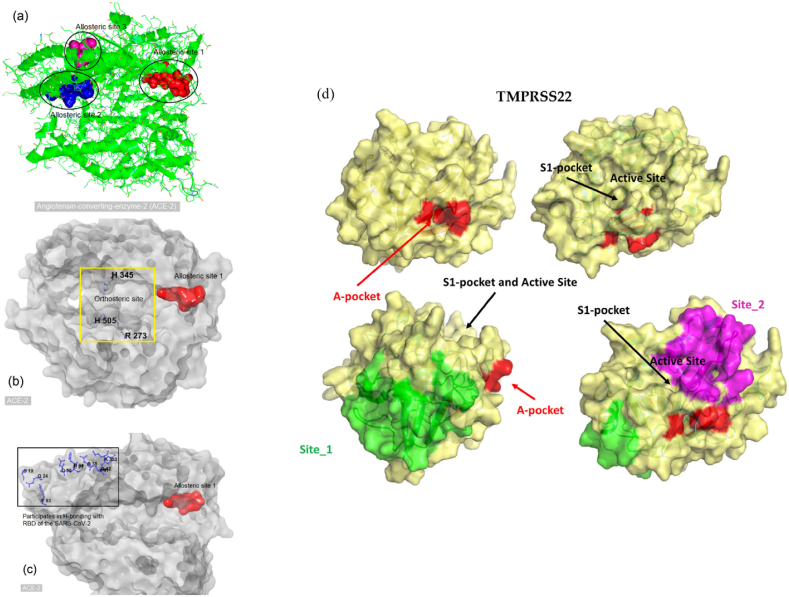
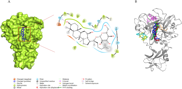
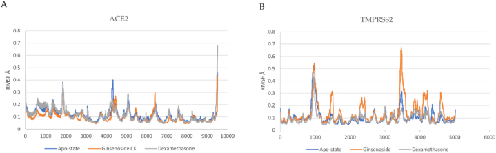
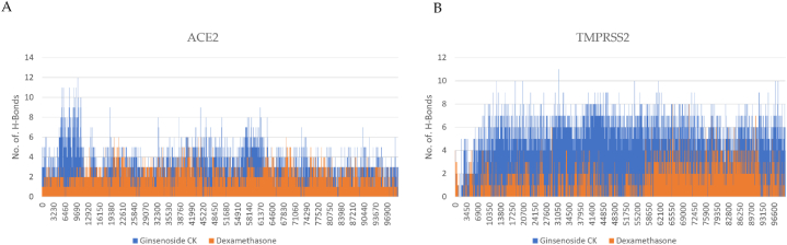
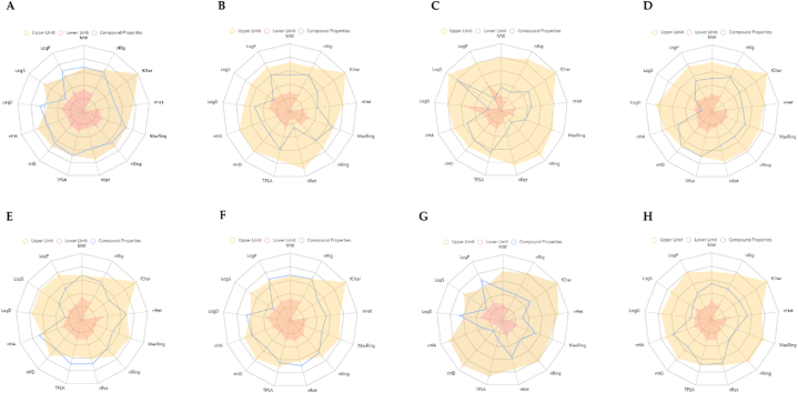
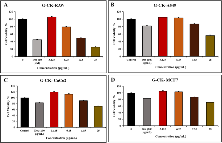
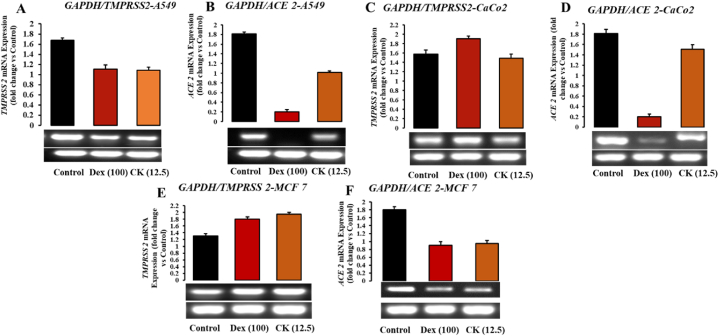
SARS-CoV-2 is a novel coronavirus that emerged as an epidemic, causing a respiratory disease with multiple severe symptoms and deadly consequences. ACE-2 and TMPRSS2 play crucial and synergistic roles in the membrane fusion and viral entry of SARS-CoV-2 (COVID-19). The spike (S) protein of SARS-CoV-2 binds to the ACE-2 receptor for viral entry, while TMPRSS2 proteolytically cleaves the S protein into S1 and S2 subunits, promoting membrane fusion. Therefore, ACE-2 and TMPRSS2 are potential drug targets for treating COVID-19, and their inhibition is a promising strategy for treatment and prevention. This study proposes that ginsenoside compound K (G-CK), a triterpenoid saponin abundant in Panax Ginseng, a dietary and medicinal herb highly consumed in Korea and China, effectively binds to and inhibits ACE-2 and TMPRSS2 expression. We initially conducted an in-silico evaluation where G-CK showed a high affinity for the binding sites of the two target proteins of SARS-CoV-2. Additionally, we evaluated the stability of G-CK using molecular dynamics (MD) simulations for 100 ns, followed by MM-PBSA calculations. The MD simulations and free energy calculations revealed that G-CK has stable and favorable energies, leading to strong binding with the targets. Furthermore, G-CK suppressed ACE2 and TMPRSS2 mRNA expression in A549, Caco-2, and MCF7 cells at a concentration of 12.5 μg/mL and in LPS-induced RAW 264.7 cells at a concentration of 6.5 μg/mL, without significant cytotoxicity.ACE2 and TMPRSS2 expression were significantly lower in A549 and RAW 264.7 cells following G-CK treatment. These findings suggest that G-CK may evolve as a promising therapeutic against COVID-19.

Keywords: A549; ACE-2; CaCo-2; Compound K; Ginsenoside; MCF7; Molecular docking; Molecular dynamics simulation; Raw 264.7; TMPRSS2.


Conflict of interest statement

The authors declare that they have no known competing financial interests or personal relationships that could have appeared to influence the work reported in this paper.

Figures

Image 1
Graphical abstract
Fig. 1
Fig. 1
Illustration of the mechanism of action of G-CK against the main drug targets (ACE2 and TMPRSS2) of COVID-19.
Fig. 2
Fig. 2
The allosteric sites of angiotensin-converting enzyme 2. (a) Cartoon presentation of the human angiotensin-converting enzyme 2(3SCJ). Different black shapes highlight potential allosteric areas. (b) A yellow square highlights the orthosteric site of ACE-2. Essential amino acid residues are H345, H505, and R273. (c) Allosteric site 1 (red) and amino acid residues (blue) of ACE-2 participating in hydrogen bonding (H-bond) with the receptor-binding domain (RBD) of SARS-CoV-2 (d) Visual summary of all the possible binding sites of TMPRSS2. Adapted from Refs. [71,73].
Fig. 3
Fig. 3
(A) 2D & 3D docking interactions of G-CK with ACE2 (PDB ID: 6M0J). (B) Docking interactions of G-CK (blue) and control drug inhibitors (Dexamethasone (Orange), NAG(Green), Remdesivir (magenta), Indinavir (cyan), Chloroquine (yellow), and Camostat (red)) with ACE2.
Fig. 4
Fig. 4
(A) 2D & 3D docking interactions of G-CK with TMPRSS2 (PDB ID: 7MEQ). (B) Docking interactions of G-CK (blue) and control drug inhibitors (Dexamethasone (Orange), Nafamostat (Green), Remdesivir (magenta), Indinavir (cyan), Chloroquine (yellow), and Camostat (red)) with TMPRSS2.
Fig. 5
Fig. 5
3D docking interactions of G-CK with (A) ACE2 (B) TMPRSS2.
Fig. 6
Fig. 6
(A) RMSD of G-CK and Dexamethasone in complex with SARS-Cov-2 ACE2 as a function of MD simulation time. (B) RMSD of G-CK in complex with SARS-Cov-2 TMPRSS2 as a function of MD simulation time.
Fig. 7
Fig. 7
(A) RMSF of G-CK and Dexamethasone in complex with SARS-Cov-2 ACE2 as a function of MD simulation time. (B) RMSF of G-CK in complex with SARS-Cov-2 ACE2 as a function of MD simulation time.
Fig. 8
Fig. 8
(A) The radius of gyration plots of molecular dynamics (MD) simulation of ACE2 receptor- G-CK and Dexamethasone complexes. (B) The radius of gyration plots of molecular dynamics (MD) simulation of ACE2 receptor G-CK and Dexamethasone complexes.
Fig. 9
Fig. 9
(A) Line plots of Ligand-protein H bonds for ACE2 with G-CK and Dexamethasone. (B) Line plots of Ligand-protein H bonds for ACE2 with G-CK and Dexamethasone.
Fig. 10
Fig. 10
ADMET properties of G-CK and the control drugs (A) G-CK, (B) Dexamethasone, (C) NAG, (D) Nafamostat, (E) Remdesivir, (F) Indinavir, (G) Chloroquine, and (H) Camostat. Abbreviations: MW: Molecular weight; nRig: number of rigid bonds; fChar: formal charge; nHet: number of heteroatoms; MaxRing: number of atoms in the biggest ring; nRing number of rings; nRot: number of rotatable bonds; TPSA: topological polar surface area; nHD: number of hydrogen bond donors; nHA: number of hydrogen bond acceptor; LogD: logP at physiological pH 7.4; logS: log of the aqueous solubility; and LogP: log of the octanol/water partition coefficient.
Fig. 11
Fig. 11
In vitro, cell cytotoxicity evaluation for G-CK (A) RAW 264.7, (B) A549 cells, (C) Caco-2, and (D) MCF-7 cells, cells. Every value is expressed as the mean ± standard error of three independent experiments. ***p < 0.001 compared with control.
Fig. 12
Fig. 12
Effects of G-CK on mRNA expression levels of Anti-covid related genes in (A–B) A549 cells, (C–D) Caco-2 cells, and (E–F) MCF-7 cells.
Fig. 13
Fig. 13
Effects of G-CK on mRNA expression levels of Anti-covid (A–B) and Cytokine storm (C–G) related genes on Raw 264.7 cells compared with LPS.

References

    1. Wu Y.C., Chen C.S., Chan Y.J. The outbreak of COVID-19: an overview. J. Chin. Med. Assoc. 2020;83(3):217–220. - PMC - PubMed
    1. Peiris J.S., et al. Clinical progression and viral load in a community outbreak of coronavirus-associated SARS pneumonia: a prospective study. Lancet. 2003;361(9371):1767–1772. - PMC - PubMed
    1. Perlman S. Another decade, another coronavirus. N. Engl. J. Med. 2020;382(8):760–762. - PMC - PubMed
    1. Jackson C.B., et al. Mechanisms of SARS-CoV-2 entry into cells. Nat. Rev. Mol. Cell Biol. 2022;23(1):3–20. - PMC - PubMed
    1. Li F. Structure, function, and evolution of coronavirus spike proteins. Ann. Rev. Virol. 2016;3(1):237–261. - PMC - PubMed