Supraorbital artery: Anatomical variations and neurosurgical applications
- PMID: 37810326
- PMCID: PMC10559381
- DOI: 10.25259/SNI_597_2023
Supraorbital artery: Anatomical variations and neurosurgical applications
Abstract
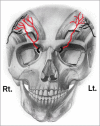
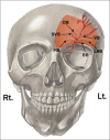
Background: The supraorbital artery (SOA) originates from the ophthalmic artery in a superomedial aspect of the orbit, exiting through the supraorbital groove to emerge onto the forehead. The SOA has important neurosurgical considerations regarding different approaches and bypasses. The SOA is poorly described in the standard anatomical textbooks. Therefore, we present this article to describe the anatomical variations of the SOA and their implications on the neurosurgical field.
Methods: We conducted a literature review in PubMed and Google Scholar databases to review the existing literature describing the SOA anatomy and its neurosurgical applications.
Results: While reading the available articles and original works regarding SOA, we identified 22 studies that discuss the SOA. We noticed the anatomical variations of the SOA in terms of origin, course, diameter, branches, depth, and distance in relation to the midline and vertical glabellar line. We also discussed certain applications of SOA and its importance in neurosurgical approaches, bypass, photoplethysmography, aneurysms, and reconstruction of cranial fossa defects.
Conclusion: The variable anatomy of the SOA has a paramount impact on performing different neurosurgical approaches. Therefore, cadaveric studies of the SOA are important to explore potential methods for the preservation of the artery in different neurosurgical applications.
Keywords: Anatomical variation; Neuroanatomy; Supraorbital artery.
Copyright: © 2023 Surgical Neurology International.
Conflict of interest statement
There are no conflicts of interest.
Figures


References
-
- Carstens MH, Greco RJ, Hurwitz DJ, Tolhurst DE. Clinical applications of the subgaleal fascia. Plast Reconstr Surg. 1991;87:615–26. - PubMed
-
- Cavalcanti DD, García-González U, Agrawal A, Crawford NR, Tavares PL, Spetzler RF, et al. Quantitative anatomic study of the transciliary supraorbital approach: Benefits of additional orbital osteotomy? Neurosurgery. 2010;66(suppl_2 Operative):205–10. - PubMed
-
- Cong LY, Phothong W, Lee SH, Wanitphakdeedecha R, Koh I, Tansatit T, et al. Topographic analysis of the supratrochlear artery and the supraorbital artery: Implication for improving the safety of forehead augmentation. Plast Reconstr Surg. 2017;139:620e-7. - PubMed
-
- Cotofana S, Lachman N. Arteries of the face and their relevance for minimally invasive facial procedures: An anatomical review. Plast Reconstr Surg. 2019;143:416–26. - PubMed
-
- Delashaw JB, Jr, Jane JA, Kassell NF, Luce C. Supraorbital craniotomy by fracture of the anterior orbital roof. J Neurosurg. 1993;79:615–8. - PubMed
Publication types
LinkOut - more resources
Full Text Sources
