Case Reports
. 2023 Sep 4:20:33.
doi: 10.25259/Cytojournal_26_2022. eCollection 2023.

Unusual case of parietal scalp swelling without palpable swelling in head and neck region

Affiliations
Case Reports

Unusual case of parietal scalp swelling without palpable swelling in head and neck region

Priyanka Gautam et al. Cytojournal. .
No abstract available


Conflict of interest statement

The authors declare that they have no competing interests.

Figures
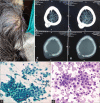
Figure 1:
Figure 1:
(a) Swelling over the left parietal scalp region; (b) contrast-enhanced computed tomography brain shows destructive mildly enhancing bony mass lesion; (c) aspirate from scalp swelling showing round cells with moderate amount of cytoplasm in sheets and groups with mild nuclear atypia (× 400, Papanicolaou stain); and (d) round cells arranged in small groups and clusters with central nuclei and moderate amount of granular cytoplasm (×400, May–Grunwald– Giemsa stain).
Figure 2:
Figure 2:
(a) May–Grunwald–Giemsa stained smear from scalp swelling showing the cohesive sheet of round cells with large central nuclei and moderate granular cytoplasm. Occasional binucleation also noted (×400) and (b) Papanicolaou stained smear showing round cells with moderate amount of granular cytoplasm in sheet with well-defined cell border (×400).
Figure 3:
Figure 3:
(a and b) Aspirate from thyroid showing the sheet of Hurthle cells with moderate amount of granular cytoplasm in sheet with well-defined cell border with central round nucleus and occasional binucleation (×400).

References

    1. Harel G, Hadar T, Segal K, Levy R, Sidi J. Hurthle cell carcinoma of the thyroid gland. A tumor of moderate malignancy. Cancer. 1986;57:1613–7. doi: 10.1002/1097-0142(19860415)57:8<1613::AID-CNCR2820570829>3.0.CO;2-3. - DOI - PubMed
    1. Caturegli P, Ruggere C. Karl Hurthle! Now, who was he? Thyroid. 2005;15:121–3. doi: 10.1089/thy.2005.15.121. - DOI - PubMed
    1. LIoyd R, Osamura R, Kloppel G, Rosai J. 4th ed. Lyon: International Agency for Research on Cancer; 2017. WHO Classification of Tumours of Endocrine Organs.
    1. Pu RT, Yang J, Wasserman PG, Bhuiya T, Griffith KA, Michael CW. Does Hurthle cell lesion/neoplasm predict malignancy more than follicular lesion/neoplasm on thyroid fine-needle aspiration? Diagn Cytopathol. 2006;34:330–4. doi: 10.1002/dc.20440. - DOI - PubMed
    1. Stojadinovic A, Hoos A, Ghossein R, Urist MJ, Leung DH, Spiro RH, et al. Hurthle cell carcinoma: A 60-year experience. Ann Surg Oncol. 2002;9:197–203. doi: 10.1245/aso.2002.9.2.197. - DOI - PubMed

Publication types

LinkOut - more resources