. 2023 Sep 21:11:1267563.
doi: 10.3389/fchem.2023.1267563. eCollection 2023.

Hierarchical self-assembly of a reflectin-derived peptide

Affiliations

Hierarchical self-assembly of a reflectin-derived peptide

Ana Margarida Gonçalves Carvalho Dias et al. Front Chem. .

Abstract

Reflectins are a family of intrinsically disordered proteins involved in cephalopod camouflage, making them an interesting source for bioinspired optical materials. Understanding reflectin assembly into higher-order structures by standard biophysical methods enables the rational design of new materials, but it is difficult due to their low solubility. To address this challenge, we aim to understand the molecular self-assembly mechanism of reflectin's basic unit-the protopeptide sequence YMDMSGYQ-as a means to understand reflectin's assembly phenomena. Protopeptide self-assembly was triggered by different environmental cues, yielding supramolecular hydrogels, and characterized by experimental and theoretical methods. Protopeptide films were also prepared to assess optical properties. Our results support the hypothesis for the protopeptide aggregation model at an atomistic level, led by hydrophilic and hydrophobic interactions mediated by tyrosine residues. Protopeptide-derived films were optically active, presenting diffuse reflectance in the visible region of the light spectrum. Hence, these results contribute to a better understanding of the protopeptide structural assembly, crucial for the design of peptide- and reflectin-based functional materials.

Keywords: bio-based materials; films; hydrogels; optical materials; peptides; reflectins; self-assembly; supramolecular.


Conflict of interest statement

The authors declare that the research was conducted in the absence of any commercial or financial relationships that could be construed as a potential conflict of interest. The author(s) declared that they were an editorial board member of Frontiers, at the time of submission. This had no impact on the peer review process and the final decision.

Figures

FIGURE 1
FIGURE 1
Protopeptide—the basic unit of reflectins. (A) Proposed origin of the basic unit of reflectins—the protopeptide. The symbiosis between a bacterium Vibrio fischeri and cephalopods resulted in a gene transfer that led to the formation of the reflectin gene. The expressed reflectin protein incorporates several copies of the protopeptide (blue). (B) Reflectin sequences are composed by a highly conserved N-terminal sequence (bold) and several repeating motifs (orange) connected by linkers (dark orange). Several truncated sequences have been generated to study reflectin’s self-assembly, with D. pealeii reflectin A1 (Uniprot: D3UA43) being one of the most studied sequences, and the classification of repeating motifs and linkers was adapted from Umerani et al. (2020). Repeating motifs contain the protopeptide sequence or its variants (blue): (Y/W/G)MD(M/F) X(G/N)X2, (X = S, Y, Q, W, H, R), adapted from Guan et al. (2017). (C) Protopeptide chemical structure YMDMSGYQ used in this work, with highlighted amino acid side chains in different colors—Tyr (green), Met (yellow), Asp (red), and Gln, Gly, and Ser (blue). (D) The distinct hierarchical levels approached in this work to understand protopeptide self-assembly into a macroscopic hydrogel.
FIGURE 2
FIGURE 2
Characterization of macroscopic protopeptide hydrogels. (A) Hydrogels at 36 mg/mL were prepared in Gel7.5_Im (buffer A), Gel4_Im (buffer B), Gel7.5 (buffer C), and Gel4 (buffer D). The four hydrogels were characterized by AFM analysis. (B) All hydrogels show nanofibers. In addition, by rheology (C), for all gels, the storage modulus (G′) is one order of magnitude higher than the loss modulus (G″) (first graph—gels with imidazole and second graph—gels without imidazole). The mean G′ for each hydrogel is compared between samples (third graph, n = 3 with significance levels and p-value below 0.05 as determined by Tukey’s test using Origin 2023 software).
FIGURE 3
FIGURE 3
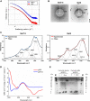
Characterization of hydrogels Gel7.5 and Gel4 without imidazole. (A) The small angle X-ray scattering results were fitted to a “gel model”; (B) visualization of X-ray scattering in an area detector shows three main scattering rings; (C) ATR-FTIR deconvolution results of the Amide I band (1700–1600 nm-1) of protopeptide gels produced using different pH conditions (black line: original spectrum; red or blue line: fitted spectra; gray lines: deconvoluted peaks). (D) Far-UV circular dichroism spectra of peptide hydrogels (36 mg/mL) at 25°C—Gel7.5 in buffer C and Gel4 in buffer D. (E) Native Tris-tricine gel 16.5% with samples from Gel4 incubated with curcumin and without curcumin, showing two main bands 10 kDa and 2 kDa. Marker: Precision Plus Dual Xtra protein ladder (Bio-Rad).
FIGURE 4
FIGURE 4
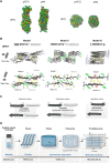
In silico studies with the protopeptide and hierarchical assembly models. The amino acid side-chains of the peptide are showed in different colors—Tyr (green), Met (yellow), Asp (red), and Gln, Gly, and Ser (blue). (A) Coarse-grained molecular dynamics simulations of 130 molecules of the peptide in a water box in pH 4 (neutral charge) and pH 7.5 (negatively charged) at 25°C and 90°C. At both pHs, at 25°C, there is formation of fibers, and at 90°C, there is formation of aggregates. (B) All-atom molecular dynamics simulations: input structures at the top and front view, where different peptide sheets are identified. In bold are identified the amino acids with the highest propensity for aggregation by using ZipperDB (Goldschmidt et al., 2010), in black and gray are the amino acids from two different peptide sheets, and the amino acids with lower aggregation propensity are underlined. (C) Schematics of models I, II, and III and the possible properties of each interface region. Images designed in VMD (Humphrey et al., 1996) and PyMOL (DeLano, 2002) visualization software. (D) Protopeptide hierarchical assembly model. At the macroscale, peptide hydrogels were studied by rheology. At the microscale, the hydrogels were analyzed by AFM and SEM, revealing nanofibers with the diameter of 200–400 Å. At the nanoscale, SAXS revealed that within these fibers, there were repeated units with a short correlation length between 30 and 50 Å. Furthermore, it was determined in a native protein gel that the peptide forms aggregates with 10 kDa. At the molecular level, the X-ray scattering revealed diffraction characteristics of β-sheet organization, which was further complemented by in silico studies. The coarse-grain simulations confirmed the aggregation propensity of the peptide into fibers, and the atomistic level simulations allowed creating a putative model for the aggregation pattern of the protopeptide.
FIGURE 5
FIGURE 5
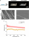
Optical characterization of peptide films. (A) Protopeptide and A1H1 peptide films deposited over a glass slide. (B) SEM characterization of the protopeptide (left panel) and A1H1 (right panel) films. (C) Diffuse reflectance plots of the protopeptide (in buffer D) and A1H1 peptide with the sequence AATAVSHTTHHA (in 95:5 (v/v) water in acetonitrile) films in the visible region of the light spectrum. The lines represent the average signal for each sample (n = 2), and the standard deviation is represented as a shade.

References

    1. Alakpa E. V., Jayawarna V., Burgess K. E. V., West C. C., Péault B., Ulijn R. V., et al. (2017). Improving cartilage phenotype from differentiated pericytes in tunable peptide hydrogels. Sci. Rep. 7, 6895. 10.1038/s41598-017-07255-z - DOI - PMC - PubMed
    1. Bastos P., Fracalossi D. M., Chimal M. E., Sánchez A., Rosas C. (2020). Digestive enzymes and timing of digestion in Octopus vulgaris type II. Aquac. Rep. 16, 100262. 10.1016/j.aqrep.2019.100262 - DOI
    1. Berendsen H. J. C., Postma J. P. M., Gunsteren W. F. V., DiNola A., Haak J. R. (1984). Molecular Dynamics with coupling to an external bath. J. Chem. Phys. 81, 3684–3690. 10.1063/1.448118 - DOI
    1. Chen T., Li M., Liu J. (2018). π-π stacking interaction: A nondestructive and facile means in material engineering for bioapplications. Cryst. Growth Des. 18, 2765–2783. 10.1021/acs.cgd.7b01503 - DOI
    1. Chen Y. L., Chang Y. H., Huang J. L., Chen I., Kuo C. (2012). Light scattering and enhanced photoactivities of electrospun titania nanofibers. J. Phys. Chem. C 116, 3857–3865. 10.1021/jp2117246 - DOI

LinkOut - more resources