. 2023 Dec 1;78(12):2830-2839.
doi: 10.1093/jac/dkad306.

Optimization of the EUCAST reference broth microdilution method for echinocandin susceptibility testing of Aspergillus fumigatus

Affiliations

Optimization of the EUCAST reference broth microdilution method for echinocandin susceptibility testing of Aspergillus fumigatus

Maria Siopi et al. J Antimicrob Chemother. .

Abstract

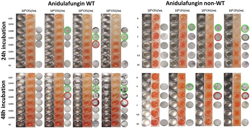
Background: Because of the high inoculum (105 cfu/mL) used in the EUCAST susceptibility testing of Aspergillus spp., determination of the minimal effective concentration (MEC) of echinocandins is challenging as the morphological differences are subtle.

Methods: The MECs of 10 WT and 4 non-WT Aspergillus fumigatus isolates were determined with the EUCAST E.Def 9.4. Plates were inoculated with increasing inocula (102-105 cfu/mL) and after 24 and 48 h of incubation, MECs were determined macroscopically (magnifying mirror) and microscopically (inverted microscope) by two observers, spectrophotometrically (OD at 405 nm) and colorimetrically (absorbance at 450/630 nm after 2 h incubation with 400 mg/L XTT/6.25 μM menadione). The interobserver (between observers)/intermethod (compared with the microscopic method) essential agreement (EA, ±1 2-fold dilution) and categorical agreement (CA) were determined for each inoculum.

Results: Echinocandin-induced microscopic hyphal alterations or macroscopic changes in turbidity were subtle with a 105 cfu/mL inoculum compared with the lower inocula of 103 and 102 cfu/mL, where more distinct changes in turbidity and formation of characteristic rosettes were obvious at the MEC after 48 h. A 105 cfu/mL inoculum resulted in wider MEC distributions (3-6 dilutions) and lower interobserver EA (69%), macroscopic-microscopic EA (26%) and CA (71%) compared with a 103 cfu/mL inoculum (2-3 dilutions, 100%, 100% and 100%, respectively). Spectrophotometric readings using a 103 cfu/mL inoculum showed good EA (57-93%) and excellent CA (86%-100%), while the XTT assay demonstrated excellent EA (93%) and CA (100%).

Conclusions: A 48 h incubation using a 103 cfu/mL inoculum improved echinocandin MEC determination for A. fumigatus with the EUCAST method, while the colorimetric assay could allow automation.


Figures

Figure 1.
Figure 1.
Macroscopic (first column), XTT (second column) and microscopic (third column) evaluation of growth of a WT A. fumigatus (left) and a non-WT A. fumigatus (right) exposed to 2-fold seral dilutions of anidulafungin (0.0005–8 mg/L) using different inoculum sizes and time of incubation. Green and red circles represent microscopic MEC (mycelial rosettes appear as black dots) and mMEC (mycelial rosettes and healthy hyphae), respectively. This figure appears in colour in the online version of JAC and in black and white in the print version of JAC.
Figure 2.
Figure 2.
Macroscopic (first row), microscopic (second row) and XTT (third row) evaluation of growth of the atypical non-WT A. fumigatus isolate DPL32458 exposed to 2-fold seral dilutions of anidulafungin (0.008–8 mg/L) using different inoculum sizes and time of incubation. This figure appears in colour in the online version of JAC and in black and white in the print version of JAC.
Figure 3.
Figure 3.
Spectrophotometric (OD at 405 nm) and colorimetric (ABS after XTT conversion by viable fungi measured at 450/630 nm, 2 h incubation with 400 mg/L XTT/6.25 μM menadione) concentration–effect curves of a WT A. fumigatus (left) and a non-WT A. fumigatus (right) exposed to anidulafungin (AFG) using different inoculum sizes and time of incubation. Green and red arrows represent microscopic MEC (mycelial rosettes) and mMEC (mycelial rosettes and healthy hyphae), respectively. The microscopic and macroscopic evaluations of growth of the same isolates are presented in Figure 1. Horizontal dotted lines corresponds to 50% growth. This figure appears in colour in the online version of JAC and in black and white in the print version of JAC.
Figure 4.
Figure 4.
Spectrophotometric (OD at 405 nm) and colorimetric (ABS after XTT conversion by viable fungi measured at 450/630 nm, 2 h incubation with 400 mg/L XTT/6.25 μM menadione) concentration–effect curves of the non-WT A. fumigatus isolate DPL32458 exposed to anidulafungin (AFG) for 24 h (left) and 48 h (right) using different inoculum sizes. Horizontal dotted lines corresponds to 50% growth.

References

    1. Ullmann AJ, Aguado JM, Arikan-Akdagli S et al. Diagnosis and management of Aspergillus diseases: executive summary of the 2017 ESCMID-ECMM-ERS guideline. Clin Microbiol Infect 2018; 24: e1–e38. 10.1016/j.cmi.2018.01.002 - DOI - PubMed
    1. Patterson TF, Thompson GR, Denning DW et al. Practice guidelines for the diagnosis and management of aspergillosis: 2016 update by the Infectious Diseases Society of America. Clin Infect Dis 2016; 63: e1–e60. 10.1093/cid/ciw444 - DOI - PMC - PubMed
    1. Aruanno M, Glampedakis E, Lamoth F. Echinocandins for the treatment of invasive aspergillosis: from laboratory to bedside. Antimicrob Agents Chemother 2019; 63: e00399-19. 10.1128/AAC.00399-19 - DOI - PMC - PubMed
    1. Arastehfar A, Gabaldón T, Garcia-Rubio R et al. Drug-resistant fungi: an emerging challenge threatening our limited antifungal armamentarium. Antibiotics 2020; 9: 877. 10.3390/antibiotics9120877 - DOI - PMC - PubMed
    1. Jiménez-Ortigosa C, Moore C, Denning DW et al. Emergence of echinocandin resistance due to a point mutation in the fks1 gene of Aspergillus fumigatus in a patient with chronic pulmonary aspergillosis. Antimicrob Agents Chemother 2017; 61: e01277-17. 10.1128/AAC.01277-17 - DOI - PMC - PubMed