Review
. 2023 Oct 1;150(19):dev202077.
doi: 10.1242/dev.202077. Epub 2023 Oct 9.

Craniofacial developmental biology in the single-cell era

Affiliations
Review

Craniofacial developmental biology in the single-cell era

Kuo-Chang Tseng et al. Development. .

Abstract

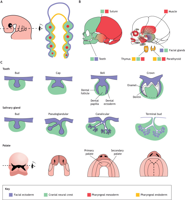
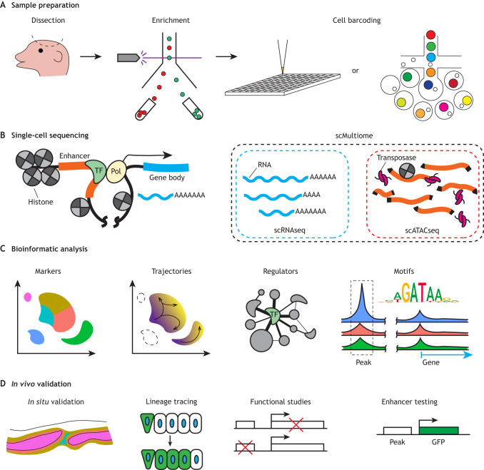
The evolution of a unique craniofacial complex in vertebrates made possible new ways of breathing, eating, communicating and sensing the environment. The head and face develop through interactions of all three germ layers, the endoderm, ectoderm and mesoderm, as well as the so-called fourth germ layer, the cranial neural crest. Over a century of experimental embryology and genetics have revealed an incredible diversity of cell types derived from each germ layer, signaling pathways and genes that coordinate craniofacial development, and how changes to these underlie human disease and vertebrate evolution. Yet for many diseases and congenital anomalies, we have an incomplete picture of the causative genomic changes, in particular how alterations to the non-coding genome might affect craniofacial gene expression. Emerging genomics and single-cell technologies provide an opportunity to obtain a more holistic view of the genes and gene regulatory elements orchestrating craniofacial development across vertebrates. These single-cell studies generate novel hypotheses that can be experimentally validated in vivo. In this Review, we highlight recent advances in single-cell studies of diverse craniofacial structures, as well as potential pitfalls and the need for extensive in vivo validation. We discuss how these studies inform the developmental sources and regulation of head structures, bringing new insights into the etiology of structural birth anomalies that affect the vertebrate head.

Keywords: Congenital anomalies; Cranial neural crest; Craniofacial; Single-cell genomics; Vertebrate head development.


Conflict of interest statement

Competing interests The authors declare no competing or financial interests.

Figures

Fig. 1.
Fig. 1.
Overview of human craniofacial development and organ systems. (A) Pharyngeal arch stage (human 4-5 weeks) with cross-sectional diagrams illustrating the major germ layer contributions. Facial ectoderm, purple; cranial neural crest, green; pharyngeal mesoderm, red; pharyngeal endoderm, orange. (B) Contribution of germ layers to the bones and teeth of the face and skull (left), muscles and glands (right). (C) Developmental stages of a typical tooth, facial gland and palate. Germ layer contributions are shown for the tooth and gland.
Fig. 2.
Fig. 2.
Flow chart of single-cell studies from sample preparation to validation. (A) Craniofacial tissues can be dissected, and specific cell types can be enriched by fluorescence-activated cell sorting (FACS) based on transgenes or cell surface markers. Cell barcoding can be performed by sequential pipetting into multi-well plates with unique barcodes or by emulsion with barcode-containing lipid droplets. (B) Diagram of a transcriptional locus showing sources of RNA for scRNAseq and accessible chromatin for scATACseq. (C) Bioinformatics packages can be used to generate cell clusters and marker genes, to infer potential cell lineage trajectories, and to identify potential master regulatory genes and potential enhancers. (D) In vivo validation includes mRNA in situ hybridization, lineage tracing, loss- and gain-of-function studies, and transgenic testing of enhancers.

References

    1. Aibar, S., González-Blas, C. B., Moerman, T., Huynh-Thu, V. A., Imrichova, H., Hulselmans, G., Rambow, F., Marine, J.-C., Geurts, P., Aerts, J.et al. (2017). SCENIC: single-cell regulatory network inference and clustering. Nat. Methods 14, 1083-1086. 10.1038/nmeth.4463 - DOI - PMC - PubMed
    1. Alghadeer, A., Hanson-Drury, S., Patni, A. P., Ehnes, D. D., Zhao, Y. T., Li, Z., Phal, A., Vincent, T., Lim, Y. C., O'Day, D.et al. (2023). Single-cell census of human tooth development enables generation of human enamel. Dev. Cell 10.1016/j.devcel.2023.07.013 - DOI - PMC - PubMed
    1. Amsterdam, A., Nissen, R. M., Sun, Z., Swindell, E. C., Farrington, S. and Hopkins, N. (2004). Identification of 315 genes essential for early zebrafish development. Proc. Natl. Acad. Sci. USA 101, 12792-12797. 10.1073/pnas.0403929101 - DOI - PMC - PubMed
    1. Anderson, T., Mo, J., Gagarin, E., Sherwood, D., Blumenkrantz, M., Mao, E., Leon, G., Chen, H.-J., Tseng, K.-C., Fabian, P.et al. (2023). Ligament injury in adult zebrafish triggers ECM remodeling and cell dedifferentiation for scar-free regeneration. npj Regen. Med. 8, 51. 10.1038/s41536-023-00329-9 - DOI - PMC - PubMed
    1. Arnold, C. D., Gerlach, D., Stelzer, C., Boryń, L. M., Rath, M. and Stark, A. (2013). Genome-wide quantitative enhancer activity maps identified by STARR-seq. Science 339, 1074-1077. 10.1126/science.1232542 - DOI - PubMed

Publication types

LinkOut - more resources