. 2023 Nov 1;80(11):1209-1221.
doi: 10.1001/jamaneurol.2023.3530.

Tau Oligomer-Containing Synapse Elimination by Microglia and Astrocytes in Alzheimer Disease

Affiliations

Tau Oligomer-Containing Synapse Elimination by Microglia and Astrocytes in Alzheimer Disease

Raquel N Taddei et al. JAMA Neurol. .

Abstract

Importance: Factors associated with synapse loss beyond amyloid-β plaques and neurofibrillary tangles may more closely correlate with the emergence of cognitive deficits in Alzheimer disease (AD) and be relevant for early therapeutic intervention.

Objective: To investigate whether accumulation of tau oligomers in synapses is associated with excessive synapse elimination by microglia or astrocytes and with cognitive outcomes (dementia vs no dementia [hereinafter termed resilient]) of individuals with equal burdens of AD neuropathologic changes at autopsy.

Design, setting, and participants: This cross-sectional postmortem study included 40 human brains from the Massachusetts Alzheimer Disease Research Center Brain Bank with Braak III to IV stages of tau pathology but divergent antemortem cognition (dementia vs resilient) and cognitively normal controls with negligible AD neuropathologic changes. The visual cortex, a region without tau tangle deposition at Braak III to IV stages, was assessed after expansion microscopy to analyze spatial relationships of synapses with microglia and astrocytes. Participants were matched for age, sex, and apolipoprotein E status. Evidence of Lewy bodies, TDP-43 aggregates, or other lesions different from AD neuropathology were exclusion criteria. Tissue was collected from July 1998 to November 2020, and analyses were conducted from February 1, 2022, through May 31, 2023.

Main outcomes and measures: Amyloid-β plaques, tau neuropil thread burden, synapse density, tau oligomers in synapses, and internalization of tau oligomer-tagged synapses by microglia and astrocytes were quantitated. Analyses were performed using 1-way analysis of variance for parametric variables and the Kruskal-Wallis test for nonparametric variables; between-group differences were evaluated with Holm-Šídák tests.

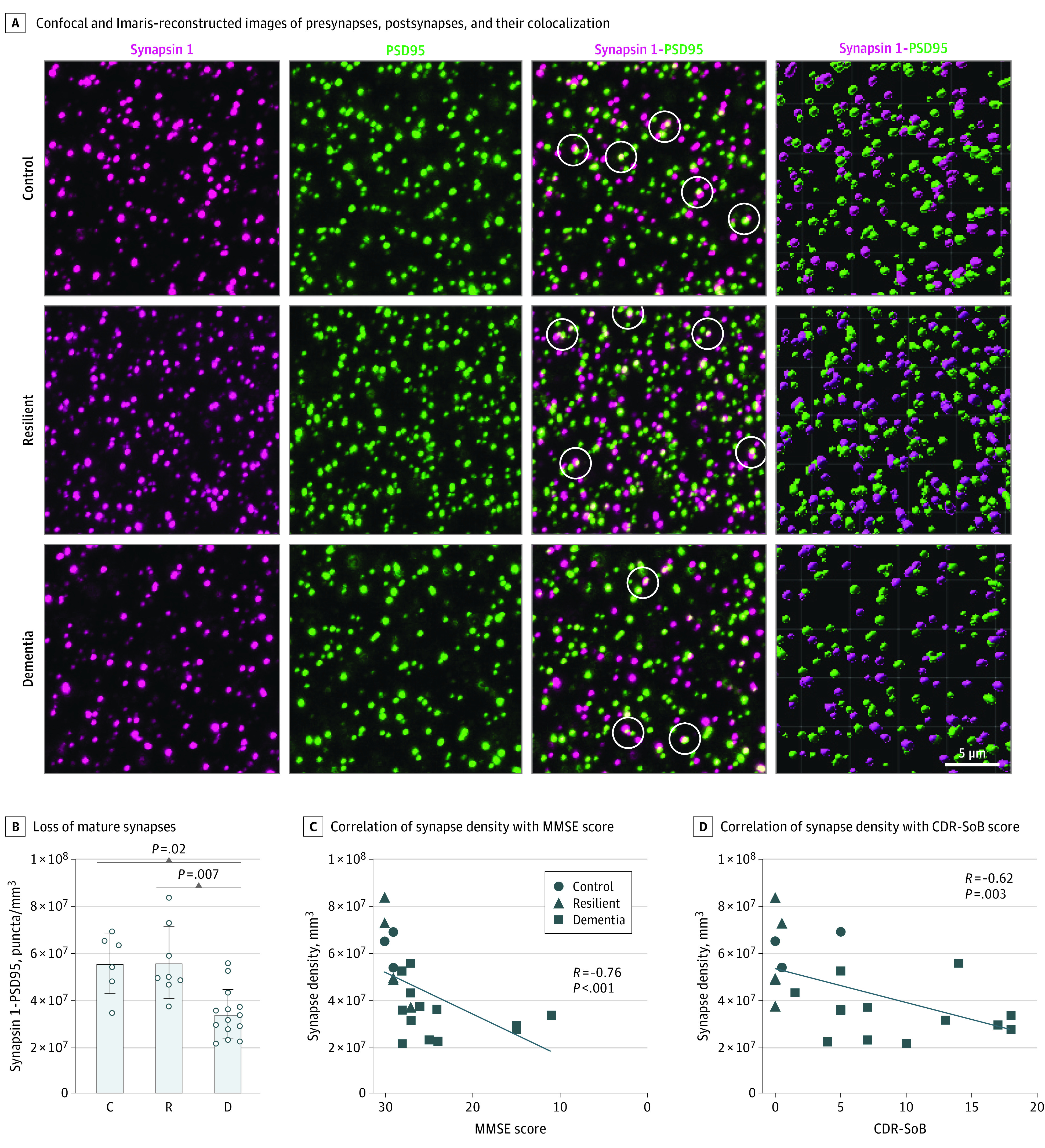
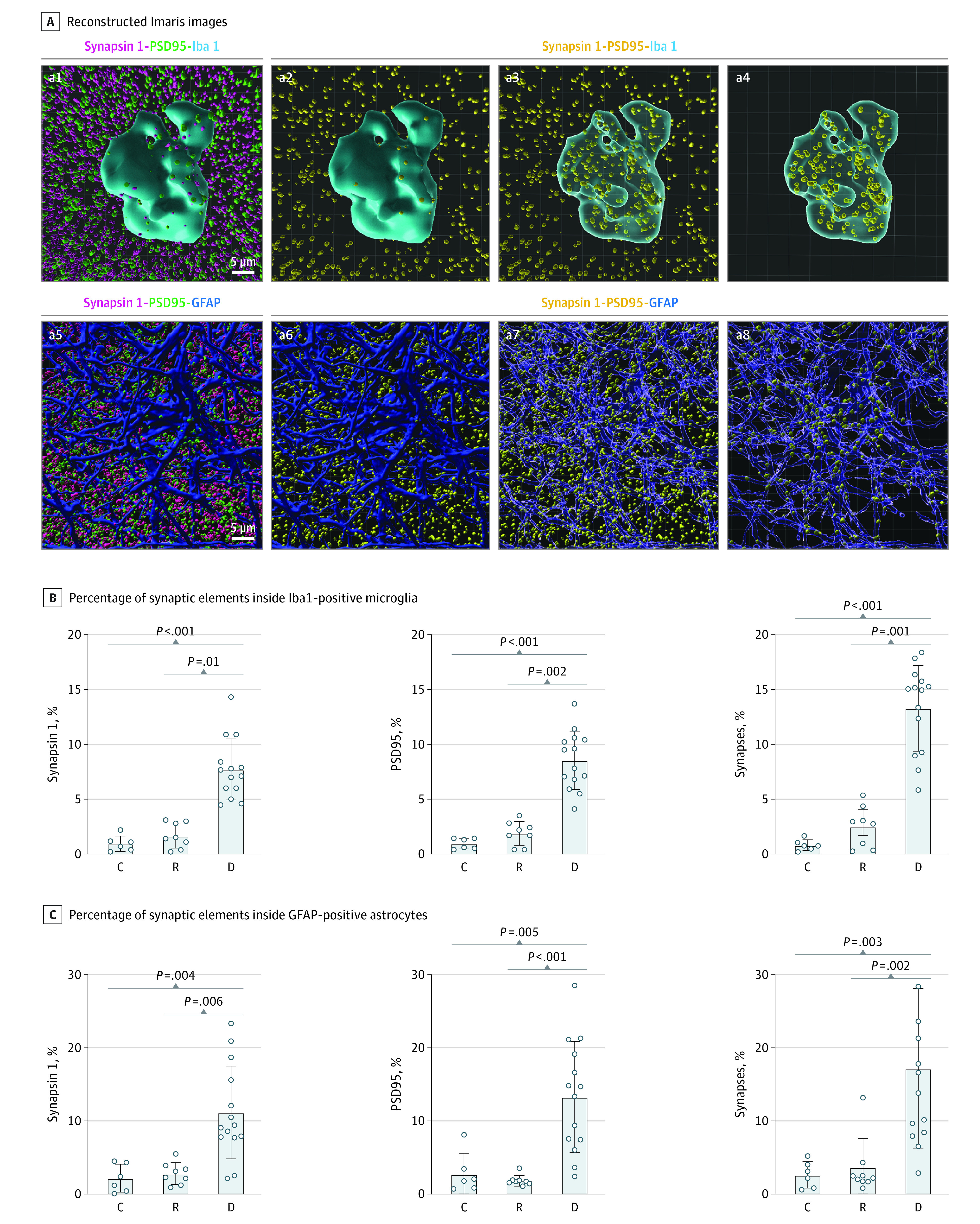
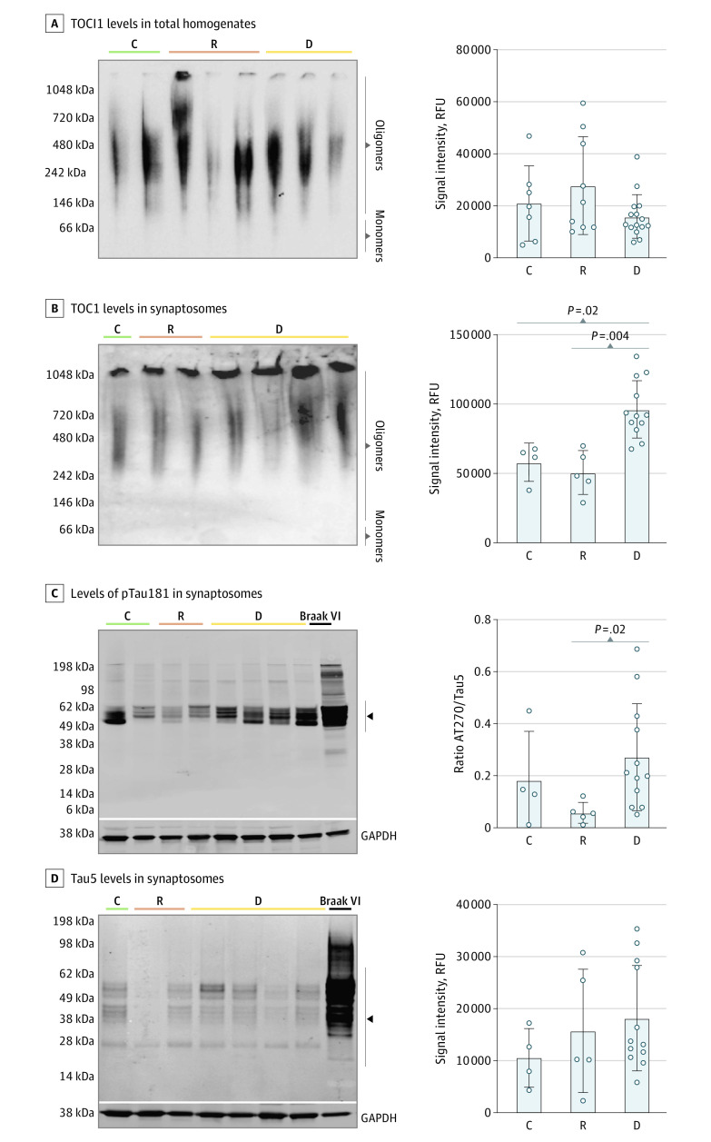
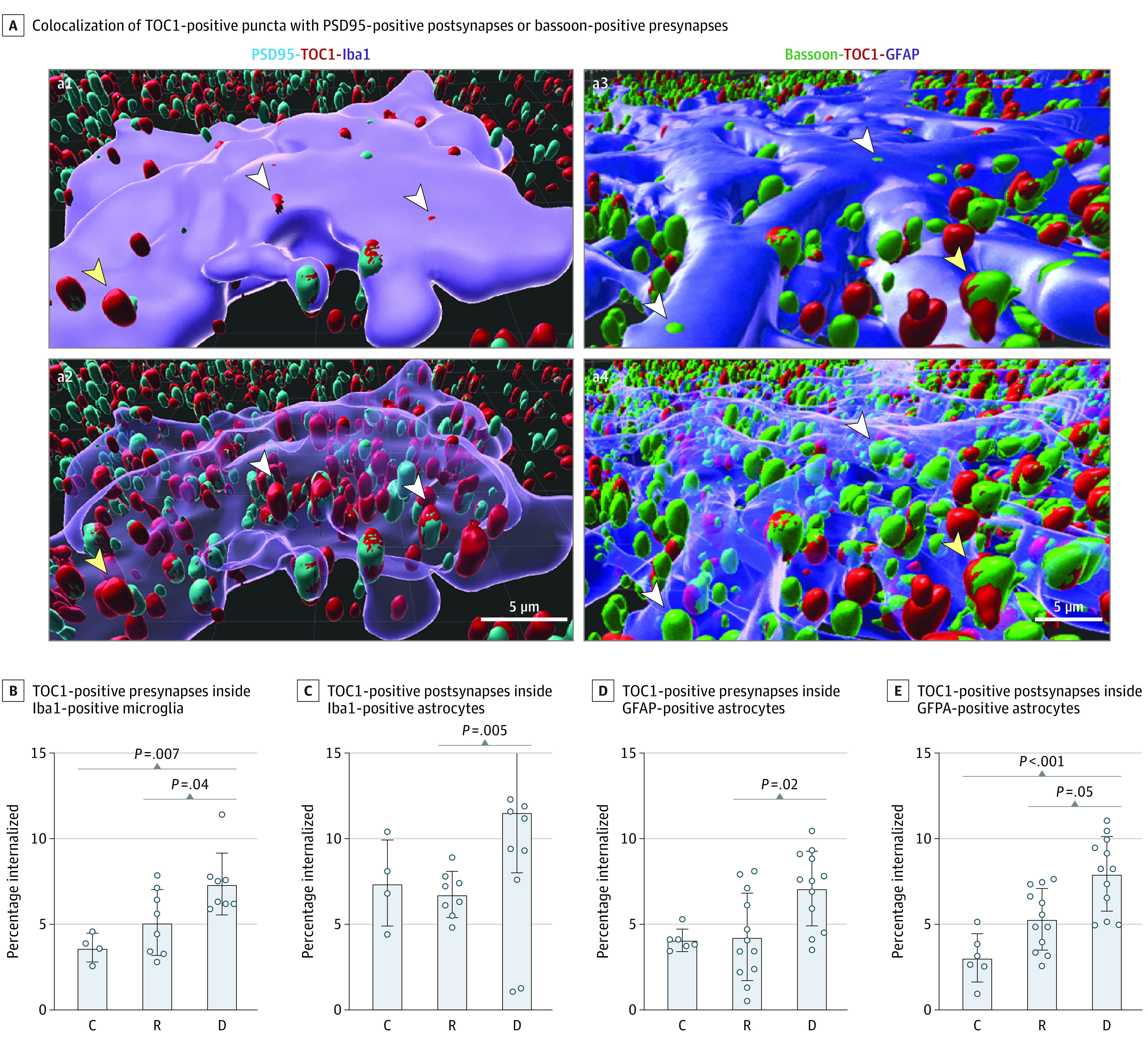
Results: Of 40 included participants (mean [SD] age at death, 88 [8] years; 21 [52%] male), 19 had early-stage dementia with Braak stages III to IV, 13 had resilient brains with similar Braak stages III to IV, and 8 had no dementia (Braak stages 0-II). Brains with dementia but not resilient brains had substantial loss of presynaptic (43%), postsynaptic (33%), and colocalized mature synaptic elements (38%) compared with controls and significantly higher percentages of mature synapses internalized by IBA1-positive microglia (mean [SD], 13.3% [3.9%] in dementia vs 2.6% [1.9%] in resilient vs 0.9% [0.5%] in control; P < .001) and by GFAP-positive astrocytes (mean [SD], 17.2% [10.9%] in dementia vs 3.7% [4.0%] in resilient vs 2.7% [1.8%] in control; P = .001). In brains with dementia but not in resilient brains, tau oligomers more often colocalized with synapses, and the proportions of tau oligomer-containing synapses inside microglia (mean [SD] for presynapses, mean [SD], 7.4% [1.8%] in dementia vs 5.1% [1.9%] resilient vs 3.7% [0.8%] control; P = .006; and for postsynapses 11.6% [3.6%] dementia vs 6.8% [1.3%] resilient vs 7.4% [2.5%] control; P = .001) and astrocytes (mean [SD] for presynapses, 7.0% [2.1%] dementia vs 4.3% [2.2%] resilient vs 4.0% [0.7%] control; P = .001; and for postsynapses, 7.9% [2.2%] dementia vs 5.3% [1.8%] resilient vs 3.0% [1.5%] control; P < .001) were significantly increased compared with controls. Those changes in brains with dementia occurred in the absence of tau tangle deposition in visual cortex.

Conclusion and relevance: The findings from this cross-sectional study suggest that microglia and astrocytes may excessively engulf synapses in brains of individuals with dementia and that the abnormal presence of tau oligomers in synapses may serve as signals for increased glial-mediated synapse elimination and early loss of brain function in AD.


Conflict of interest statement

Conflict of Interest Disclosures: Dr Gómez-Isla reported receiving personal fees from Eli Lilly and Company outside the submitted work. No other disclosures were reported.

Figures

Figure 1.
Figure 1.. Synapse Densities Across Groups and Correlation Analyses Between Synapse Densities and Cognitive Measures
A, Representative images of synapsin 1–positive presynapses, postsynaptic density protein–positive postsynapses, and their colocalization after expansion microscopy (ExM) with confocal imaging in the first 3 columns and in Imaris 3-dimensional reconstructed images in the fourth column. White circles in the third column indicate colocalization. Loss of mature synapses (B) is significantly correlated with antemortem Mini-Mental State Examination (MMSE) score (C) and Clinical Dementia Rating-Sum of Boxes (CDR-SoB) score (D) in the visual cortex. Analyses shown were performed on 28 brains (6-8 fields of view per case). Synapse densities in quantification plots correspond to values obtained in expanded tissue sections and must be multiplied by approximately 100 (4.63) to account for the volume expansion factor of 4.6 achieved by the ExM protocol to extrapolate to pre-expanded tissue material. Control (indicated by C in panel B) comprised 6 brains with Braak stages 0 to II; resilient (R), 8 brains with Braak stages III to IV; dementia (indicated by D in panel B), 14 brains with Braak stages III to IV.
Figure 2.
Figure 2.. Analyses of Engulfment of Synaptic Elements by Microglia and Astrocytes
Representative Imaris 3-dimensional image reconstructions (A) showing internalized synaptic elements inside Iba1-positive ameboid microglial cells and glial fibrillary acidic protein (GFAP)–positive astrocytes, and quantification of engulfed synaptic elements inside Iba1-positive microglia (B) and GFAP-positive astrocytes (C). Reconstructed 3-dimensional Imaris images (A) showing glial cells in blue, synapsin 1–positive presynapses in magenta (a1, a5), postsynaptic density protein 95 (PSD95)–positive postsynapses in green (a1, a5), and colocalized synapsin 1–positive and PSD95-positive puncta in yellow (a2-a4, a6-a8), displaying colocalized synapses in yellow inside and outside of a microglial cell (a3) and an astrocyte (a7), and colocalized synapses in yellow only inside the microglia (a4) and astrocyte (a8). C indicates control; D, dementia; and R, resilient.
Figure 3.
Figure 3.. Measurement Oligomeric and Hyperphosphorylated Tau Species in Total Brain Homogenates and in Synaptosome Fractions
Synaptosomes derived from brains with dementia show a significant increase in tau oligomeric complex 1 (TOC1)–positive tau oligomers and of hyperphosphorylated AT270 phosphorylated (p)Thr181–positive tau compared with resilient and control brains. Western blot (WB) analyses of TOC1 plus tau oligomers in total brain tissue homogenates (A) and synaptosome extractions (B) were conducted by quantifying the signal intensity of the full lane labeled oligomers for each case in the native TOC1 WB. The WB analyses of synaptosome-enriched fractions from the visual cortex (B-D) total tau, as measured with the middle domain total tau antibody (Tau5), did not differ across dementia, resilient, and control brains (D). Analyses were performed for 31 total homogenates (6 controls, 10 resilient, 15 dementia) and 21 synaptosome extractions (4 controls, 5 resilient, 12 dementia). Black arrowheads indicate the bandwidth quantified for each WB analysis and respective antibodies. C indicates control brain (Braak stage 0-II); D, dementia brain (Braak stage III-IV); and R, resilient brain (Braak stage III-IV); RFU, relative fluorescence unit.
Figure 4.
Figure 4.. Glial-Mediated Engulfment of Tau Oligomer–Tagged Synapses
A, Representative 3-dimensional Imaris reconstructed images of postsynaptic density 95 (PSD95)–positive with tau oligomeric complex 1 (TOC1)–positive synapses inside an ionized calcium-binding adaptor protein molecule 1 (Iba1)–positive ameboid microglia before (a1) and after (a2) making the cell body transparent, and bassoon-positive with TOC1-positive synapses inside a glial fibrillary acidic protein (GFAP)–positive astrocyte before (a3) and after (a4) making the cell body transparent. White arrowheads indicate engulfed; yellow arrowheads, not engulfed. Assessments of percentages of internalized TOC1-positive presynapses (B) and TOC1-positive postsynapses (C) inside Iba1-positive ameboid microglia and GFAP-positive astrocytes (D and E). Analyses were performed for 20 ionized calcium-binding adaptor molecule 1–positive and 30 GFAP-positive cells from 10 brains (2 controls, 4 resilient, 4 dementia). C indicates control (Braak stage 0-II); D, dementia (Braak stage III-IV); and R, resilient (Braak stage III-IV).

Comment in

References

    1. DeKosky ST, Scheff SW. Synapse loss in frontal cortex biopsies in Alzheimer’s disease: correlation with cognitive severity. Ann Neurol. 1990;27(5):457-464. doi:10.1002/ana.410270502 - DOI - PubMed
    1. Henstridge CM, Tzioras M, Paolicelli RC. Glial contribution to excitatory and inhibitory synapse loss in neurodegeneration. Front Cell Neurosci. 2019;13:63. doi:10.3389/fncel.2019.00063 - DOI - PMC - PubMed
    1. Vogels T, Murgoci AN, Hromádka T. Intersection of pathological tau and microglia at the synapse. Acta Neuropathol Commun. 2019;7(1):109. doi:10.1186/s40478-019-0754-y - DOI - PMC - PubMed
    1. Crystal H, Dickson D, Fuld P, et al. . Clinico-pathologic studies in dementia: nondemented subjects with pathologically confirmed Alzheimer’s disease. Neurology. 1988;38(11):1682-1687. doi:10.1212/WNL.38.11.1682 - DOI - PubMed
    1. Katzman R, Terry R, DeTeresa R, et al. . Clinical, pathological, and neurochemical changes in dementia: a subgroup with preserved mental status and numerous neocortical plaques. Ann Neurol. 1988;23(2):138-144. doi:10.1002/ana.410230206 - DOI - PubMed

Publication types

Substances