. 2023 Oct 18;34(10):1873-1881.
doi: 10.1021/acs.bioconjchem.3c00368. Epub 2023 Oct 9.

Development of Prodrug-Payloads for Targeted Therapeutic Applications of Platinum-Acridine Anticancer Agents

Affiliations

Development of Prodrug-Payloads for Targeted Therapeutic Applications of Platinum-Acridine Anticancer Agents

Ikeer Y Mancera-Ortiz et al. Bioconjug Chem. .

Abstract

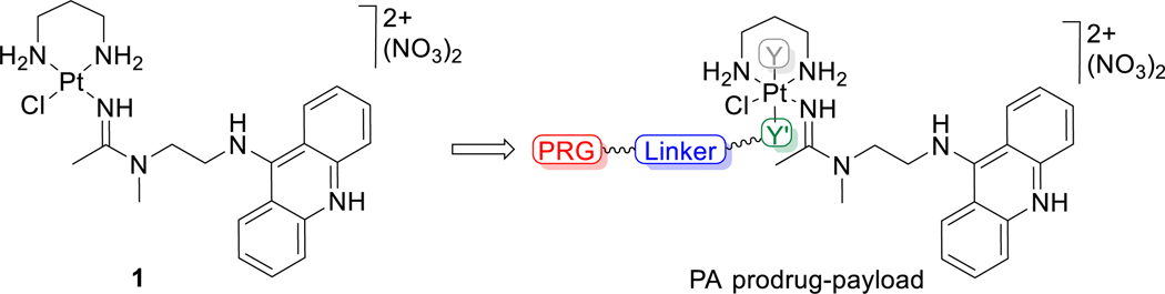
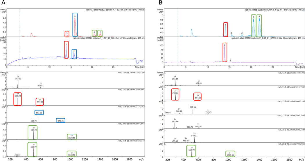
A synthetic platform has been developed that provides access to platinum(IV) prodrugs of highly cytotoxic platinum-acridine anticancer agents and allows them to be incorporated into conjugation-ready prodrug-payloads (PPLs). The PPLs can be conveniently assembled in highly efficient microscale reactions utilizing strain-promoted azide-alkyne cycloaddition chemistry. Model reactions were performed to study the stability of the PPLs in buffers and media and to assess their compatibility with cysteine-maleimide Michael addition chemistry. Amide coupling was a successful strategy to generate a conjugate containing integrin-targeted cyclo[RGDfK] peptide. Reactions with ascorbate were performed to mimic the reductive activation of the PPLs and the latter conjugate, and a cyanine (Cy5) fluorophore-labeled PPL was used to probe the reduction of platinum(IV) in cancer cells by confocal microscopy. The PPL concept introduced here should be evaluated for treating solid tumors with PAs using cancer-targeting vehicles, such as antibody-drug conjugates.


Figures

Figure 1.
Figure 1.
Structure of platinum−acridine agent 1 and schematic of platinum(IV) prodrug−payloads developed in this study. Y and Y′ are axial ligands, with an extendable linker featuring a protein-reactive group (PRG) installed on Y′.
Figure 2.
Figure 2.
Crystal structures of compounds 2 (A) and 5 (B) with the lattice solvent molecules and counterions omitted, confirming the stereoselective introduction of axial ligands in 1. Nonhydrogen atoms are represented by 50% probability ellipsoids, and hydrogen atoms are represented by arbitrarily small spheres. Only heteroatoms of the asymmetric unit are labeled. For a summary of crystal parameters and a list of bond distances and angles, see the SI.
Figure 3.
Figure 3.
Reduction of bioconjugate 14 by ascorbic acid monitored by LC-MS after ∼10 min (A) and ∼40 min (B) of incubation in PBS (pH 7.4) at 37 °C. The top panels in parts A and B show base peak chromatograms (top) and UV−vis chromatograms (bottom) monitored at an acridine-specific wavelength (413 nm). The bottom panels in A and B show positive-ion mode electrospray mass spectra of HPLC fractions. Characteristic HPLC peaks and the corresponding MS features are highlighted with boxes for intact 14 (blue; calc. m/z for [M + H]2+ = 873.37, [M+2H]3+ = 582.25), the reduction product 1 (red; calc. m/z for [M]+ = 597.19, [M + H]2+ = 299.09), and the released axial cyclo[RGDfK]-DBCO-carboxylato ligand (green, two SPAAC regioisomers; calc. m/z for [M]+ = 1020.47). See Scheme 5, reaction c, for color coding.
Figure 4.
Figure 4.
Colocalization assay in NCI-H460 lung cancer cells. (A) Structure of the cyanine dye-modified probe, Cy5-DBCO-7 (15), generated from 7 and Cy5-DBCO, with the blue emitting acridine and red emitting Cy5 chromophores highlighted. (B) Confocal microscopy images captured of NCI-H460 cells (top panel) treated with 15 (20 μM, 12 h). Fluorescence associated with the acridine and Cy5 fluorophores was recorded in the blue and red channels, respectively. The arrow in the blue channel of a single cell (bottom panel) indicates blue fluorescence localized to the nucleolus, consistent with the accumulation of compound 1 in this region of the nucleus.
Scheme 1.
Scheme 1.. Synthesis of Click Chemistry-Ready Pt(IV) Derivatives of 1a
aReagents and conditions: (a) PhICl2 (dropwise), MeOH, −78 °C to rt over 12 h; (b) 30% H2O2 (excess), H2O/DMF, 6 h, rt; (c) 30% H2O2/ butyric acid (excess), DMF, 24 h, rt; (d) Ac2O (excess), DMF, 12 h, rt; (e) 2-azidoacetic acid NHS ester, DMF, 1 h, rt; (f) Ac2O (excess), DMF, 24 h, rt; (g) pentyl isocyanate (excess), DMF, 24 h, rt.
Scheme 2.
Scheme 2.. Preparation of Prodrug-Payloads (PPLs)a
aConditions: 1:1 stoichiometry, dry DMF, 24 h, dark, rt.
Scheme 3.
Scheme 3.. Reactivity of PPLs in Media and Buffered Solutionsa
aBased on product analysis by LC-MS after 72 h of continuous incubation in buffers at 37 °C. (i) PPL 9 in 1 × PBS (pH 7.4), saline (0.9% NaCl, pH 7.0), (ii) PPL 11 in 50 mM ammonium bicarbonate (ABC, pH 7.4), (iii) PPL 11 in 1 × PBS (pH 7.4).
Scheme 4.
Scheme 4.. Model Reactions in Selected PPLsa
aReagents and Conditions: (a) 100 μM, GSH (1 equiv), pH 7.4 (PBS), rt. (b) 10 equiv. sodium ascorbate, pH 7.4 (pH-adjusted water, PBS), 37 °C. Reactions were monitored by LC-MS.
Scheme 5.
Scheme 5.. Preparation and Reductive Activation of an RGD Peptide Conjugatea
aReagents and Conditions: (a) 1. DIPEA, DMF, 6 h, rt; 2. purification by SEC. (b) 8, DMF, 15 h, rt. (c) 10 equiv sodium ascorbate, PBS (pH 7.4), 37 °C. Reactions were monitored by LC-MS.

Similar articles

References

    1. Reuvers TGA; Kanaar R; Nonnekens J. DNA Damage-Inducing Anticancer Therapies: From Global to Precision Damage. Cancers 2020, 12, 2098. - PMC - PubMed
    1. Zhong L; Li Y; Xiong L; Wang W; Wu M; Yuan T; Yang W; Tian C; Miao Z; Wang T; Yang S. Small molecules in targeted cancer therapy: advances, challenges, and future perspectives. Signal Transduct. Target. Ther. 2021, 6, 201. - PMC - PubMed
    1. Conilh L; Sadilkova L; Viricel W; Dumontet C. Payload diversification: a key step in the development of antibody-drug conjugates. J. Hematol. Oncol. 2023, 16, 3. - PMC - PubMed
    1. Yaghoubi S; Karimi MH; Lotfinia M; Gharibi T; Mahi-Birjand M; Kavi E; Hosseini F; Sineh Sepehr K; Khatami M; Bagheri N; Abdollahpour-Alitappeh M. Potential drugs used in the antibody-drug conjugate (ADC) architecture for cancer therapy. J. Cell. Physiol. 2020, 235, 31–64. - PubMed
    1. Joubert N; Beck A; Dumontet C; Denevault-Sabourin C. Antibody-Drug Conjugates: The Last Decade. Pharmaceuticals 2020, 13, 245. - PMC - PubMed

MeSH terms