. 2023 Oct 9;14(1):6243.
doi: 10.1038/s41467-023-41893-4.

GLP-1R signaling neighborhoods associate with the susceptibility to adverse drug reactions of incretin mimetics

Affiliations

GLP-1R signaling neighborhoods associate with the susceptibility to adverse drug reactions of incretin mimetics

Shane C Wright et al. Nat Commun. .

Abstract

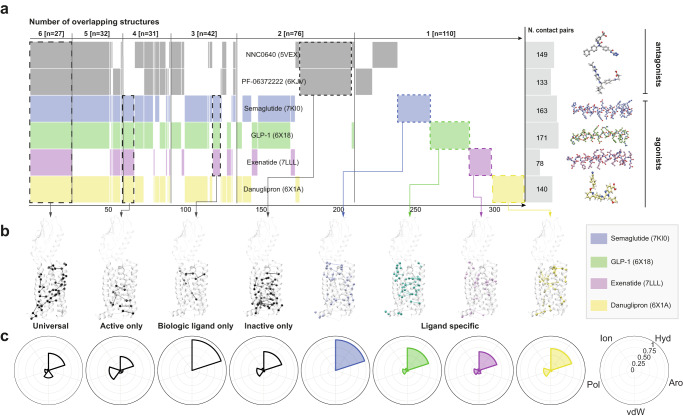
G protein-coupled receptors are important drug targets that engage and activate signaling transducers in multiple cellular compartments. Delineating therapeutic signaling from signaling associated with adverse events is an important step towards rational drug design. The glucagon-like peptide-1 receptor (GLP-1R) is a validated target for the treatment of diabetes and obesity, but drugs that target this receptor are a frequent cause of adverse events. Using recently developed biosensors, we explored the ability of GLP-1R to activate 15 pathways in 4 cellular compartments and demonstrate that modifications aimed at improving the therapeutic potential of GLP-1R agonists greatly influence compound efficacy, potency, and safety in a pathway- and compartment-selective manner. These findings, together with comparative structure analysis, time-lapse microscopy, and phosphoproteomics, reveal unique signaling signatures for GLP-1R agonists at the level of receptor conformation, functional selectivity, and location bias, thus associating signaling neighborhoods with functionally distinct cellular outcomes and clinical consequences.


Conflict of interest statement

M.B. is the president of the scientific advisory board for Domain Therapeutics. M.B. and C.L.G. have filed patent application (US20200256869A1) related to some of the biosensors used in this work and the technology has been licensed to Domain Therapeutics. All biosensors are available for academic research through regular material transfer agreement. V.M.L. is co-founder, the CEO, and shareholder of HepaPredict AB, as well as co-founder and shareholder of PersoMedix AB. A.M., S.K., P.S., C.M.B., A.B., E.B.-T., K.M., I.P., D-C.S., N.A.L., J.V.O., R.A.Z., A.S.H. and S.C.W. declare no competing interests.

Figures

Fig. 1
Fig. 1. Comparative structure analysis identifies differences in residue contact pairs for GLP-1R agonists.
a Shared contact pairs based on comparative structure analysis, represented by a Venn-like diagram (Supervenn), where each row is a set of PDB-specific contact pairs, and the overlapping parts (groupings) correspond to shared residue contact pairs among a set, sorted by the number of overlapping structures [from six structures (left) to a single structure (right)]. The columns on the right represent the total set sizes (number of residue contact pairs), and the colored ligand structures. The x-axis numbers represent a reference scale as a proportion of the total number of residue contacts shared among all structure combinations (n = 318). b Group-specific and unique shared residue contacts mapped onto a representative GLP-1R structure [PDB: 5NX2]. Residues are denoted as circles (Cα) and the noncovalent contacts between residues are denoted as lines. c Polar area diagrams for types of residue contacts (hydrophobic, aromatic, van der Waals, polar and ionic) across ligand-bound structures.
Fig. 2
Fig. 2. Pharmacological characterization of GLP-1R agonist signaling profiles at the plasma membrane.
a Illustration depicting the ebBRET approach to monitoring compartmentalized signaling. In brief, donor-tagged, pathway-selective biosensors are co-expressed with acceptor-tagged, compartment-specific markers and the GPCR of interest is stimulated with an agonist to monitor pathway activation by measuring BRET. b Concentration-response curves of GLP-1R agonists across 15 pathways monitoring biosensor recruitment to the plasma membrane using rGFP-CAAX. Data are represented by the nonlinear fit and scaled according to the highest responding drug (n = 3-6 biologically independent experiments). Drug-specific polar area diagrams for efficacy (normalized to the highest responding drug) (c) and potency (logEC50) (d) of 15 pathways at the plasma membrane. Drugs were deemed to activate a given pathway after comparing the top and bottom parameters from nonlinear regression by one-sided extra sum-of-squares F test followed by Bonferroni correction for 8 compounds (P < 0.00625). Jaccard similarity index for efficacy (e) and potency (f) quantifies similarities and differences across drug responses.
Fig. 3
Fig. 3. Peptide-activated GLP-1R traffics to vesicles and the Golgi apparatus.
a Schematic of the live cell imaging experiment monitoring agonist-induced mRuby2-mGs translocation to different cellular compartments where rGFP is expressed (PM – rGFP-CAAX; EE – rGFP-FYVE; GA – tdrGFP-Giantin). b Confocal images of HEK 293 cells expressing GLP-1R, mRuby2-mGs, and rGFP-CAAX and exposed to semaglutide (1 μM) for less than 1 min. mRuby2-mGs is recruited to active GLP-1R at the plasma membrane (closed arrowhead). c Confocal images of HEK293 cells expressing GLP-1R, mRuby2-mGs and rGFP-FYVE and exposed to semaglutide (1 μM) for 15 min. mRuby2-mGs is recruited to active GLP-1R at early endosomes (closed arrowhead). d Confocal images of HEK293 cells expressing GLP-1R, mRuby2-mGs and tdrGFP-Giantin and exposed to semaglutide (1 μM) for 30 min. mRuby2-mGs is recruited to active GLP-1R at the Golgi apparatus (closed arrowhead). Images are representative of three independent experiments. Insets depict merged images of the semaglutide-treated condition. (Scale bars, 10 μm [top and middle] and 5 μm [bottom].) PM plasma membrane, EE early endosomes, GA Golgi apparatus.
Fig. 4
Fig. 4. Stimulation of GLP-1R with peptide and small molecule agonists results in selective subcellular activation.
a Schematic of the live cell experiment monitoring agonist-induced Rluc8-mGs translocation to different cellular compartments where rGFP is expressed (EE – rGFP-FYVE; GA – tdrGFP-Giantin; ER – tdrGFP-PTP1B) based on BRET. b Concentration-response curves for Gs pathway activation of GLP-1R agonists at early endosomes, Golgi apparatus and endoplasmic reticulum. Data are represented as the mean ± SEM (n = 3 biologically independent experiments). EE early endosomes, GA Golgi apparatus, ER endoplasmic reticulum.
Fig. 5
Fig. 5. Changes in the phosphoproteome depend on functional selectivity and location bias.
a Phosphoproteomics workflow (n = 3 biologically independent samples). b 2-dimensional plots of mean log2-scaled fold change (FC) (time-matched) of the relative intensity of phosphopeptides in samples treated with danuglipron (x-axis) and semaglutide (y-axis) after 5, 15, 30 and 60 min of drug exposure (both compared to vehicle) with regression lines (grey bands represent 95% confidence interval) of proteins significantly regulated in each treatment. P values were calculated by a two-sided unpaired t-test. 2D plots are accompanied by violin plots of the mean log2 FC for both danuglipron and semaglutide at each time point (both compared to vehicle; internal box plots depict the 25th and 75th percentiles). Exact p-values were calculated using the Kruskal-Wallis one way analysis of variance. c Upset plot showing shared significantly regulated phosphopeptides after treatment with either danuglipron or semaglutide compared to vehicle at each time point. d Heatmap of significant phosphopeptides (+GLP-1R) in control or GLP-1R-expressing cells treated with vehicle, danuglipron or semaglutide in the presence or absence of the internalization inhibitor Dyngo-4A. Experimental conditions are normalized to the corresponding vehicle and phosphopeptide changes are represented as log2 fold change. e Kinase substrate enrichment analysis of the significantly regulated phosphopeptides for each drug and time point.
Fig. 6
Fig. 6. Hierarchical clustering of GLP-1R agonist fingerprints correlates with ADRs.
a Schematic of the experimental workflow for analyzing the responses of 8 GLP-1R agonists in 15 pathways and 4 cellular compartments by ebBRET. Efficacy and potency were extracted from concentration-response curves and aggregated before k-means clustering. b Hierarchical clustering of GLP-1R agonists based on signaling neighborhood profiles. c Cluster centers of distinct drug clusters identified by k-means clustering. Defining cluster features are listed by rank order, and the top and bottom five features are highlighted for each cluster. Positive k-means cluster centers reflected compartmentalized pathway engagement with high potency or efficacy, while negative k-means cluster centers referred to pathways that were not activated or less activated by a given drug cluster. d Significant adverse reactions (ADRs) have been estimated from reports submitted to the FDA Adverse Event Reporting database (FAERS) by transformation into a log likelihood ratio (LLR). All 48 unique low-level ADRs are mapped to the Medical Dictionary for Regulatory Activities (MedDRA), and aggregated “System Organ Class” (SOC) ontology levels.

References

    1. Jameson JL, Longo DL. Precision medicine–personalized, problematic, and promising. N. Engl. J. Med. 2015;372:2229–2234. doi: 10.1056/NEJMsb1503104. - DOI - PubMed
    1. Wright SC, Bouvier M. Illuminating the complexity of GPCR pathway selectivity - advances in biosensor development. Curr. Opin. Struct. Biol. 2021;69:142–149. doi: 10.1016/j.sbi.2021.04.006. - DOI - PubMed
    1. Hauser AS, et al. GPCR activation mechanisms across classes and macro/microscales. Nat. Struct. Mol. Biol. 2021;28:879–888. doi: 10.1038/s41594-021-00674-7. - DOI - PMC - PubMed
    1. Hauser AS, et al. Common coupling map advances GPCR-G protein selectivity. Elife. 2022;11:e74107. doi: 10.7554/eLife.74107. - DOI - PMC - PubMed
    1. Avet C, et al. Effector membrane translocation biosensors reveal G protein and βarrestin coupling profiles of 100 therapeutically relevant GPCRs. Elife. 2022;11:e74101. doi: 10.7554/eLife.74101. - DOI - PMC - PubMed

Publication types

MeSH terms