Review
. 2023 Oct 9;21(1):703.
doi: 10.1186/s12967-023-04393-z.

Fibrosis and bone marrow: understanding causation and pathobiology

Affiliations
Review

Fibrosis and bone marrow: understanding causation and pathobiology

Kanjaksha Ghosh et al. J Transl Med. .

Abstract

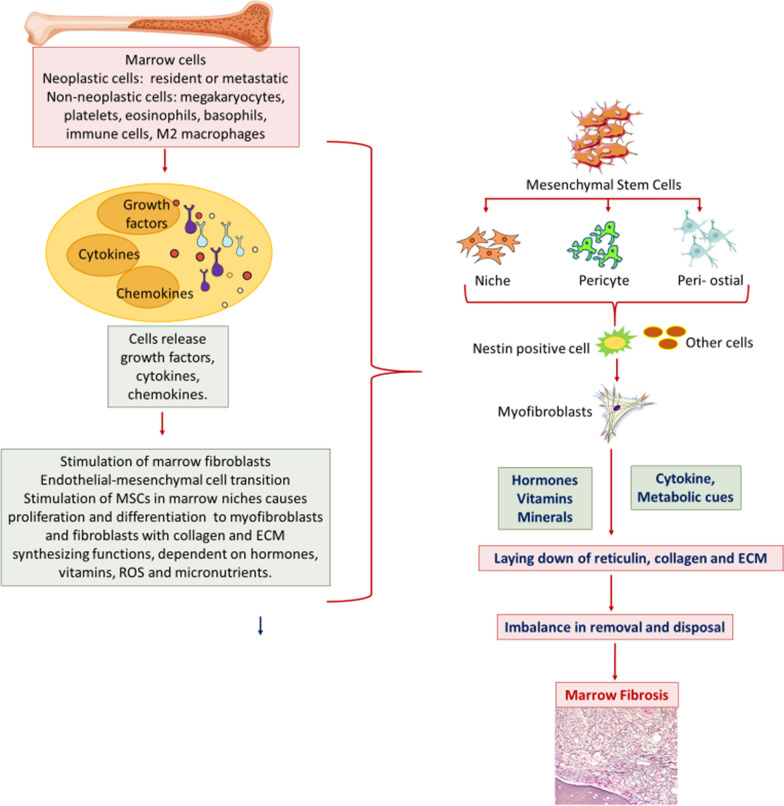
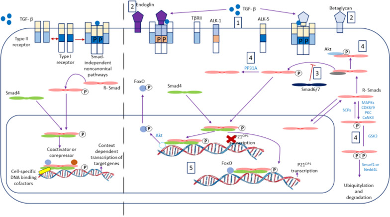
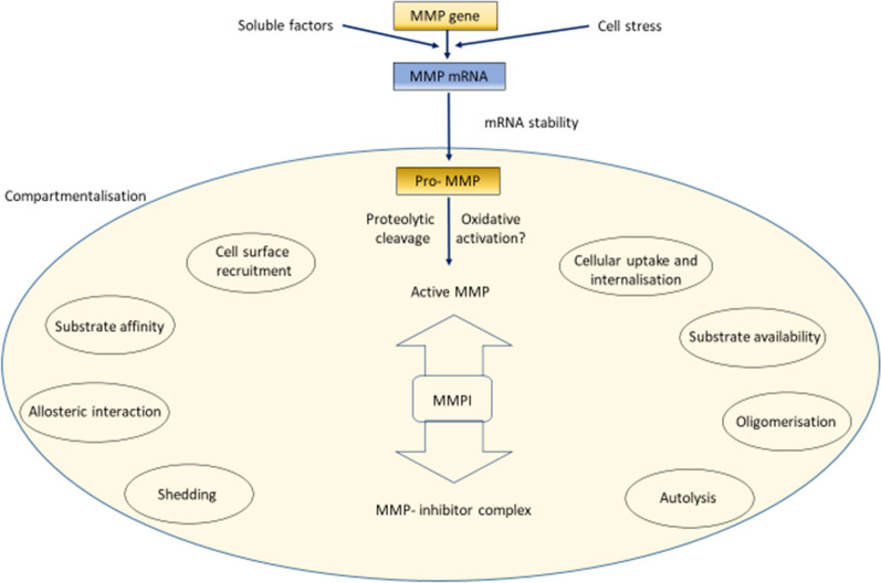
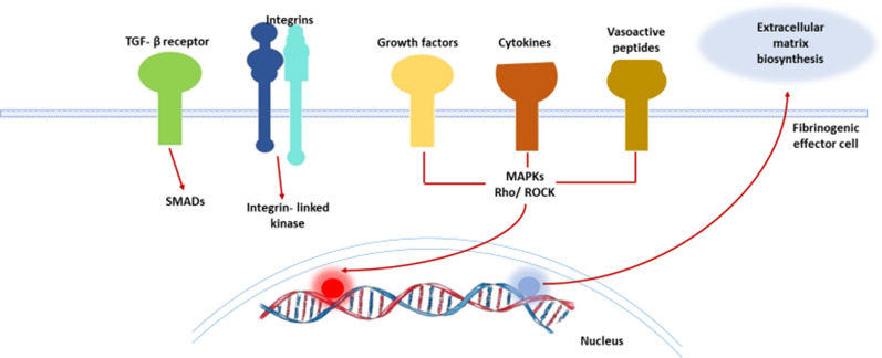
Bone marrow fibrosis represents an important structural change in the marrow that interferes with some of its normal functions. The aetiopathogenesis of fibrosis is not well established except in its primary form. The present review consolidates current understanding of marrow fibrosis. We searched PubMed without time restriction using key words: bone marrow and fibrosis as the main stem against the terms: growth factors, cytokines and chemokines, morphology, megakaryocytes and platelets, myeloproliferative disorders, myelodysplastic syndrome, collagen biosynthesis, mesenchymal stem cells, vitamins and minerals and hormones, and mechanism of tissue fibrosis. Tissue marrow fibrosis-related papers were short listed and analysed for the review. It emerged that bone marrow fibrosis is the outcome of complex interactions between growth factors, cytokines, chemokines and hormones together with their facilitators and inhibitors. Fibrogenesis is initiated by mobilisation of special immunophenotypic subsets of mesenchymal stem cells in the marrow that transform into fibroblasts. Fibrogenic stimuli may arise from neoplastic haemopoietic or non-hematopoietic cells, as well as immune cells involved in infections and inflammatory conditions. Autoimmunity is involved in a small subset of patients with marrow fibrosis. Megakaryocytes and platelets are either directly involved or are important intermediaries in stimulating mesenchymal stem cells. MMPs, TIMPs, TGF-β, PDGRF, and basic FGF and CRCXL4 chemokines are involved in these processes. Genetic and epigenetic changes underlie many of these conditions.

Keywords: Epigenetics; Haemopoietic stem cells; Megakaryocytes; Mesenchymal stem cells; Myelofibrosis; Parathormone; Signal transduction; Targeted therapy.


Conflict of interest statement

Authors declare no conflict of Interest.

Figures

Fig. 1
Fig. 1
Chronic myeloid leukemia: A hypercellular marrow; granulocytic hyperplasia (H&E, × 200); B occasional, scattered reticulin fibres, MF Grade 0 (Gomori reticulin, × 200)
Fig. 2
Fig. 2
Chronic myeloid leukemia: A hypercellular, packed marrow; granulocytic hyperplasia and many micromegakaryocytes (H&E, × 200 B focally increased loose reticulin network in paratrabecular region and C in perivascular location, MF Grade 1 (Gomori reticulin, × 200)
Fig. 3
Fig. 3
Breast carcinoma, bone marrow metastasis: A total replacement of normal marrow by tumor cells (H&E, × 200) B markedly increased reticulin with extensive intersections, MF Grade 2 (Gomori reticulin, × 200). C tumor cells, stained strongly positive for cytokeratin AE1/AE3 (immunohistochemistry, × 200)
Fig. 4
Fig. 4
Myelodysplastic syndrome, MDS-unclassifiable: A Focal cellularity, abnormal small megakaryocytes with hypolobated nuclei and hyperchromatic bare nuclei (H&E, × 200) B lymphoid aggregates were present (H&E, × 200). C focally increased loose reticulin network in cellular areas and in the lymphoid aggregate, MF Grade 1 (Gomori reticulin, × 400)
Fig. 5
Fig. 5
Primary myelofibrosis with osteomyelosclerosis: A sclerotic marrow, increased vascularity and woven bone (H&E, × 100) B markedly sclerotic marrow with prominent focal proliferation of abnormal megakaryocytes, high vascularity and dilated vascular channels (H&E, × 200). C diffusely increased, dense reticulin network with many intersections; trapped megakaryocytes (Gomori reticulin, × 200) D diffusely increased collagen, particularly surrounding foci of abnormal megakaryocytic proliferation (Masson trichrome, × 200)
Fig. 6
Fig. 6
Scheme of cellular interactions involved in myelofibrosis. The left half of the figure broadly depicts cells and cell-derived factors that collaborate to stimulate fibrosis. The right half depicts the sequence of events that follow stimulation of mesenchymal stem cells (MSCs) in marrow niches culminating in myelofibrosis
Fig. 7
Fig. 7
The transforming growth factor-β (TGF-β) signaling pathway and its context-dependent regulation. (Left) A schematic diagram of TGF-β signaling. (Right) TGF-β signaling is regulated at several levels by context-dependent factors: (1) Different combinations of paired type I and type II receptors allow for diverse ligand binding as well as intracellular signaling. (2) Accessory proteins at the plasma membrane that regulate the binding efficiency and specificity of TGF-β to their receptors influence downstream responses. (3) Proteins that regulate the recruitment and access of R-Smads to TGF-β receptors. (4) Several proteins regulate TGF-β signaling by posttranslational modification of R-Smads or by preventing their association with TGF-β receptors. (5) A specific TGF-β response can be determined by the expression and activity of transcription cofactors
Fig. 8
Fig. 8
Mechanism of induction of fibrosis by release of active mediators from mast cells promoting interactions with inflammatory cells and fibroblasts. Mast cell infiltration of the marrow may be primary (neoplastic) or a secondary reactive feature of a variety of conditions. Mast cell activation by cytokines (TNFα), IgE or C5a leads to release of their granule contents (tryptase, histamine, TNFα, chymase). Activation may be blocked by oestrogen and other pharmacological agents. Mast cell products stimulate fibroblasts directly and also indirectly through immune effector cells
Fig. 9
Fig. 9
Biochemical cycles linked to proline and hydoxyproline generation for collagen synthesis
Fig. 10
Fig. 10
Compartmentalisation of matrix metalloproteinase function leading to its specific action on specific substrates. Final availability of matrix metalloproteinase (MMP) for degradation of collagen fibres depends on the balance between its availability, reactivity to specific substrate and its abundance over its inhibitor TIMP (tissue inhibitors of matirix metalloproteinases). There are more than thirty MMPs with different substrate specificities and more than one TIMP to inhibit its activity. The enzyme is synthesised from its gene in response to various complete transcription factors and cellular stress. Translation happens through Pro MMP, specific proteolysis and activation by activation or release from its binding proteins. MMPs may be secreted or remain on the cell membrane for localisation of its action
Fig. 11
Fig. 11
Role of exogenous proline in regulating HIF1 alpha and collagen synthesis in culture. Proline plays an important role in regulation of gene expression, transcription factors, mTOR cell signaling, cellular redox reactions, synthesis of ornithine, arginine, polyamines, glutamate and collagen. Proline is formed via glutamine metabolism from amino acid pool or via tricarboxylic acid cycle via alpha keto-glutarate. Proline is hydroxylated into hydroxyproline by proline hydroxylase. Hydroxyproline is the key amino acid of collagen biosynthesis. Copper ions and vitamin C are required for collagen biosynthesis. [POX, proline hydroxylase; CDP, cytidine diphosphate; HIF1a, hypoxia inducing factor 1alpha; red line with sidebar indicates inhibition]
Fig. 12
Fig. 12
Molecular pathways leading to fibrosis of tissue including bone marrow. TGF beta is the most important growth factor causing fibrosis in the marrow. This growth factor is released by a subset of megakaryocytes and mesenchymal stem cells as well as macrophages and activated lymphocytes and metastatic malignant cells. Release in the marrow stroma is assisted by integrin ligands. Growth factors like fibroblast growth factor, cytokines and vasoactive peptides activate their cognate receptors and via MAP kinase, Rho kinase and other intracellular kinases induce gene transcription for synthesis of collagen. Some of these factors also activate either MMPs or its inhibitor TIMP
Fig. 13
Fig. 13
Druggable pathways to prevent fibrosis of marrow

References

    1. Greenberger JS. The hematopoietic microenvironment. Crit Rev Oncol Hematol. 1991;11:65–84. doi: 10.1016/1040-8428(91)90018-8. - DOI - PubMed
    1. Ramakrishnan A, Deeg HJ. A novel role for the marrow microenvironment in initiating and sustaining hematopoietic disease. Expert Opin Biol Ther. 2009;9:21–28. doi: 10.1517/14712590802603093. - DOI - PMC - PubMed
    1. Bauermeister DE. Quantitation of bone marrow reticulin–a normal range. Am J Clin Pathol. 1971;56:24–31. doi: 10.1093/ajcp/56.1.24. - DOI - PubMed
    1. Thiele J, Kvasnicka HM, Facchetti F, Franco V, van der Walt J, Orazi A. European consensus on grading bone marrow fibrosis and assessment of cellularity. Haematologica. 2005;90:1128–1132. - PubMed
    1. Yamazaki K, Allen TD. The structure and function of the blood-marrow barrier. Early ultrastructural changes in irradiated bone marrow sinus endothelial cells detected by vascular perfusion fixation. Blood Cells. 1992;18(2):215–21. - PubMed

LinkOut - more resources