

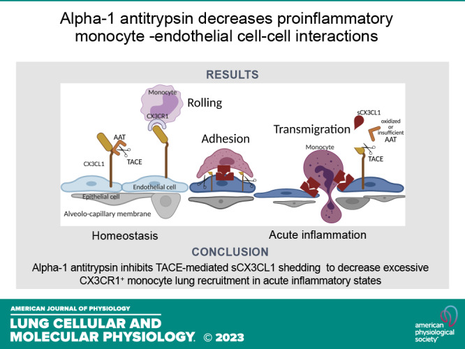
Alpha-1 antitrypsin inhibits fractalkine-mediated monocyte-lung endothelial cell interactions
- PMID: 37814796
- PMCID: PMC11068395
- DOI: 10.1152/ajplung.00023.2023
Alpha-1 antitrypsin inhibits fractalkine-mediated monocyte-lung endothelial cell interactions
Abstract
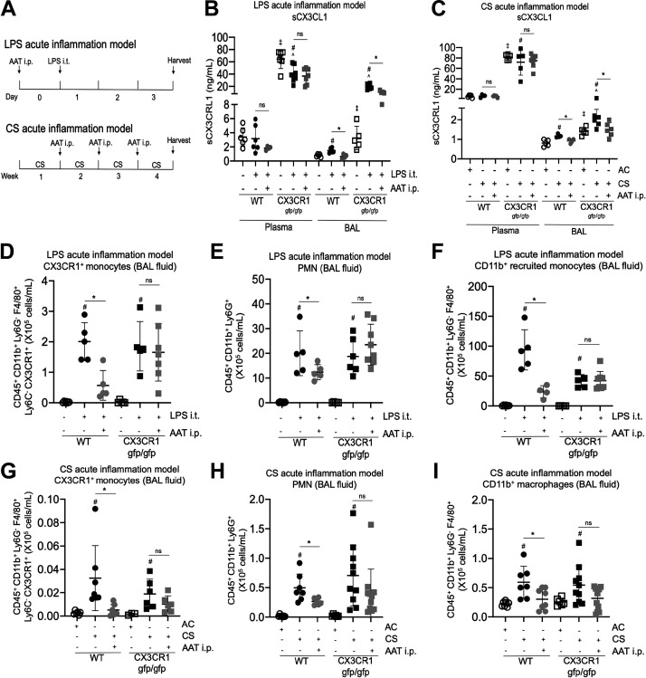
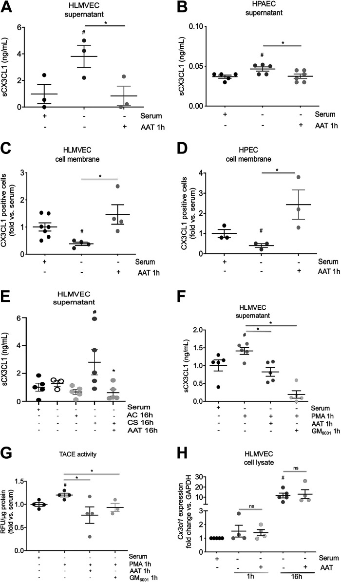
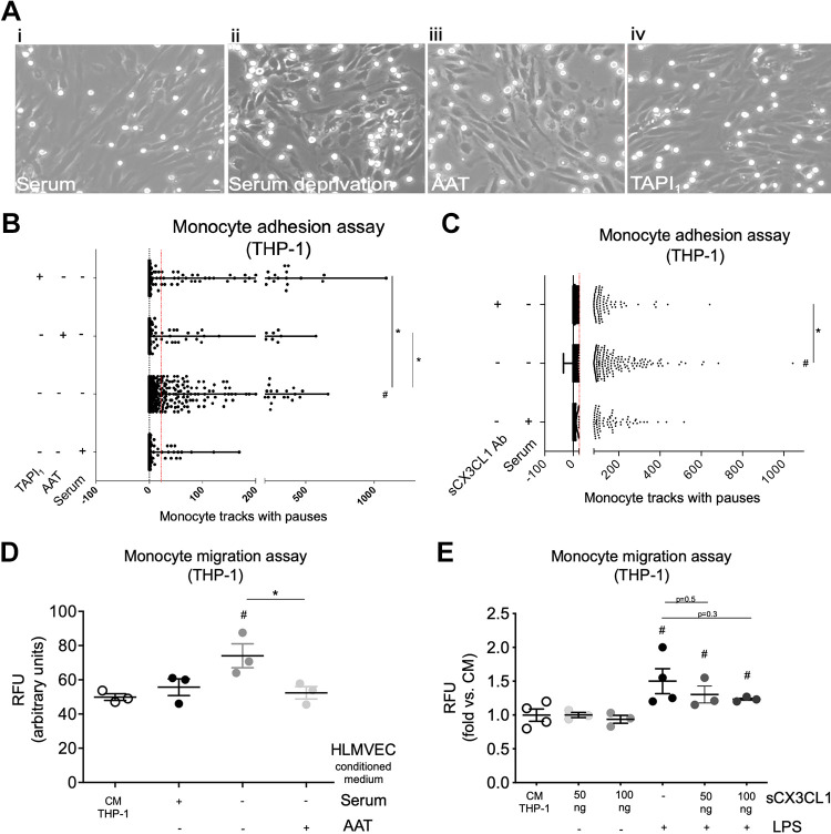
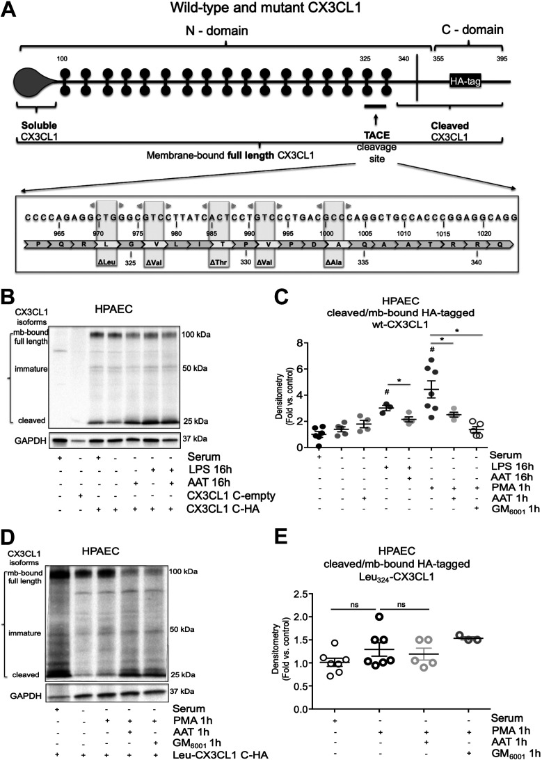
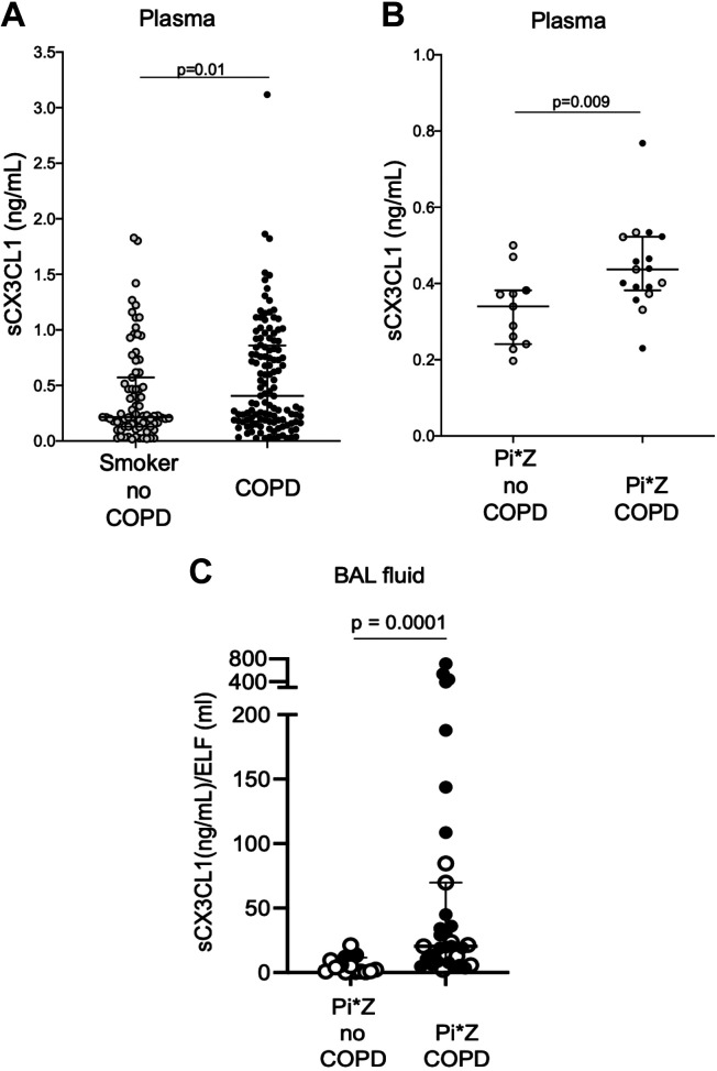
Chronic obstructive pulmonary disease (COPD) is characterized by nonresolving inflammation fueled by breach in the endothelial barrier and leukocyte recruitment into the airspaces. Among the ligand-receptor axes that control leukocyte recruitment, the full-length fractalkine ligand (CX3CL1)-receptor (CX3CR1) ensures homeostatic endothelial-leukocyte interactions. Cigarette smoke (CS) exposure and respiratory pathogens increase expression of endothelial sheddases, such as a-disintegrin-and-metalloproteinase-domain 17 (ADAM17, TACE), inhibited by the anti-protease α-1 antitrypsin (AAT). In the systemic endothelium, TACE cleaves CX3CL1 to release soluble CX3CL1 (sCX3CL1). During CS exposure, it is not known whether AAT inhibits sCX3CL1 shedding and CX3CR1+ leukocyte transendothelial migration across lung microvasculature. We investigated the mechanism of sCX3CL1 shedding, its role in endothelial-monocyte interactions, and AAT effect on these interactions during acute inflammation. We used two, CS and lipopolysaccharide (LPS) models of acute inflammation in transgenic Cx3cr1gfp/gfp mice and primary human endothelial cells and monocytes to study sCX3CL1-mediated CX3CR1+ monocyte adhesion and migration. We measured sCX3CL1 levels in plasma and bronchoalveolar lavage (BALF) of individuals with COPD. Both sCX3CL1 shedding and CX3CR1+ monocytes transendothelial migration were triggered by LPS and CS exposure in mice, and were significantly attenuated by AAT. The inhibition of monocyte-endothelial adhesion and migration by AAT was TACE-dependent. Compared with healthy controls, sCX3CL1 levels were increased in plasma and BALF of individuals with COPD, and were associated with clinical parameters of emphysema. Our results indicate that inhibition of sCX3CL1 as well as AAT augmentation may be effective approaches to decrease excessive monocyte lung recruitment during acute and chronic inflammatory states.NEW & NOTEWORTHY Our novel findings that AAT and other inhibitors of TACE, the sheddase that controls full-length fractalkine (CX3CL1) endothelial expression, may provide fine-tuning of the CX3CL1-CX3CR1 axis specifically involved in endothelial-monocyte cross talk and leukocyte recruitment to the alveolar space, suggests that AAT and inhibitors of sCX3CL1 signaling may be harnessed to reduce lung inflammation.
Keywords: TACE; cell-cell interaction; cigarette smoking; soluble fractalkine; α-1 antitrypsin.
Conflict of interest statement
No conflicts of interest, financial or otherwise, are declared by the authors.
Figures
References
-
- Nahrendorf M, Swirski FK, Aikawa E, Stangenberg L, Wurdinger T, Figueiredo JL, Libby P, Weissleder R, Pittet MJ. The healing myocardium sequentially mobilizes two monocyte subsets with divergent and complementary functions. J Exp Med 204: 3037–3047, 2007. doi:10.1084/jem.20070885. - DOI - PMC - PubMed
Publication types
MeSH terms
Substances
Grants and funding
LinkOut - more resources
Full Text Sources
Molecular Biology Databases
Research Materials
Miscellaneous