

A Suture-specific Photo Score for Metopic Synostosis
- PMID: 37815380
- PMCID: PMC10749672
- DOI: 10.1097/SCS.0000000000009773
A Suture-specific Photo Score for Metopic Synostosis
Abstract
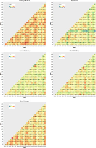
Head shape assessments in children with metopic synostosis are a relevant outcome measure in addition to functional measures, such as neurocognitive outcomes, behavioral outcomes, and visual functioning outcomes. However, consensus on head shape assessments in children with metopic synostosis is lacking. The aim of this study is to develop a reproducible and reliable suture-specific photo score that can be used for cross-center comparison of phenotypical severity of metopic synostosis and evaluation of esthetic outcome of treatment later in childhood. We conducted a retrospective study among nonsyndromic metopic synostosis patients aged <18 years. Preoperative and postoperative photosets of patients with metopic synostosis from 6 expert centers were included. The photo score was discussed in the group of expert craniofacial plastic surgeons and pediatric neurosurgeons. Interrater reliability was determined with modified weighted Fleiss' kappa and intraclass correlation coefficients. Correlation between individual photo score items with overall phenotype was assessed using Spearman correlation analyses. The metopic synostosis photo score contained the following items: "wedging of the forehead", "hypotelorism", "temporal hollowing", "biparietal widening,"and an assessment of "overall phenotype". Items were scored on a 4-point ordinal scale ranging from normal to severe. We found moderate interrater reliability for all items, but substantial agreement for the summed scores. Correlation with overall phenotype was lowest for biparietal widening. To conclude, although agreement on individual photo score items was suboptimal, the agreement on the summed score was substantial, which indicates there is consensus on the overall severity of the metopic synostosis phenotype.
Copyright © 2023 The Author(s). Published by Wolters Kluwer Health, Inc. on behalf of Mutaz B. Habal, MD.
Conflict of interest statement
The authors report no conflicts of interest.
Figures


References
-
- Mathijssen IM, van Splunder J, Vermeij-Keers C, et al. Tracing craniosynostosis to its developmental stage through bone center displacement. J Craniofac Genet Dev Biol 1999;19:57–63 - PubMed
-
- Kellogg R, Allori AC, Rogers GF, et al. Interfrontal angle for characterization of trigonocephaly: part 1: development and validation of a tool for diagnosis of metopic synostosis. J Craniofac Surg 2012;23:799–804 - PubMed
-
- Linden OE, Baratta VM, Gonzalez JA, et al. Surgical correction of metopic craniosynostosis: a 3-D photogrammetric analysis of cranial vault outcomes. Cleft Palate Craniofac J 2019;56:231–235 - PubMed
-
- Bottero L, Lajeunie E, Arnaud E, et al. Functional outcome after surgery for trigonocephaly. Plast Reconstr Surg 1998;102:952–958; discussion 9-60 - PubMed
-
- Posnick JC, Lin KY, Chen P, et al. Metopic synostosis: quantitative assessment of presenting deformity and surgical results based on CT scans. Plast Reconstr Surg 1994;93:16–24 - PubMed
MeSH terms
LinkOut - more resources
Full Text Sources
