. 2023 Oct 31;42(10):113221.
doi: 10.1016/j.celrep.2023.113221. Epub 2023 Oct 9.

MYC is a regulator of androgen receptor inhibition-induced metabolic requirements in prostate cancer

Affiliations

MYC is a regulator of androgen receptor inhibition-induced metabolic requirements in prostate cancer

Preston D Crowell et al. Cell Rep. .

Abstract

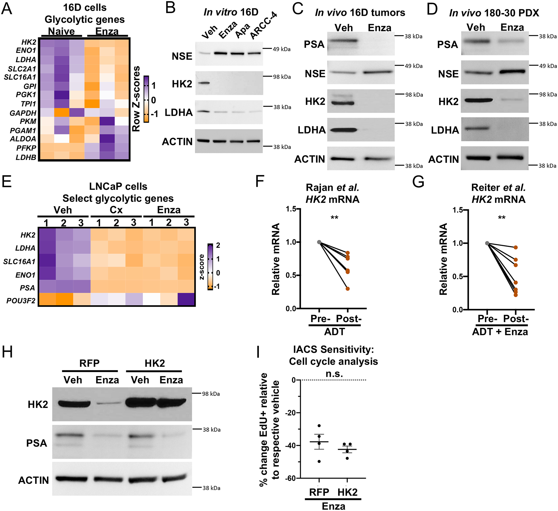
Advanced prostate cancers are treated with therapies targeting the androgen receptor (AR) signaling pathway. While many tumors initially respond to AR inhibition, nearly all develop resistance. It is critical to understand how prostate tumor cells respond to AR inhibition in order to exploit therapy-induced phenotypes prior to the outgrowth of treatment-resistant disease. Here, we comprehensively characterize the effects of AR blockade on prostate cancer metabolism using transcriptomics, metabolomics, and bioenergetics approaches. The metabolic response to AR inhibition is defined by reduced glycolysis, robust elongation of mitochondria, and increased reliance on mitochondrial oxidative metabolism. We establish DRP1 activity and MYC signaling as mediators of AR-blockade-induced metabolic phenotypes. Rescuing DRP1 phosphorylation after AR inhibition restores mitochondrial fission, while rescuing MYC restores glycolytic activity and prevents sensitivity to complex I inhibition. Our study provides insight into the regulation of treatment-induced metabolic phenotypes and vulnerabilities in prostate cancer.

Keywords: CP: Cancer; CP: Metabolism.


Conflict of interest statement

Declaration of interests P.C.B. sits on the scientific advisory boards of Sage Bionetworks, BioSymetrics, Inc., and Intersect Diagnostics, Inc.

Figures

Figure 1.
Figure 1.. Transcriptomic and metabolic profiling identify AR inhibition-induced metabolic reprogramming.
(A) Top 30 significantly enriched pathways identified by KEGG PATHWAY analysis on differentially expressed (fold change 2, FDR < 0.2) Rajan et al. pre-androgen deprivation therapy (Pre-ADT) and post-androgen deprivation therapy (Post-ADT) genes. Metabolism-related pathways highlighted in red. Also see Supplementary Table 1. (B) Naïve, 24hr Enzalutamide-treated (Enza), 48hr Enza, and LTenza 16D transcriptomics data projected onto principle component analysis (PCA) plot of pre-androgen deprivation therapy (pre-ADT) and post-ADT samples from Rajan et al. data. (C) Top 30 significantly enriched pathways identified by KEGG PATHWAY analysis on differentially expressed genes (fold change 2, FDR < 0.05) in naïve and LTenza 16D cells. Metabolism-related pathways highlighted in red. Also see Supplementary Table 2. (D) Heatmap of differentially abundant metabolites (fold change 1.25, FDR < 0.2) in 1-week enzalutamide-treated 16D tumors compared to vehicle-treated 16D tumors. (E-F) PCA of U13C-glucose (E) or U13C-glutamine (F) fractional contribution data from 16D cells treated with vehicle, 10μM Enzalutamide, 10μM Apalutamide, or 0.5μM ARCC-4 for nine days prior to addition of U13C-glucose or U13C-glutamine for 24 hours. (G) Venn diagram illustrating overlap in metabolites with increased abundance (fold change 1.5) in Enzalutamide-, Apalutamide-, or ARCC-4-treated 16D cells relative to vehicle-treated 16D cells. (H) Heatmap of metabolite abundances in 16D cells treated with vehicle, 10μM Enzalutamide, 10μM Apalutamide, or 0.5μM ARCC-4 for nine days.
Figure 2.
Figure 2.. AR blockade maintains oxidative mitochondrial metabolism and reduces glycolysis.
(A and B) Representative kinetic trace plots of the Oxygen Consumption Rate (OCR) (A) and Extracellular Acidification Rate (ECAR) (B) of naïve and LTenza 16D cells. Treatment with Oligomycin (O), FCCP (F), Rotenone and Antimycin A (R/A) are indicated with arrows. Data represent mean +/− SEM. (C and D) ATP-linked respiration (C) and maximal respiration (D) of naïve and LTenza 16D cells from 10 biological replicate experiments. (E and F) Mitochondrial (Mito) ATP production (E) and glycolytic ATP production (F) of naïve and LTenza 16D cells from 10 biological replicate experiments. (G) Total ATP production as the sum of mitochondrial ATP production (Mito ATP) and glycolytic ATP production (Glyco ATP) of naïve and LTenza 16D cells from 10 biological replicate experiments. Statistics refer to comparison of total ATP levels. Data represent mean + SEM. (H) Percentage of total ATP production from mitochondrial ATP production (% ATP from Mito) of naïve and LTenza 16D cells from 10 biological replicate experiments. (I) Lactate abundance in naïve and LTenza 16D cells from 3 biological replicate experiments. P-values were calculated from a ratio paired t-test. *p < 0.05, **p < 0.01, ****p < 0.0001, n.s. = not significant, p 0.05.
Figure 3.
Figure 3.. AR blockade enhances sensitivity to complex I inhibition.
(A) ATP-linked respiration of naïve and LTenza 16D cells treated with vehicle (DMSO) or 30nM IACS-010759 (IACS) for 24 hrs. Data represent the mean +/− SEM of 5 technical replicates. (B) Cell cycle analysis to quantify the relative sensitivity of naïve and LTenza 16D cells to 30nM IACS. Data represent the mean +/− SEM of 4 technical replicates from a representative experiment (n=3). (C) Apoptosis analysis to identify the percentage of Annexin V-positive cells (% Annexin V+) in naïve and LTenza 16D cells treated with DMSO or 30nM IACS for 48 hours. Data represent the mean +/− SEM of 8 technical replicates. (D) Cell cycle analysis to quantify the relative sensitivity of 16D cells treated with 10μM Enzalutamide, 10μM Apalutamide, or 0.1μM ARCC-4 to 30nM IACS. Data represent the mean +/− SEM of 3 technical replicates. (E) Waterfall plot indicating the ex vivo sensitivity of 180–30 PDX tumor tissue from veh- and 1w Enza-treated tumors to 30nM IACS. Data represent the percent change in EdU positivity (% change EdU+) relative to the respective vehicle. (F-G) Cell cycle analysis of the sensitivity of vehicle-treated (F) or Enza-treated (G) 180–30 PDX tumor tissue to ex vivo culture +/− 30nM IACS. Data represent the mean +/− SEM of 5 tumor samples per treatment group. (H) Immunohistochemical analysis of representative 173–2 PDX tumors grown in intact or castrated mice and treated −/+ 7.5 mg/kg/day IACS for five days stained for Ki67. Scale bars, 50 μm. (I) Quantification of Ki67 staining area from six representative images of 173–2 PDX tumors grown in intact or castrated mice and treated −/+ 7.5 mg/kg/day IACS for five days. P-values were calculated from an unpaired t-test with Welch’s correction (A-D) and a ratio paired t-test (F and G). *p < 0.05, **p < 0.01, ****p < 0.0001, n.s. = not significant, p 0.05.
Figure 4.
Figure 4.. AR blockade elongates mitochondria via reduced DRP1 activity.
(A) Representative immunofluorescent images of naïve and LTenza 16D cells stained for TUFM (green) and DAPI (blue). Scale bars, 5 μm. (B) Schematic illustrating calculation of aspect ratio and form factor. (C and D) Quantification of mitochondrial aspect ratio (C) and mitochondrial form factor (D) from TUFM stains from 20 images per treatment group. Data represent the mean +/− SEM. (E and F) Western blots detecting DRP1 phosphorylation at S616 (DRP1-P616), DRP1, PSA, and Actin (loading control) in 16D cells treated with vehicle, 10μM Enzalutamide, 10μM Apalutamide, or 0.5μM ARCC-4 for nine days (E), and 183-A PDX organoids treated with vehicle or 10μM Enzalutamide for one week (F). (G) Representative immunofluorescent images of RFP- and DRP1S616E-transduced LTenza 16D cells stained for TUFM (green) and DAPI (blue). Scale bars, 10μm. (H) Quantification of mitochondrial aspect ratio from TUFM stains from at least 28 cells per treatment group. Data represent the mean +/− SEM. (I) Western blot indicating expression of DRP1-P616, DRP1, NSE, PSA, and Actin (loading control) in RFP- and DRP1S616E-transduced 16D cells maintained +/− 10μM Enzalutamide for 1 week. (J) Cell cycle analysis to quantify the relative sensitivity of RFP- and DRP1S616E-transduced 16D cells maintained in 10μM Enzalutamide for 1 week to 30nM IACS. Data represent the mean +/− SEM of 4 technical replicates from a representative experiment (n=2). P-values were calculated from an unpaired t-test with Welch’s correction. *p < 0.05, **p < 0.01, ***p < 0.001, ****p < 0.0001, n.s. = not significant, p 0.05.
Figure 5.
Figure 5.. Downregulation of key glycolytic enzymes following AR inhibition.
(A) Heatmap showing the mRNA expression of glycolytic genes from RNA sequencing of 3 technical replicates of naïve and LTenza 16D cells. (B-D) Western blots indicating the expression of PSA, NSE, HK2, LDHA, and Actin (loading control) in lysates from 16D cells treated with vehicle, 10μM Enzalutamide, 10μM Apalutamide, or 0.5μM ARCC-4 for nine days (B), 16D tumors treated with vehicle (veh) or Enzalutamide (Enza) for one week in vivo (C), and 180–30 PDX tumors treated with vehicle (veh) or Enzalutamide (Enza) for one week in vivo (D). (E) Heatmap showing the mRNA expression of select glycolytic genes from RNA sequencing of 3 technical replicates of veh, 72-hour castrated (Cx), and 72-hour Enzalutamide-treated (Enza) LNCaP cells. (F-G) HK2 mRNA expression in matched pre- and post-AR blockade biopsies from the Rajan et al. (F) and Reiter et al. (G) datasets. (H) Western blot indicating expression of HK2, PSA, and Actin (loading control) in RFP- and HK2-transduced 16D cells maintained +/− 10μM Enzalutamide for 1 week. (I) Cell cycle analysis to quantify the relative sensitivity of RFP- and HK2-transduced 16D cells maintained in 10μM Enzalutamide for 1 week to 30nM IACS. Data represent the mean +/− SEM of 4 technical replicates from a representative experiment (n=2). P-values were calculated from a ratio paired t-test (F and G) and an unpaired t-test with Welch’s correction (I). **p < 0.01, ***p < 0.001, n.s. = not significant, p 0.05.
Figure 6.
Figure 6.. Decreased Myc signaling following AR inhibition mediates complex I inhibition sensitivity.
(A-D) GSEA of Hallmark_Myc_targets in Rajan et al. pre-ADT and post-ADT samples (A), Akamatsu et al. pre-castration (pre-cx) and 12 weeks post castration (post-cx) (B), naïve and LTenza 16D cells (C), and RFP- and MYC-transduced LTenza cells (D) showing normalized enrichment scores (NES) and false discovery rates (FDR). (E) Western blot indicating expression of MYC, HK2, LDHA, PSA, and Actin (loading control) in naïve, RFP-transduced LTenza, and MYC-transduced LTenza 16D lysates. (F) Cell cycle analysis to quantify the relative sensitivity of naïve and RFP- and MYC-transduced LTenza 16D cells to 30nM IACS. Data represent the mean +/− SEM of 4 technical replicates from a representative experiment (n=3). (G) mRNA expression of RB1 and TP53 in naïve, shScr-transduced (shScr) LTenza, and shRB1_shTP53-transduced (DKD) LTenza 16D cells. Data represent the mean +/− SEM of 3 technical replicates. (H) GSEA of Hallmark_Myc_targets in naïve and DKD LTenza cells. (I) Cell cycle analysis to quantify the relative sensitivity of naïve and DKD LTenza 16D cells to 30nM IACS. Data represent the mean +/− SEM of 4 technical replicates from a representative experiment (n=2). P-values were calculated from an unpaired t-test with Welch’s correction. *p < 0.05, **p < 0.01, ****p < 0.0001, n.s. = not significant, p 0.05.
Figure 7.
Figure 7.. Sustained MYC expression promotes antiandrogen resistance.
(A) Western blot indicating expression of MYC, HK2, LDHA, DRP1-P616, DRP1, and Actin (loading control) in RFP- and MYC-transduced 16D cells maintained +/− 10μM Enzalutamide for 1 week. (B-C) Glycolytic ATP production (B) and percentage of total ATP production from mitochondrial ATP production (% ATP from Mito) (C) of RFP- and MYC-transduced 16D cells maintained +/− 10μM Enzalutamide for 1 week. Data represent the mean +/− SEM of 4 technical replicates. (D-E) PCA of U13C-glucose (D) or U13C-glutamine (E) fractional contribution data from RFP- and MYC-transduced 16D cells maintained +/− 10μM Enzalutamide for six days prior to addition of U13C-glucose or U13C-glutamine for 24 hours. (F-G) Fractional contribution for M3 serine, M3 lactate, M2 citrate, and M2 malate from U13C-glucose (F) or M5 glutamate, M5 alpha-ketoglutarate, M4 succinate, and M4 malate (G) in RFP- and MYC-transduced 16D cells maintained +/− 10μM Enzalutamide for six days prior to addition of U13C-glucose or U13C-glutamine for 24 hours. (H) Heatmap of differentially abundant metabolites (fold change 1.5, p < 0.05) in RFP- and MYC-transduced 16D cells maintained +/− 10μM Enzalutamide for seven days. (I) Cell cycle analysis to quantify the relative sensitivity of RFP- and Myc-transduced 16D cells to 10μM Enzalutamide. Data represent the mean +/− SEM of 4 technical replicates from a representative experiment (n=2). (J) GSEA of Hallmark_Myc_targets in 21 mCRPC tumors before and after Enzalutamide treatment from Westbrook et al. dataset. P-values were calculated from an unpaired t-test with Welch’s correction. **p < 0.01, ***p < 0.001, ****p < 0.0001, n.s. = not significant, p 0.05.

References

    1. Siegel RL, Miller KD, Wagle NS, and Jemal A (2023). Cancer statistics, 2023. CA Cancer J Clin 73, 17–48. 10.3322/caac.21763. - DOI - PubMed
    1. Sayegh N, Swami U, and Agarwal N (2022). Recent Advances in the Management of Metastatic Prostate Cancer. JCO Oncol Pract 18, 45–55. 10.1200/OP.21.00206. - DOI - PubMed
    1. Nakazawa M, Paller C, and Kyprianou N (2017). Mechanisms of Therapeutic Resistance in Prostate Cancer. Curr Oncol Rep 19, 13. 10.1007/s11912-017-0568-7. - DOI - PMC - PubMed
    1. Tran C, Ouk S, Clegg NJ, Chen Y, Watson PA, Arora V, Wongvipat J, Smith-Jones PM, Yoo D, Kwon A, et al. (2009). Development of a second-generation antiandrogen for treatment of advanced prostate cancer. Science 324, 787–790. 10.1126/science.1168175. - DOI - PMC - PubMed
    1. Beer TM, Armstrong AJ, Rathkopf DE, Loriot Y, Sternberg CN, Higano CS, Iversen P, Bhattacharya S, Carles J, Chowdhury S, et al. (2014). Enzalutamide in metastatic prostate cancer before chemotherapy. N Engl J Med 371, 424–433. 10.1056/NEJMoa1405095. - DOI - PMC - PubMed

Publication types

MeSH terms