Association of EEG Background and Neurodevelopmental Outcome in Neonates With Hypoxic-Ischemic Encephalopathy Receiving Hypothermia
- PMID: 37816642
- PMCID: PMC10727206
- DOI: 10.1212/WNL.0000000000207744
Association of EEG Background and Neurodevelopmental Outcome in Neonates With Hypoxic-Ischemic Encephalopathy Receiving Hypothermia
Abstract
Background and objectives: Predicting neurodevelopmental outcome for neonates with hypoxic-ischemic encephalopathy (HIE) is important for clinical decision-making, care planning, and parent communication. We examined the relationship between EEG background and neurodevelopmental outcome among children enrolled in a trial of erythropoietin or placebo for neonates with HIE treated with therapeutic hypothermia.
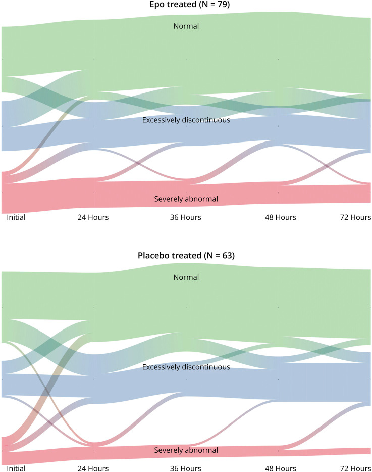
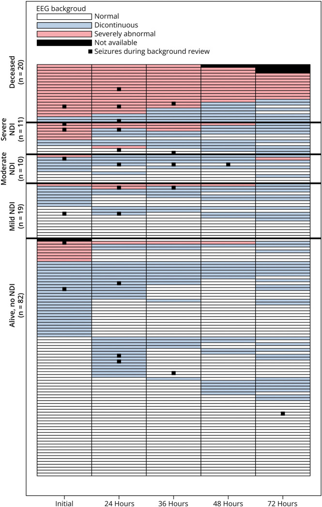
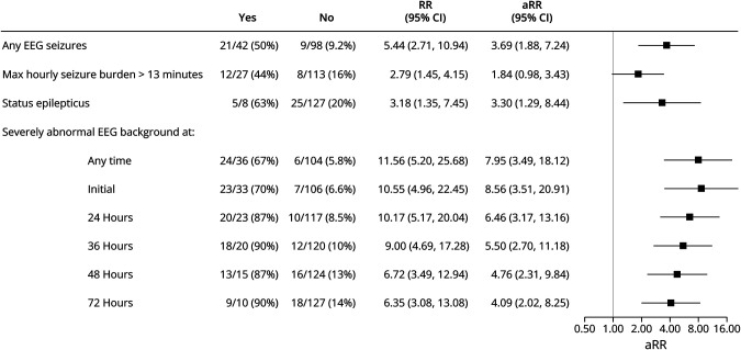
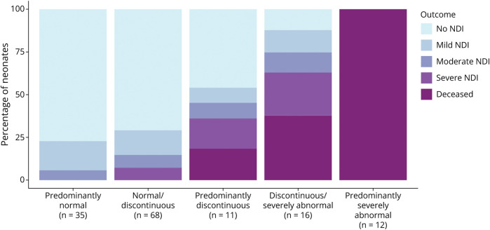
Methods: Participants had EEG recorded throughout hypothermia. EEG background was classified as normal, discontinuous, or severely abnormal (defined as burst suppression, low voltage suppressed, or status epilepticus) at 5 1-hour epochs: onset of recording, 24, 36, 48, and 72 hours after birth. The predominant background pattern during the entire continuous video EEG monitoring recording was calculated using the arithmetic mean of the 5 EEG background ratings (normal = 0; discontinuous = 1; severely abnormal = 2) as follows: "predominantly normal" (mean = 0), "normal/discontinuous" (0 < mean<1), "predominantly discontinuous" (mean = 1), "discontinuous/severely abnormal" (1 < mean<2), or "predominantly severely abnormal" (mean = 2). Primary outcome was death or neurodevelopmental impairment (NDI) defined as cerebral palsy, Gross Motor Function Classification Score ≥1, or cognitive score <90 on Bayley Scales of Infant Toddler Development, third edition at age 2 years. Neurodevelopment was also categorized into a 5-level ordinal measure: no, mild, moderate, severe NDI, or death for secondary analysis. We used generalized linear regression models with robust standard errors to assess the relative risk of death or NDI by EEG background in both unadjusted and adjusted analyses controlling for the effects of treatment group, sex, HIE severity, and study recruitment site.
Results: Among 142 neonates, the predominant background EEG pattern was predominantly normal in 35 (25%), normal/discontinuous in 68 (48%), predominantly discontinuous in 11 (7.7%), discontinuous/severely abnormal in 16 (11%), and predominantly severely abnormal in 12 (8.5%). Increasing severity of background across monitoring epochs was associated with increasingly worse clinical outcomes. Children with severe EEG background abnormality at any time point (n = 36, 25%) were significantly more likely to die or have severe NDI at 2 years (adjusted relative risk: 7.95, 95% CI 3.49-18.12).
Discussion: EEG background is strongly associated with NDI at age 2 years. These results can be used to assist health care providers to plan follow-up care and counsel families for decision-making related to goals of care.
© 2023 American Academy of Neurology.
Conflict of interest statement
K.A. Ahmad, S.L. Bonifacio, B.A. Comstock report no disclosures; H.C. Glass is funded by NIH R01NS104322, R01NS111166, R01NS124051, and her spouse holds shares in Elemeno Health; F.F. Gonzalez, P.J. Heagerty, S.E. Juul, N. Maitre, S.L. Massey, D.E. Mayock, U. Mietzsch, N. Natarajan, A.L. Numis, G.M. Sokol, C. Thomas, K.P. Van Meurs, Y.W. Wu report no disclosures; C.J. Wusthoff reports: disclosures: Scientific Advisory Boards (1) PRA Health Sciences- Data And Safety Monitoring (2) ICON- Data And Safety Monitoring Editorial Boards (1) Journal of Clinical Neurophysiology, Associate Editor, 2020–2022; editorial board 2022-current (2) Neurology, Associate Editor, 2022-current Research Support, Government Entities (1) NIH/NINDS, 1K02NS102598, PI, 2017–2020 (2) PCORI 1507–31187, study neurophysiologist, 2016–2020 (3) NIH/NINDS, 1R01NS104322-01A1, co-investigator, 2018–2022 (4) NIH/NINDS, 1R01NS111166-01A1, co-investigator, 2020–2024 Research Support, Foundations and Societies (1) Thrasher Research Fund 2020-2023. Go to
Figures