. 2023 Oct 10;14(1):6135.
doi: 10.1038/s41467-023-41308-4.

An Influenza A virus can evolve to use human ANP32E through altering polymerase dimerization

Affiliations

An Influenza A virus can evolve to use human ANP32E through altering polymerase dimerization

Carol M Sheppard et al. Nat Commun. .

Abstract

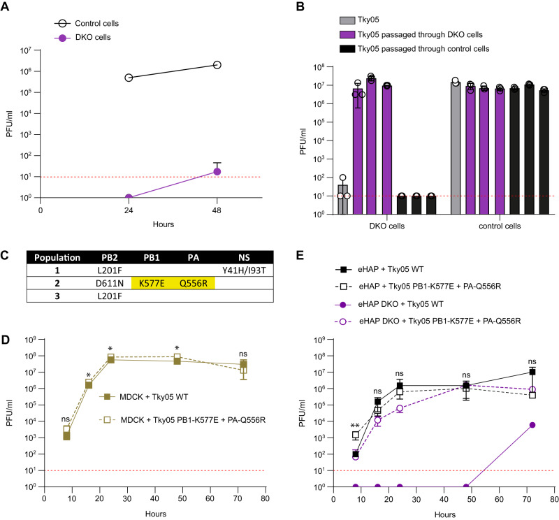
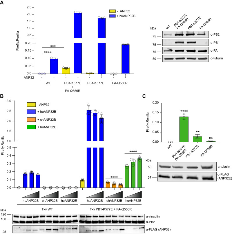
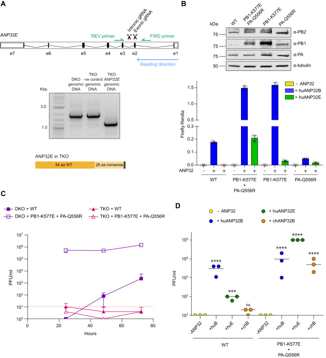
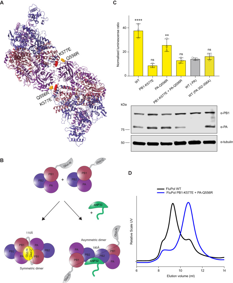
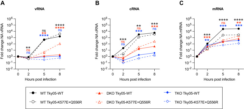
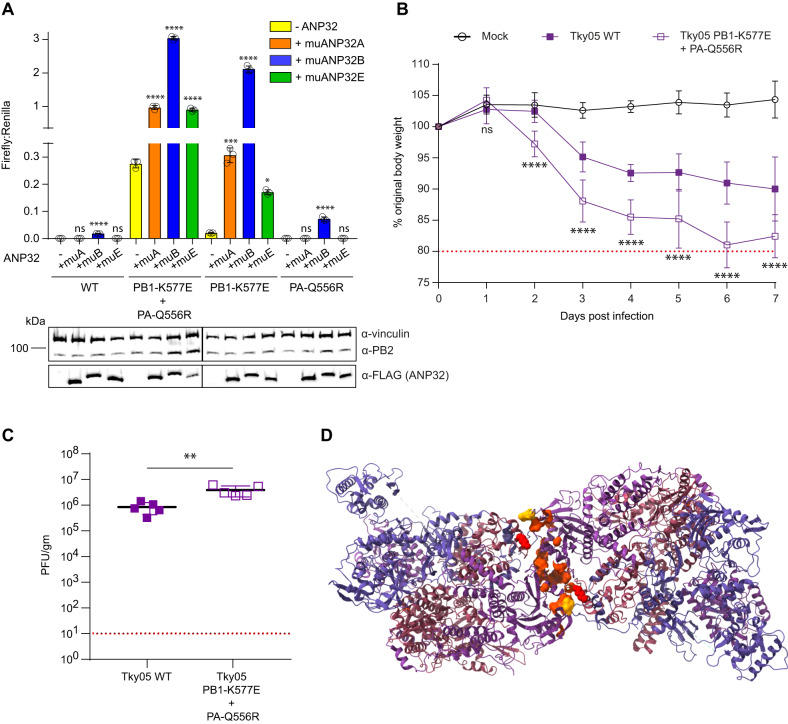
Human ANP32A and ANP32B are essential but redundant host factors for influenza virus genome replication. While most influenza viruses cannot replicate in edited human cells lacking both ANP32A and ANP32B, some strains exhibit limited growth. Here, we experimentally evolve such an influenza A virus in these edited cells and unexpectedly, after 2 passages, we observe robust viral growth. We find two mutations in different subunits of the influenza polymerase that enable the mutant virus to use a novel host factor, ANP32E, an alternative family member, which is unable to support the wild type polymerase. Both mutations reside in the symmetric dimer interface between two polymerase complexes and reduce polymerase dimerization. These mutations have previously been identified as adapting influenza viruses to mice. Indeed, the evolved virus gains the ability to use suboptimal mouse ANP32 proteins and becomes more virulent in mice. We identify further mutations in the symmetric dimer interface which we predict allow influenza to adapt to use suboptimal ANP32 proteins through a similar mechanism. Overall, our results suggest a balance between asymmetric and symmetric dimers of influenza virus polymerase that is influenced by the interaction between polymerase and ANP32 host proteins.


Conflict of interest statement

The authors declare no competing interests.

Figures

Fig. 1
Fig. 1. An influenza A virus evolved to grow in human cells lacking ANP32A and ANP32B proteins.
A Tky05 was inoculated on 12-well plates of eHAP (control) and DKO cells at an MOI of 0.0005. Samples were titred on MDCK cells via plaque assay at 24 and 48 h post infection. Data are presented as mean values +/− SD. B Following two passages through either DKO cells or WT eHAP cells (control), 1000 PFU from each population as well as the ancestor (Tky05) were inoculated onto six-well plates of DKO and eHAP cells. After 48 h, viruses were titred on MDCK cells via plaque assay. n = 3 wells, data are presented as mean values +/− SD. C Mutations from populations passaged through DKO cells were discovered using Sanger sequencing. Highlighted mutations were found in the plaque-purified virus, which was used for future experiments. Growth curves of Tky05 compared to plaque-purified virus containing PB1 K577E, PA Q556R on MDCK (D) or eHAP (E) cells. 12-well plates were infected at MOI 0.001 (D) or 0.0005 (E), and viral titres were measured on MDCK cells at 8, 16, 24, 48 and 72 h. n = 3 wells, data are presented as mean values +/− SD. Statistical significance was determined by multiple t-test of log-transformed data, *P < 0.05, **P < 0.01, ***P < 0.001, ****P < 0.0001, (1D; 8 hpi p = 0.238, 16 hpi p = 0.206, 24 hpi p = 0.035, 48 hpi p = 0.017, 72 hpi p = 0.339. 1E; 8 hpi p = 0.003, 16 hpi p = 0.101, 24 hpi p = 0.802, 48 hpi p = 0.600, 72 hpi p = 0.066) Source data are provided as a Source Data file.
Fig. 2
Fig. 2. Tky05 PB1 K577E and PA Q556R mutations enhance influenza polymerase activity in the absence of human ANP32A/B.
Minigenome assays were performed in 24-well plates of eHAP DKO cells transfected with pCAGGS Tky05 PB2 (0.04 µg), PB1 or K577E (0.04 µg), PA or Q556R (0.02 µg), NP (0.08 µg), reporter pPolI-luc (0.08 µg) and control pCAGGS-Renilla luciferase (0.04 µg) and (A) +/− huANP32B-FLAG (0.04 µg). Western blot showing expression of PB2, PB1, PA and tubulin. B Minigenome with 0, 0.02, 0.04 or 0.08 µg of huANP32B, chANP32B and huANP32E. Accompanying Western blot showing expression of vinculin, PB2 and ANP32-FLAG. C Minigenome with 0.08 µg of huANP32E. All data presented is representative of n = 3 biological repeats each conducted with n = 3 wells, presented as mean values +/− SD. Statistical significance was determined by multiple comparisons of a one-way ANOVA (A, C) or test for trend of a one-way ANOVA (B) *P < 0.05, **P < 0.01, ***P < 0.001, ****P < 0.0001 (A; Tky WT -ANP32 vs Tky WT +huANP32B P < 0.0001, Tky WT -ANP32 vs Tky PB1-K577E + PA-Q556R -ANP32 p = 0.00015, B; trend for Tky PB1-K577E + PA-Q556R chANP32B titration P < 0.0001, trend for Tky PB1-K577E + PA-Q556R huANP32E titration P < 0.0001, C; Tky WT vs Tky PB1-K577E + PA-Q556R p < 0.0001, Tky WT vs PB1-K577E p = 0.0063, Tky WT vs PA-Q556R p = 0.878). Source data are provided as a Source Data file.
Fig. 3
Fig. 3. PB1 K577E and PA Q556R allow appropriation of human ANP32E.
A Schematic showing the location of the CRISPR guide RNAs and PCR primers in huANP32E. Gel showing PCR product from genomic DNA for WT, −ve control and TKO cells and a schematic illustrating the resulting truncated ANP32E in TKO cells. A single TKO clone was obtained. B Minigenome assay conducted in 24-well plates of eHAP TKO cells transfected with pCAGGS Tky05 PB1 or K577E (0.04 µg), PB2 (0.04 µg), PA or Q556R (0.02 µg), NP (0.08 µg), reporter pPolI-luc (0.08 µg) and control pCAGGS-Renilla luciferase (0.04 µg) and +/− huANP32B-FLAG (0.04 µg). Data presented is representative of n = 3 biological repeats each conducted with n = 3 wells, presented as mean values +/− SD. Western blot showing expression of PB2, PB1, PA and tubulin. C Viral Growth curve in DKO and TKO cells for WT and PB1 K577E + PA Q556R virus. Six-well plates were infected with virus at MOI of 0.01 and samples plaqued at 24, 48 and 72 h, n = 3 wells presented as mean values +/− SD, limit of detection = 10 PFU/ml. D TKO cells were transfected in a 12-well plate with -ANP32, huANP32B, huANP32E or chANP32B (0.32 µg). After 24 h, cells were infected with either WT (Tky05) or PB1 K577E + PA Q556R virus at MOI of 1. Virus was titred at 72 h, n = 3 wells. Statistical significance was determined by one-way ANOVA of log transformed data. Samples were compared to -ANP32 of the appropriate virus, *P < 0.05, **P < 0.01, ***P < 0.001, ****P < 0.0001 3D; Tky WT -ANP32 vs Tky WT +huANP32B p < 0.0001, Tky WT -ANP32 vs Tky WT +huANP32E p = 0.0005, Tky WT -ANP32 vs Tky WT +chANP32B p = 0.2189, Tky PB1-K577E + PA-Q556R -ANP32 vs Tky PB1-K577E + PA-Q556R +huANP32B p < 0.0001, Tky PB1-K577E + PA-Q556R -ANP32 vs Tky PB1-K577E + PA-Q556R +huANP32E p < 0.0001, Tky PB1-K577E + PA-Q556R -ANP32 vs Tky PB1-K577E + PA-Q556R +chANP32B p < 0.0001. Source data are provided as a Source Data file.
Fig. 4
Fig. 4. PB1 K577E and PA Q556R increase binding to huANP32E.
A Asymmetric dimer of influenza polymerase (PDB: 6XZP) and ANP32 (green) showing PB2 627K in blue, PB1 K577E in red and PA Q556R in yellow. B Co-immunoprecipitation assays were conducted from 10 cm dishes of eHAP TKO cells transfected with pCAGGS Tky05 PB2 (5 µg), Tky05 PB1 or K577E (5 µg), Tky05 PA or Q556R (5 µg) and huANP32B-FLAG/chANP32B-FLAG/huANP32E-FLAG (5 µg). Twenty-four hours after transfection, cells were lysed, FLAG-tagged proteins were immunoprecipitated, and co-precipitation of PB2 was detected using immunoblotting. Data presented are representative of n = 3 biological repeats. C Schematic showing the reconstitution of Gaussia luciferase activity from the interaction of the C-terminus of Gaussia luciferase fused to ANP32 with the N-terminus of Gaussia luciferase fused to PB1. D Split luciferase assay measuring the interaction between influenza polymerase combinations Tky05 WT, PB1 K577E + PA Q556R, PB1-K577E and PA-Q556R (formed using PB1-Gluc1 or PB1-K577E-Gluc1 fusions) and either huANP32B-Gluc2, chANP32B-Gluc2 or huANP32E-Gluc2. Data presented are representative of n = 3 biological repeats each conducted with n = 3 wells, presented as mean values +/− SD. Accompanying Western blot showing expression of PA/Q556R, PB1/K577E-Gluc1, tubulin and ANP32-Gluc2. Source data are provided as a Source Data file.
Fig. 5
Fig. 5. PB1 K577E destabilises formation of the polymerase dimer.
A Symmetric dimer of influenza polymerase (PDB: 6QX8) showing PB1 K577E in red and PA Q556R in yellow. B Schematic showing the split-luciferase dimerization assay with C-terminus of Gaussia luciferase and the N-terminus of Gaussia luciferase attached to separate PB1 proteins. The two halves of the luciferase are 110Å in the same plane of the symmetric dimer and 180 Å on opposite sides of the asymmetric dimer. C Split luciferase complementation assay measuring dimerization of Tky05 WT, PB1 K577E + PA Q556R, PB1 K577E and PA Q556R polymerases (formed using equal amounts of PB1-Gluc1 + PB1-Gluc2 or PB1-K577E-Gluc1 + PB1-K577E-Gluc2). Data presented are representative of n = 3 biological repeats each conducted with n = 3 wells, presented as mean values +/− SD. Statistical significance was determined by multiple comparisons of one-way ANOVA, samples were compared to WT (-PA), *P < 0.05, **P < 0.01, ***P < 0.001, ****P < 0.0001 (-PA vs WT p < 0.0001, -PA vs PB1-K577E p = 0.2708, -PA vs PA-Q556R p = 0.0019, -PA vs PB1-K577E + PA-Q556R p = 0.9993, -PA vs WT(PA 352-356A) p = 0.9683). Accompanying Western blot showing expression of PB1/K577E-Gluc, PA/Q556R and tubulin. D Tky05 WT polymerase and PB1 K577E + PA Q556R were cloned into baculovirus and expressed in Sf9 cells. The polymerases were purified and concentrated (as described in the methods) then analysed by size exclusion chromatography. Lower elution volume is indicative of higher molecular weight. Source data are provided as a Source Data file.
Fig. 6
Fig. 6. PB1 K577E + PA Q556R requires ANP32E to support vRNA synthesis.
AC Segment 6 vRNA (A) cRNA (B) or mRNA (C) accumulation over time in eHAP WT, DKO and TKO cells following infection with Tky05-WT or Tky05-K577E + Q556R (MOI = 3). Fold change was calculated over input (0 h.p.i.) for each cell type. n = 3 biological replicates, plotted as mean ± s.d. Statistical significance was assessed using multiple unpaired two-sided t tests following log transformation, corrected for multiple comparisons using the false discovery rate *P < 0.05, **p < 0.01; ***p < 0.001; ****p < 0.0001. Statistical comparisons are between Tky05-WT and Tky05-K577E + Q556R within the same cell line at each timepoint (vRNA WT cells; 2 h, p = 0.0043, 5 h p = 0.7484, 8 h p < 0.0001, vRNA DKO cells; 2 h P = 0.8564, 5 h p = 0.0043, 8 h p < 0.0001, vRNA TKO cells; 2 h p = 0.0464, 5 h p < 0.0001, 8 h p = 0.0266, cRNA WT cells; 2 h p = 0.009, 5 h p = 0.0015, 8 h p = 0.0004, cRNA DKO cells; 2 h p = 0.4967, 5 h p = 0.0539, 8 h p = 0.0102, cRNA TKO cells; 2 h p = 0.0444, 5 h p = 0.0004, 8 h p = 0.0102, mRNA WT cells; 2 hrs p = 0.0003, 5 h p < 0.0001, 8 h p < 0.0001, mRNA DKO cells; 2 h p = 0.0003, 5 h p = 0.0259, 8 h p = 0.0010, mRNA TKO cells; 2 h p = 0.0035, 5 h p = 0.0004, 8 h p = 0.0002). Comparisons for WT cells are shown at the top, DKO cells in the middle and TKO cells at the bottom (indicated by matching colour to traces). Source data are provided as a Source Data file.
Fig. 7
Fig. 7. PB1 K577E + PA Q556R polymerase can use suboptimal murine ANP32 proteins.
A Minigenome assays were performed in 24-well plates of eHAP DKO cells transfected with pCAGGS Tky05 PB2 (0.04 µg), PB1 or K577E (0.04 µg), PA or Q556R (0.02 µg), NP (0.08 µg), reporter pPolI-luc (0.08 µg), control pCAGGS-Renilla luciferase (0.04 µg) and either -ANP32, muANP32A-FLAG, muANP32B-FLAG or muANP32E-FLAG (0.04 µg). Data presented are representative of n = 3 biological repeats each conducted with n = 3 wells, presented as mean values +/− SD. Statistical significance was determined by multiple comparisons of one-way ANOVA, *P < 0.05, **P < 0.01, ***P < 0.001, ****P < 0.0001 (Tky WT; -ANP32 vs +muANP32A p = 0.923, -ANP32 vs +muANP32B p > 0.0001, -ANP32 vs +muANP32E p = 0.923, Tky PB1-K577E + PA-Q556R; -ANP32 vs +muANP32A p < 0.0001, -ANP32 vs +muANP32B p < 0.0001, -ANP32 vs +muANP32E p < 0.0001, Tky PB1-K577E; -ANP32 vs +muANP32A p = 0.0003, -ANP32 vs +muANP32B p < 0.0001, -ANP32 vs +muANP32E p = 0.0135, Tky PA-Q556R; -ANP32 vs +muANP32A p < 0.9999, -ANP32 vs +muANP32B p < 0.0001, -ANP32 vs +muANP32E p > 0.9999). Accompanying western blot showing expression of vinculin, PB2 and ANP32-FLAG. B Six to eight-week-old female BALB/c mice were mock infected (10 mice) or infected intranasally with 105 PFU of Tky05 WT (10 mice) or PB1 K577E + PA Q556R virus (10 mice). 5 mice per group were culled at day 2 and 5 mice culled at day 7. Weight loss was measured each day. One mouse was excluded from the Tky05 WT group for weight loss calculation because it did not become infected after inoculation. Data are presented as mean values +/− SD. Statistical significance was determined by two-way ANOVA, *P < 0.05, **P < 0.01, ***P < 0.001, ****P < 0.0001 (displayed is a comparison between WT and PB1-K577E + PA-Q556R; day 1 p > 0.3258, day 2 p < 0.0001, day 3 p < 0.0001, day 4 p < 0.0001, day 5 p < 0.0001, day 6 p < 0.0001, day 7 p < 0.0001). C Viral titres from the homogenized lung tissue on day 2 p.i. (n = 5 murine lungs per group). Data are presented as mean values +/− SD. Statistical significance was determined by an unpaired t-test, *P < 0.05, **P < 0.01, ***P < 0.001, ****P < 0.0001 (WT vs PB1-K577E + PA-Q556R p = 0.0058). D Symmetric dimer of the influenza A polymerase (PDB: 6QX8) with mouse-adaptive mutations mapping to the interface. Highlighted residues are PA N291, L295, L336, A343, D347, E349, K353, K356, L550, T552, I554, Q556 and PB1 K577. Source data are provided as a Source Data file.
Fig. 8
Fig. 8. PB1 K577E and PA Q556R rebalance influenza polymerase dimer formation.
Schematic showing the proposed mechanism of how PB1 K577E and PA Q556R appropriate ANP32E to re-balance the equilibrium of polymerase dimers.

References

    1. Long JS, Mistry B, Haslam SM, Barclay WS. Host and viral determinants of influenza A virus species specificity. Nat. Rev. Microbiol. 2019;17:67–81. doi: 10.1038/s41579-018-0115-z. - DOI - PubMed
    1. Peacock TP, Sheppard CM, Staller E, Barclay WS. Host determinants of influenza RNA synthesis. Annu. Rev. Virol. 2019;6:215–233. doi: 10.1146/annurev-virology-092917-043339. - DOI - PubMed
    1. König R, et al. Human host factors required for influenza virus replication. Nature. 2010;463:813–817. doi: 10.1038/nature08699. - DOI - PMC - PubMed
    1. Edinger TO, Pohl MO, Stertz S. Entry of influenza A virus: host factors and antiviral targets. J. Gen. Virol. 2014;95:263–277. doi: 10.1099/vir.0.059477-0. - DOI - PubMed
    1. Han J, et al. Genome-wide CRISPR/Cas9 screen identifies host factors essential for influenza virus replication. Cell Rep. 2018;23:596–607. doi: 10.1016/j.celrep.2018.03.045. - DOI - PMC - PubMed

Publication types