. 2023 Oct 11;14(10):668.
doi: 10.1038/s41419-023-06182-4.

Macrophages originated IL-33/ST2 inhibits ferroptosis in endometriosis via the ATF3/SLC7A11 axis

Affiliations

Macrophages originated IL-33/ST2 inhibits ferroptosis in endometriosis via the ATF3/SLC7A11 axis

Qiong Wu et al. Cell Death Dis. .

Erratum in

Abstract

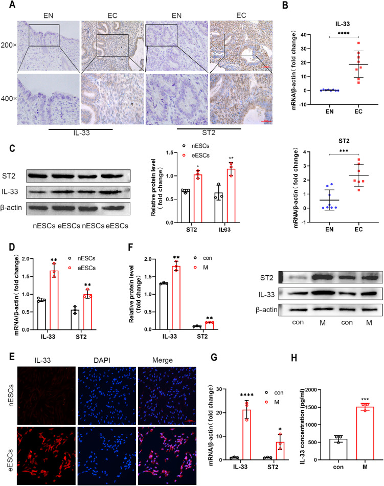
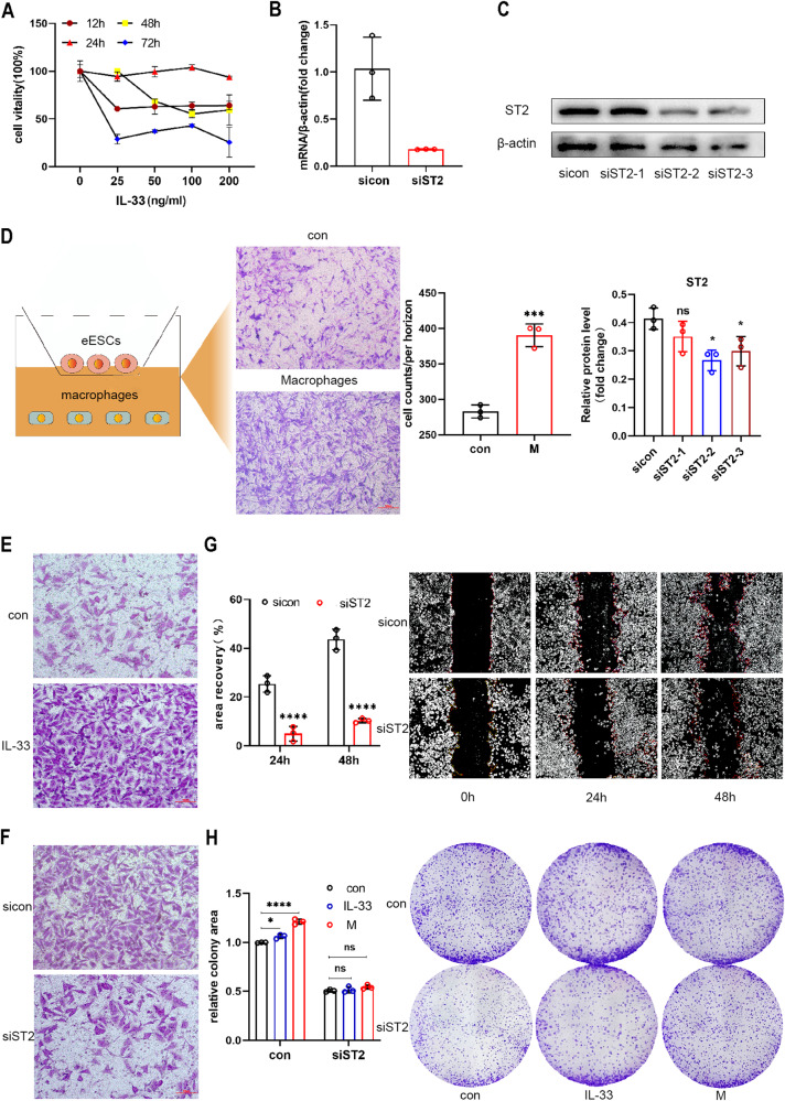
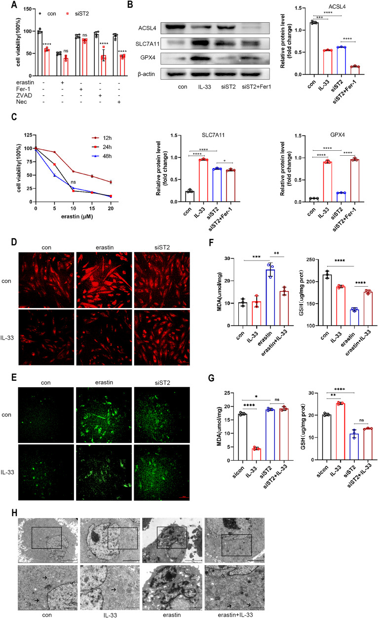
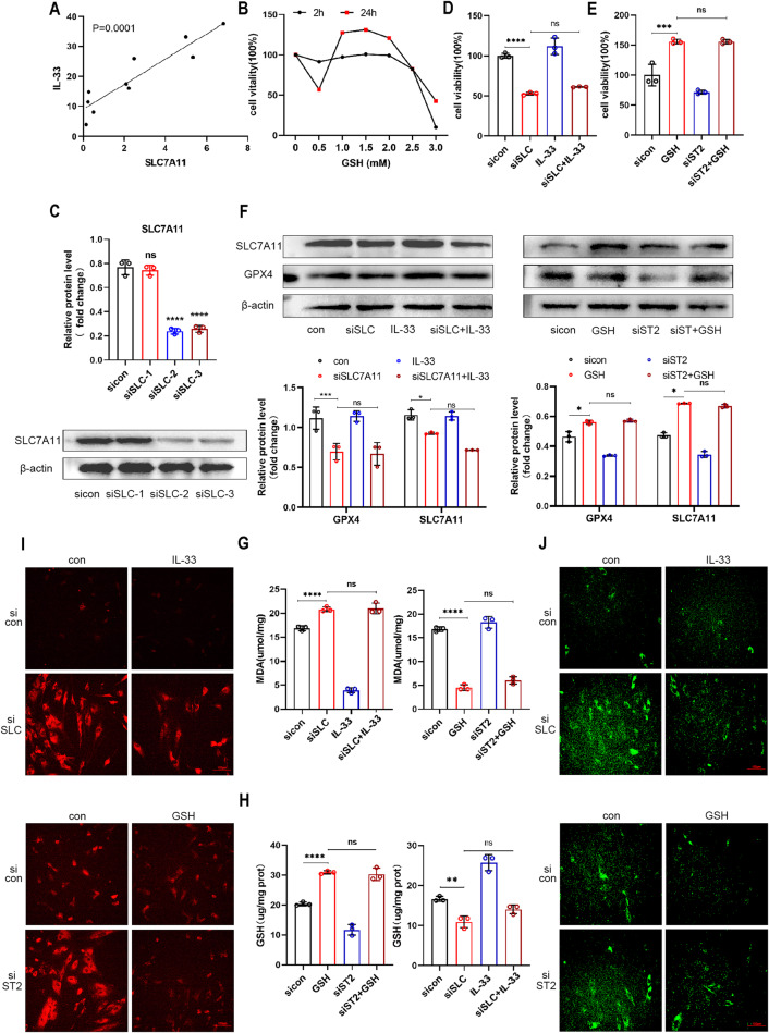
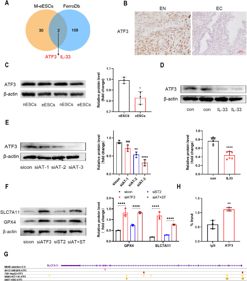
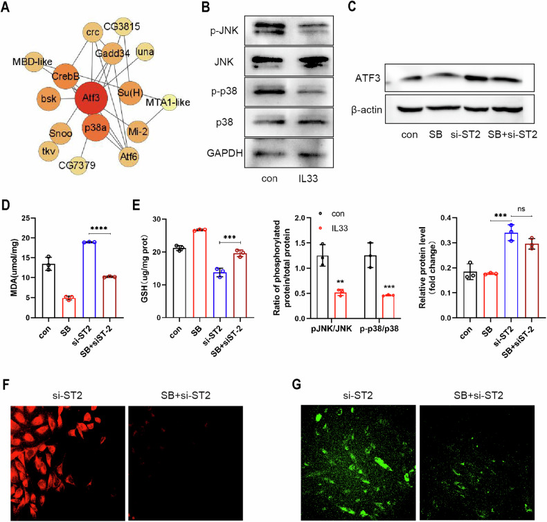
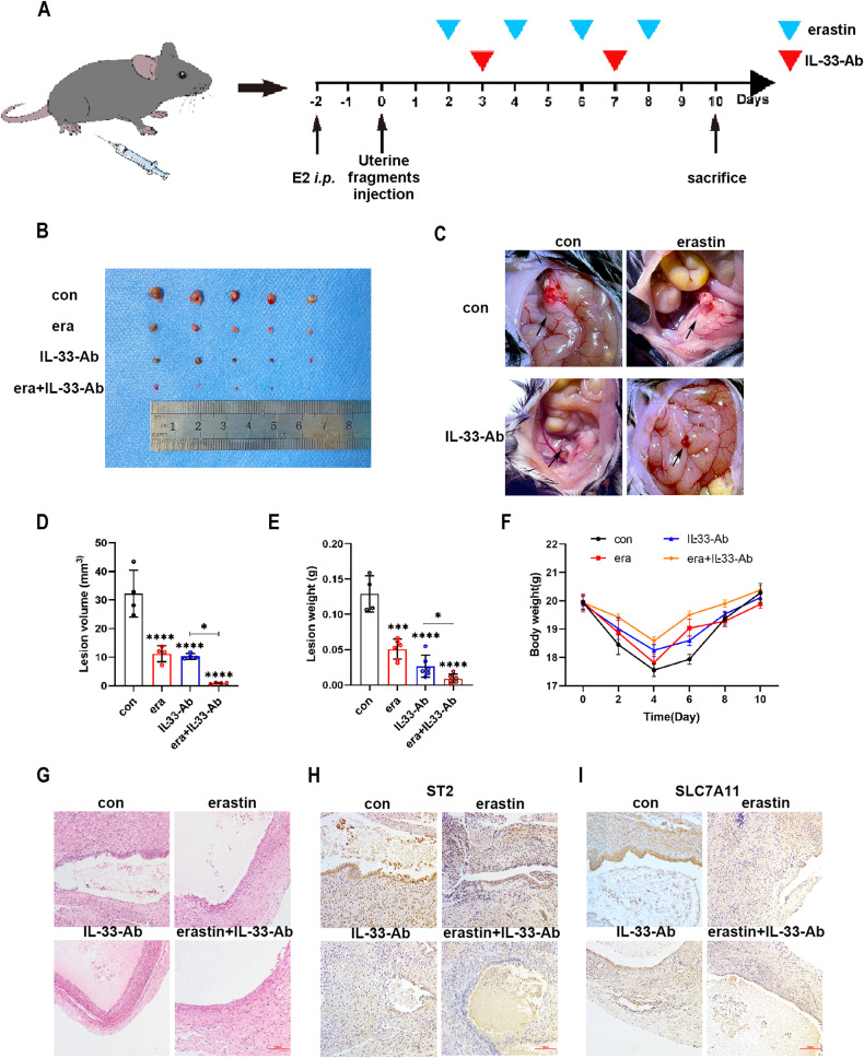
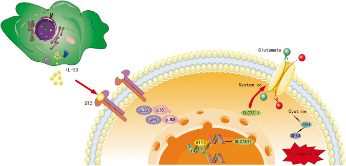
Endometriosis is a gynecological inflammatory disease that is linked with immune cells, specifically macrophages. IL-33 secreted from macrophages is known to accelerate the progression of endometriosis. The periodic and repeated bleeding that occurs in women with endometriosis leads to excess iron in the microenvironment that is conducive to ferroptosis, a process related to intracellular ROS production, lipid peroxidation and mitochondrial damage. It is suggested that eESCs may specifically be able to inhibit ferroptosis. However, it is currently unclear whether IL-33 directly regulates ferroptosis to influence the disease course in endometriosis. In this study, eESCs co-cultured with macrophages or stimulated with IL-33/ST2 were observed to have increased cell viability and migration. Additionally, IL-33/ST2 decreased intracellular iron levels and lipid peroxidation in eESCs exposed to erastin treatment. Furthermore, IL-33/ST2 treatment resulted in a notable upregulation in SLC7A11 expression in eESCs due to the downregulation of negative transcription factor ATF3, thereby suppressing ferroptosis. The P38/JNK pathway activated by IL-33/ST2 was also found to inhibit the transcription factor ATF3. Therefore, we concluded that IL-33/ST2 inhibits the ATF3-mediated reduction in SLC7A11 transcript levels via the P38/JNK pathway. The findings reveal that macrophage-derived IL-33 upregulates SLC7A11 in eESCs through the p38/JNK/ATF3 pathway, ultimately resulting in protection against ferroptosis in eESCs. Moreover, we conducted an experiment using endometriosis model mice that showed that a combination of IL-33-Ab and erastin treatment alleviated the disease, showing the promise of combining immunotherapy and ferroptosis therapy.


Conflict of interest statement

The authors declare no competing interests.

Figures

Fig. 1
Fig. 1. IL-33 from co-cultured macrophages activates ST2 in eESCs.
A Representative immunohistochemical images staining with IL-33 and ST2 in normal endometrial tissue (EN) and ectopic endometriosis lesion tissue (EC). (original magnification ×200 or ×400) (n = 5). B RT-qPCR was used to determine the mRNA levels of IL-33 and ST2 in EN and EC (n = 8). C Western blot was used to detect the protein levels of IL-33 and ST2 in normal endometrial stromal cells (nESCs) and ectopic endometrial stromal cells (eESCs). D RT-qPCR was used to determine the mRNA levels of IL-33 and ST2 in nESCs and eESCs. E Representative immunofluorescence (IF) images of IL-33 (red) in nESCs and eESCs, Nuclei were stained with DAPI (blue). (original magnification ×200). F Western blot was used to detect the protein levels of IL-33 and ST2 in eESCs with or without macrophages co-culture treatment. G RT-qPCR was used to determine the mRNA levels of IL-33 and ST2 in eESCs with or without macrophages co-culture treatment. H ELISA analysis of IL-33 concentration in eESCs culture medium with or without macrophages co-culture treatment. Data are presented as the mean ± SD, n = 3 independent experiments. Statistical analysis was performed using Student’s t test. ****p < 0.0001, ***p < 0.001, **p < 0.01, *p < 0.05. IL-33 interleukin-33, ST2 interleukin receptor-like 1 (IL1R-L1), M macrophages, con control.
Fig. 2
Fig. 2. IL-33/ST2 increased the survival rate and migration ability of eESCs.
A The eESCs were treated with specified concentrations of human recombinant IL-33 protein (rIL-33) (25, 50, 100, and 200 ng/ml) for different times (12, 24, 48, and 72 h). CCK-8 assays were performed to detect cell viability in different groups. B RT-qPCR was used to determine the relative levels of ST2 mRNA in eESCs transfected with siST2 (50 nM), sicon (50 nM) for 48 h. C Western blot was used to determine the protein levels of ST2 in eESCs transfected with siST2 (50 nM) or sicon (50 nM) for 48 h. D Transwell migration assay was performed to detect the migrant ability of eESCs treated with or without macrophages co-culture. Cartoon picture showed the experimental progress. (original magnification ×200). E, F Transwell migrantion assays were performed in rIL-33 (100 ng/ml) treated eESCs (E) and siST2 (50 nM) transfected eESCs (F). (original magnification ×200). G The wound healing assays were conducted in eESCs transfected with siST2 (50 nM) and control group. (original magnification ×40). H Colony formation assays were performed in siST2 (50 nM) transfected eESCs treated with rIL-33 (100 ng/ml) or macrophages co-culture. (original magnification ×40).Data are presented as the mean ± SD, n = 3 independent experiments. Statistical analysis was performed using Student’s t test (BD, G) or 2-way ANOVA (H). ****p < 0.0001, ***p < 0.001, *p < 0.05, ns, non-significant. sicon negative control siRNA, siST2 siRNA targeting ST2, M macrophages co-culture treatment.
Fig. 3
Fig. 3. IL-33/ST2 inhibited ferroptosis in eESCs.
A Cell viability was detected by CCK-8 assay in siST2 (50 nM) transfected eESCs with different inhibitors treatment (erasin, 10 μM; Ferrostatin-1, 1 μM; ZVAD-FMK, 10 μM; Necrostatin, 10 μM) for 24 h. B Western blot was used to detect the protein levels of ACSL4, SLC7A11, and GPX4 in different groups of eESCs treated with rIL-33 (100 ng/ml), siST2 (50 nM), or siST2 (50 nM)+Ferrostatin-1 (1 μM). C Cell viability was measured by CCK-8 assay in eESCs treated with different concentration of erasin (5, 10, 15, and 20 μM) for 12, 24, or 48 h. D Intracellular Fe2+ were detected by treating eESCs with 1 μM FerroOrange after indicated treatment: (a) we add erastin (10 μM) and rIL-33(100 ng/ml) into eESCs, eESCs treated with no treatment (con) and only with rIL-33 play as compared groups. (b) We added rIL-33 (100 ng/ml) into siST2 transfected eESCs, only siST2 transfected and rIL-33 addition only play as compared groups (original magnification ×200). E LiperFluo reagent (5 μM) were used to detect intracellular lipid peroxidation levels in eESCs with indicated treatment (same as treatment in (D)). (original magnification ×200). F, G The MDA levels (F) and GSH levels (G) were measured in eESCs with indicated treatment (same as treatment in (D)). H Transmission Electron Microscopy (TEM) was used to observe the morphological changes of eESCs mitochondria. black arrowheads: normal mitochondria. white arrowheads: shrunken mitochondria. The scale bar = 5.0 μm (Upper row). The scale bar = 2.0 μm (Lower row). Data are presented as the mean ± SD, n = 3 independent experiments. Statistical analysis was performed using Student’s t test (A) or one-way ANOVA (B, F, G). ****p < 0.0001, ***p < 0.001, **p < 0.01, *p < 0.05, ns non-significant, Fer-1 Ferrostatin-1, Nec Necrostatin-1, ZVAD-FMK, benzyloxycarbonyl-Val-Ala-Asp (OMe)-fluoromethylketone, ACSL4 acyl-CoA synthetase long-chain family member 4, SLC7A11 solute carrier family 7 member 11, GPX4 glutathione peroxidase 4, MDA malondialdehyde, GSH glutathione.
Fig. 4
Fig. 4. IL-33/ST2 inhibited ferroptosis by regulating SLC7A11 expression in eESCs.
A Pearson’s test was used to analyze the relationship between the levels of IL-33 and SLC7A11 mRNA in EC tissues (n = 8). B EESCs were treated with specified concentrations of GSH (0.5, 1.0, 1.5, 2.0, 2.5, and 3.0 mM) for 2 or 24 h. CCK-8 assay was used to detect cell viability. C Western blot was used to determine the efficiency of siRNA-mediated knockdown of SLC7A11 in eESCs (50 nM). DJ EESCs underwent indicated treatment: rIL-33(100 ng/ml) and/or transfection with siSLC7A11 (50 nM); GSH (1.5 mM) and/or transfection with siST2 (50 nM). D, E Cell viability was detected by CCK-8 assay. F Western blot was used to detect the protein levels of SLC7A11, and GPX4 in eESCs. G, H Determinations of intracellular MDA (G) and GSH levels (H). I FerroOrange reagent (1 μM) was used to measure intracellular Fe2+concentration. (original magnification ×200). J LiperFluo reagent (5 μM) was used to determine intracellular lipid peroxidation level. (original magnification ×200). Data are presented as the mean ± SD, n = 3 independent experiments. Statistical analysis was performed using Student’s t test (C) or one-way ANOVA (D, E, G, H) or Two-way ANOVA (F). ****p < 0.0001, ***p < 0.001, **p < 0.01, *p < 0.05, ns non-significant, siSLC siRNA targeting SLC7A11.
Fig. 5
Fig. 5. IL-33/ST2 upregulated SLC7A11 by regulating ATF3.
A Venn graph showed the intersection of the differential expression gene in Macrophage co-culture treated eESCs and Ferroptosis marker (data originated from GSE 19834 and FerroDb). B Representative immunohistochemical images staining with ATF3 in normal endometrial tissue (EN) (n = 8) and ectopic endometriosis lesion tissue (EC) (n = 8) (original magnification ×200). C Western blot was used to detect the protein levels of ATF3 in nESCs and eESCs. D Western blot was used to detect the protein levels of ATF3 in eESCs treated with or without rIL-33 (100 ng/ml). E Western blot was used to determine the knockdown efficiency of si-ATF3 in eESCs. F Western blot was used to detect the protein levels of SLC7A11 and GPX4 in different groups: sicontrol (50 nM), siATF3 (50 nM), siST2 (50 nM) and double transfection of siATF3 (50 nM) and siST2 (50 nM). G The ChIP-seq data previously reported were reanalyzed.(GSM1917770, ENCSR632DCH_2, GSM803508, GSM803503). H Chromatin immunoprecipitation assay (CHIP) was used to verify the binding region of ATF3 and SLC7A11 promoter. Data are presented as the mean ± SD, n = 3 independent experiments. Statistical analysis was performed using Student’s t test. ****p < 0.0001, **p < 0.01, *p < 0.05. ATF3 activating transcription factor, siATF3 siRNA targeting ATF3, siAT+siST siRNA targeting ATF and siRNA targeting ST2.
Fig. 6
Fig. 6. IL-33/ST2 inhibited ATF3 through the P38/JNK signaling pathway.
A Bubble chart showed the predicted ATF3 interaction protein (data originated from String database). B Western blot was used to detect the protein levels of JNK, P38, phosphorylated JNK, and phosphorylated P38 in eESCs treated with or without rIL-33 (100 ng/ml). C Western blot was used to detect the protein levels of ATF3 in eESCs treated with siST2 (50 nM) and/or SB202190 (P38 inhibitor) (20 μM). D, E Determination of MDA (D) and GSH (E) levels in eESCs treated with the specified treatment (SB202190 (20 μM); siST2 and siST2 + SB202190 (20 μM)). F, G FerroOrange (F) and LiperFluo (G) were used to determine intracellular Fe2+ concentration and lipid peroxidation levels in siST2 transfected eESCs treated with or without SB202190 (original magnification ×200). Data are presented as the mean ± SD, n = 3 independent experiments. Statistical analysis was performed using one-way ANOVA. ****p < 0.0001, ***p < 0.001, **p < 0.01, ns non-significant, JNK c-Jun N-terminal protein kinase, p38 p38 MAPK p38 mitogen activated protein kinases, p-JNK phosphorylated c-Jun N-terminal protein kinase, p-p38 phosphorylated p38 mitogen activated protein kinases, GAPDH glyceraldehyde-3-phosphate dehydrogenase, SB SB202190, P38 inhibitor.
Fig. 7
Fig. 7. Collaborative treatment with IL-33-Ab and erastin alleviated endometriosis in a mouse model.
A Schematic diagram showing the mouse endometriosis model establishment and therapy process. B, C Representative photo of ectopic lesions in four groups (n = 6) at day 10. D, E Comparison of the volume (D) and weight (E) of endometriosis ectopic lesions in four groups at day 10. F Line chart for the mice weight in the four groups (n = 6) at different time points, which showed no significant change. G Hematoxylin–eosin staining (H&E staining) showing that glandular and stromal structures of endometriosis ectopic lesions in four groups (original magnification ×200). H, I Representative immunohistochemical images staining with ST2 (H) and SLC7A11 (I) in four groups endometriosis ectopic lesions.(original magnification ×200). Data are presented as the mean ± SD, n = 3 independent experiments. Statistical analysis was performed using Two-way ANOVA (D) and one-way ANOVA (E, F). ****p < 0.0001, ***p < 0.001, *p < 0.05, ns non-significant, E2 17-β-estradiol-3-benzoate, i.p. intraperitoneal injection, era erastin.
Fig. 8
Fig. 8. Cartoon illustration of IL-33/ST2 derived from macrophages inhibiting ferroptosis of ESCs via p38/JNK/ATF3/SLC7A11 pathway.
Macrophage-derived IL-33 inhibits the ATF3-mediated reduction in SLC7A11 transcript levels via the P38/JNK pathway, ultimately resulting in protection against ferroptosis in eESCs.

References

    1. Zondervan KT, Becker CM, Missmer SA. Endometriosis. N Engl J Med. 2020;382:1244–56. - PubMed
    1. Ye L, Whitaker LHR, Mawson RL, Hickey M. Endometriosis. BMJ. 2022;379:e068950. - PubMed
    1. Vallvé-Juanico J, George AF, Sen S, Thomas R, Shin MG, Kushnoor D, et al. Deep immunophenotyping reveals endometriosis is marked by dysregulation of the mononuclear phagocytic system in endometrium and peripheral blood. BMC Med. 2022;20:158. - PMC - PubMed
    1. Yoshino O, Izumi G, Shi J, Osuga Y, Hirota Y, Hirata T, et al. Activin-A is induced by interleukin-1β and tumor necrosis factor-α and enhances the mRNA expression of interleukin-6 and protease-activated receptor-2 and proliferation of stromal cells from endometrioma. Fertil Steril. 2011;96:118–21. - PubMed
    1. Bersinger NA, Günthert AR, McKinnon B, Johann S, Mueller MD. Dose-response effect of interleukin (IL)-1β, tumour necrosis factor (TNF)-α, and interferon-γ on the in vitro production of epithelial neutrophil activating peptide-78 (ENA-78), IL-8, and IL-6 by human endometrial stromal cells. Arch Gynecol Obstet. 2011;283:1291–6. - PubMed

Publication types