. 2023 Oct 10;80(11):322.
doi: 10.1007/s00018-023-04967-6.

Knockout of the C3a receptor protects against renal ischemia reperfusion injury by reduction of NETs formation

Affiliations

Knockout of the C3a receptor protects against renal ischemia reperfusion injury by reduction of NETs formation

Xiaoting Wu et al. Cell Mol Life Sci. .

Abstract

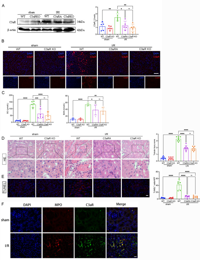
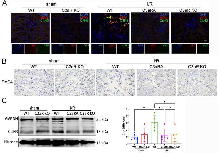
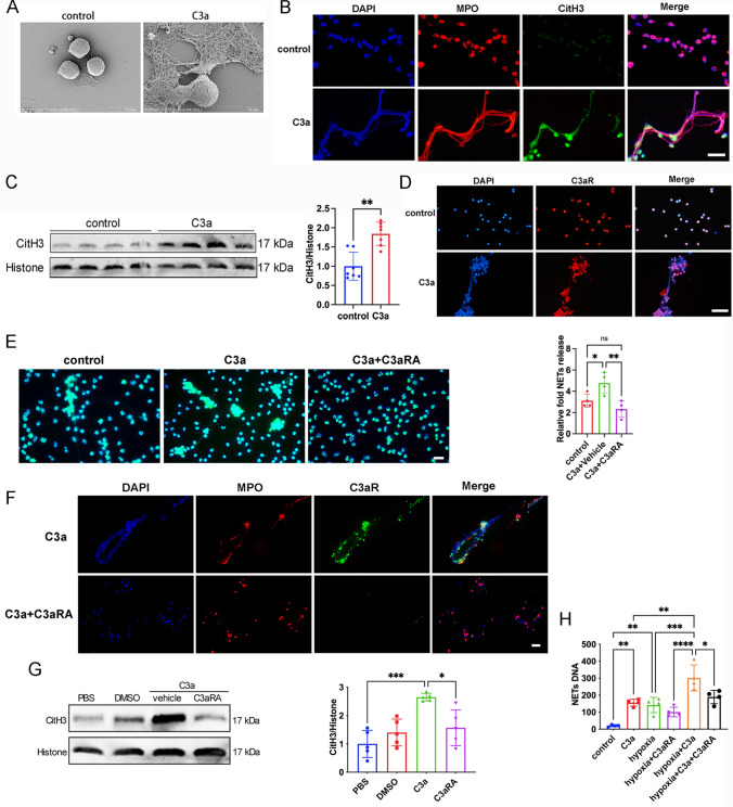
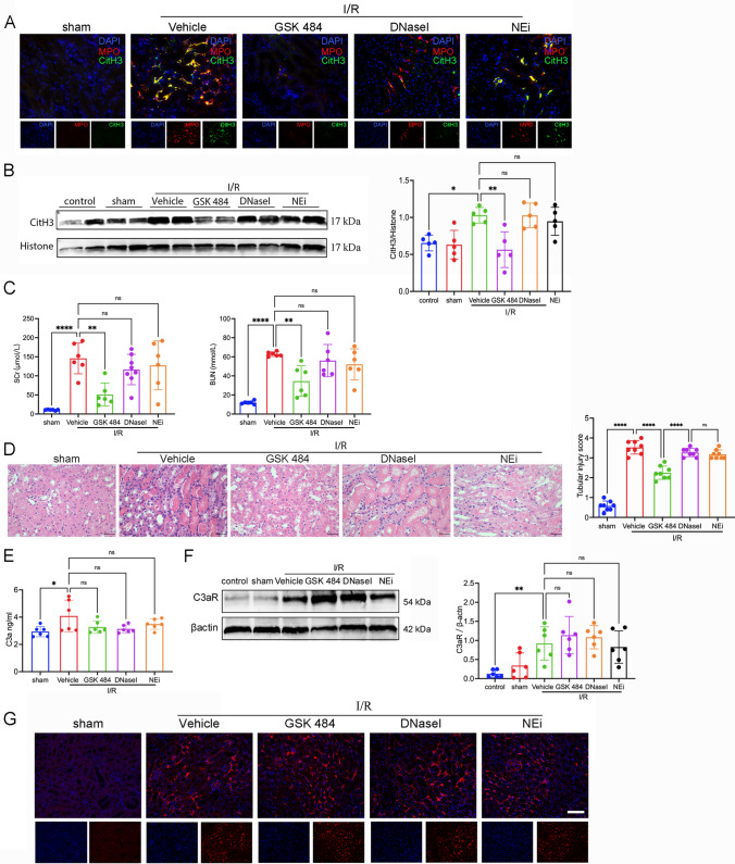
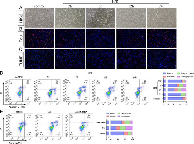
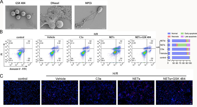
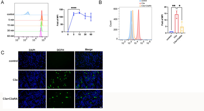
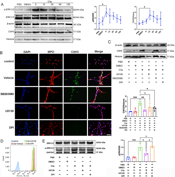
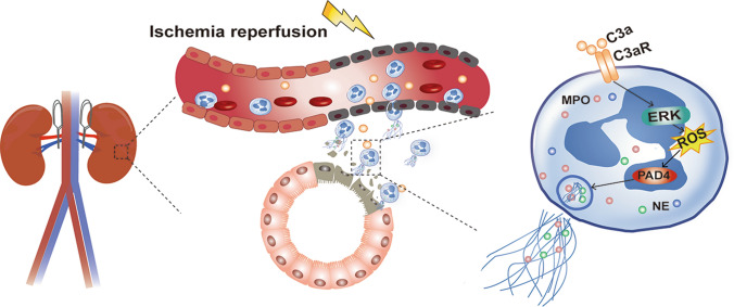
Renal ischemia/reperfusion (I/R) injury is a local sterile inflammatory response driven by innate immunity. Emerging data have revealed that complement and neutrophils contribute to hyperinflammation and oxidative stress in I/R induced acute kidney injury (AKI). However, the interplay between the C3a/C3aR axis and neutrophil extracellular traps (NETs) is imcompletelyunderstood. Here, we utilize genetically engineered mouse models and pharmacological inhibitors to investigate this association. The C3a/C3aR axis is found to promote neutrophil recruitment and NETs formation, thereby accelerating renal damage and dysfunction. Knockout of C3aR restores NETs release and improves renal function after I/R injury. Antibody-mediated blockade of NETs can also significantly ameliorate renal tubular injury and inflammation. Consistently, under stimulation by C3a, neutrophils are activated to promote NETs formation and subsequent renal tubular epithelial cell damage, and blocking C3aR rescued the injury. Interfering with reactive oxygen species (ROS) accumulation in neutrophils by antioxidant treatment significantly attenuates NETs formation. Our findings demonstrate that the C3a/C3aR-ROS-NETs axis constitutes a promising target for prevention or treatment of renal I/R injury.

Keywords: Acute kidney injury; Complement component 3a receptor; Ischemia–reperfusion injury; Neutrophil extracellular traps; Reactive oxygen species.


Conflict of interest statement

The authors declared no conflict of interest associated with this study.

Figures

Fig. 1
Fig. 1
C3aR mediates renal I/R injury. A Western blot and quantitative analysis of C3aR protein expression levels in lysates of renal tissue. B Immunofluorescence analysis of kidney sections from WT or C3aRKO or C3aRA mice after sham surgery or after 45 min of ischemia and 24 h of reperfusion. Scale bar: 100 μm. C Serum levels of creatinine and urea were measured in the respective groups. D Representative microphotographs of H&E stained histological sections of I/R-treated mice are shown, and the tubular damage score was quantified. Scale bar: above 10 μm, below 5 μm. E Representative TUNEL assay images and quantification of TUNEL positive cells in the respective groups. Scale bar: 25 μm. F Immunofluorescence staining using anti-C3aR and anti-MPO antibodies demonstrated the localization of C3aR on neutrophils. Scale bar: 25 μm. * P < 0.5, ** P < 0.01, *** P < 0.001, **** P < 0.0001, ns, no significant
Fig. 2
Fig. 2
Abolition of C3aR blocks the formation of NETs during I/R. A Immunofluorescence staining of kidney sections from WT, C3aRA, and C3aR KO mice after 45 min of ischemia and 24 h of reperfusion. DAPI (blue), myeloperoxidase (MPO, red), CitH3 (green). Colocalization of MPO and CitH3 (yellow) with DNA (DAPI, blue) indicates of NETs. Scale bar: 25 μm. B Immunohistochemical (IHC) images showing PAD4-positive cells in kidney sections. Scale bars: 10 μm. C Representative images and quantitative analysis of CitH3 in whole kidney lysates, as assessed by Western blotting. * P < 0.5, ns, no significant
Fig. 3
Fig. 3
Hypoxia induces NETs formation. A Representative images of double immunofluorescence staining for MPO (red) and CitH3 (green) in unstimulated or stimulated neutrophils isolated from the peripheral blood of healthy volunteers. Scale bars: 100 μm. B Immunofluorescence staining and quantification of neutrophils using SYTOX Green. Scale bars: 100 μm. C Representative western blots and quantitative analysis of relative C3aR protein expression levels in whole-cell lysates of neutrophils cultured under different hypoxic conditions. * P < 0.5, ** P < 0.01, *** P < 0.001, ns, no significant
Fig. 4
Fig. 4
Morphological and molecular characteristics of C3a-induced NETs. A Representative scanning electron micrographs of neutrophils 4 h after treatment with vehicle or C3a. B Representative immunofluorescence staining of MPO (red) and CitH3 (green) in neutrophils. Scale bars, 25 μm. C Western blot analysis of CitH3 in whole cell lysates. D Representative immunofluorescence staining of C3aR in neutrophils. EG: Neutrophils in the C3aRA group were pretreated with C3aRA for 30 min and then stimulated with C3a for 4 h. Neutrophils in the C3a group were stimulated with C3a for 4 h. E Immunofluorescence staining and quantification of neutrophils using SYTOX Green. Scale bars: 50 μm. F Representative immunofluorescence staining of MPO and C3aR. Scale bars: 50 μm. G Representative western blots and quantitative analysis of CitH3 in neutrophils. H Neutrophils were pretreated with or without control (PBS), vehicle (DMSO), or SB290157 (C3aRA) for 30 min and then activated with C3a (0.1 μm). * P < 0.5, ** P < 0.01, *** P < 0.001, **** P < 0.0001, ns, no significant
Fig. 5
Fig. 5
NETs mediate I/R in mouse renal cells and H/R in HK-2 cells. A Representative images of NETs staining in renal sections from WT mice injected with vehicle. Scale bar, 25 μm. B Western blot and quantitative analysis of C3aR protein expression levels in lysates of renal tissue. C Serum levels of creatinine and urea were measured in the respective groups. D Representative images and quantitative analysis of H&E staining in kidney sections. Scale bars, 10 μm. E ELISA measurement of C3a levels in WT mouse renal lysates in the respective groups. F Western blot and quantitative analysis of C3aR protein expression levels in lysates of renal tissue. G Representative immunofluorescence staining of C3aR. Scale bars: 100 μm. * P < 0.5, ** P < 0.01, **** P < 0.0001, ns, no significant
Fig. 6
Fig. 6
C3a contributes to hypoxia-induced death in HK2 cells by regulating NETs formation. A Representative images of HK-2 cells after H/R. Scale bars, 100 μm. B Representative immunofluorescence images of EdU + cells. Scale bars, 50 μm. C Representative immunofluorescence images of the TUNEL assay in HK-2 cells. Scale bars: 50 μm. D Flow cytometric analysis of HK-2 cells after H/R. The proportions of HK-2 cells exhibiting normal necrosis, early apoptosis, and late apoptosis. E Flow cytometric analysis and quantification of the percentage of apoptotic cells after treatment with C3a with or without C3aRA pretreatment
Fig. 7
Fig. 7
NETs are involved in renal epithelial injury. A Representative scanning electron micrographs of neutrophils after treatment with NETs inhibitors. B Flow cytometric analysis and quantification of the percentage of apoptotic cells in different treatment groups after H/R. C Representative immunofluorescence images of the TUNEL assay in HK-2 cells. Scale bars: 50 μm
Fig. 8
Fig. 8
ROS participates in NETs formation. A ROS production in neutrophils stimulated with C3a for the indicated times. B ROS production in neutrophils was detected after the combined treatment of C3a and C3aRA.C. Representative immunofluorescence images of neutrophils pretreated with or without C3aRA and stimulated with C3a. Scale bars: 25 μm
Fig. 9
Fig. 9
C3a activates the ERK and ROS signaling pathways in neutrophils. A p-ERK and citrullinated histone (CitH3) levels in neutrophils 5, 15, 30, 60, and 120 min after treatment with vehicle or C3a were determined and quantified by western blot analysis (n = 5). B Representative immunofluorescence staining of MPO (red) and CitH3 (green) in neutrophils. Scale bars: 25 μm. C Western blot and quantitative analysis of C3aR protein expression levels in neutrophils. D ROS content in neutrophils was determined by flow cytometric analysis after different treatments and statistical analysis of ROS levels in neutrophils from each group. E Western blot and quantitative analysis of C3aR protein expression levels in neutrophils. * P < 0.5, ** P < 0.01, *** P < 0.001, **** P < 0.0001, ns no significant
Fig. 10
Fig. 10
The proposed signaling pathways of NETs formation following AKI. Following I/R, a large amount of C3 and a large number of neutrophils infiltrate into the interstitial space. C3a binds to C3aR on the surface of neutrophils, activates the neutrophils, and induces the release of NETs via the ERK/ROS/PAD4 pathway

References

    1. Ferenbach DA, Bonventre JV. Mechanisms of maladaptive repair after AKI leading to accelerated kidney ageing and CKD. Nat Rev Nephrol. 2015;11:264–276. doi: 10.1038/nrneph.2015.3. - DOI - PMC - PubMed
    1. Matejovic M, Ince C, Chawla LS, Blantz R, Molitoris BA, Rosner MH, Okusa MD, Kellum JA, Ronco C, Grup AXW. Renal hemodynamics in AKI: in search of new treatment targets. J Am Soc Nephrol. 2016;27:49–58. doi: 10.1681/ASN.2015030234. - DOI - PMC - PubMed
    1. Jang HR, Rabb H. The innate immune response in ischemic acute kidney injury. Clin Immunol. 2009;130:41–50. doi: 10.1016/j.clim.2008.08.016. - DOI - PMC - PubMed
    1. Casiraghi F, Azzollini N, Todeschini M, Fiori S, Cavinato RA, Cassis P, Solini S, Pezzuto F, Mister M, Thurman JM, Benigni A, Remuzzi G, Noris M. Complement alternative pathway deficiency in recipients protects kidney allograft from ischemia/reperfusion injury and alloreactive T cell response. Am J Transplant. 2017;17:2312–2325. doi: 10.1111/ajt.14262. - DOI - PubMed
    1. Nakazawa D, Kumar SV, Marschner J, Desai J, Holderied A, Rath L, Kraft F, Lei Y, Fukasawa Y, Moeckel GW, Angelotti ML, Liapis H, Anders HJ. Histones and neutrophil extracellular traps enhance tubular necrosis and remote organ injury in ischemic AKI. J Am Soc Nephrol. 2017;28:1753–1768. doi: 10.1681/ASN.2016080925. - DOI - PMC - PubMed

Substances

LinkOut - more resources