. 2023 Dec;37(12):2457-2467.
doi: 10.1038/s41375-023-02055-z. Epub 2023 Oct 10.

STING activation in TET2-mutated hematopoietic stem/progenitor cells contributes to the increased self-renewal and neoplastic transformation

Affiliations

STING activation in TET2-mutated hematopoietic stem/progenitor cells contributes to the increased self-renewal and neoplastic transformation

Jiaying Xie et al. Leukemia. 2023 Dec.

Abstract

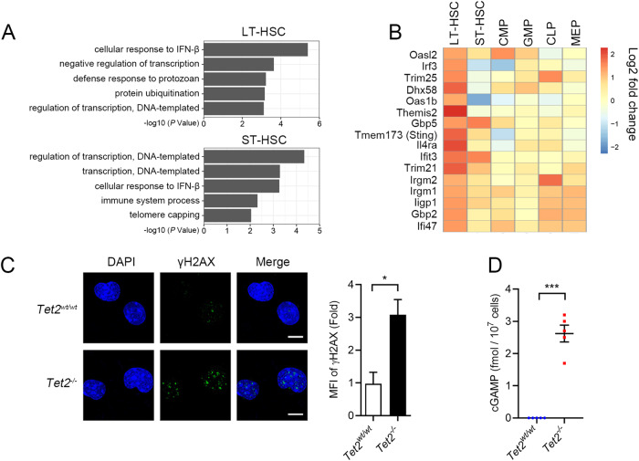
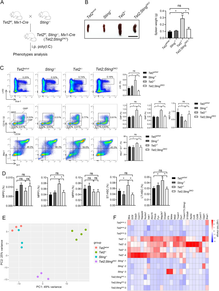
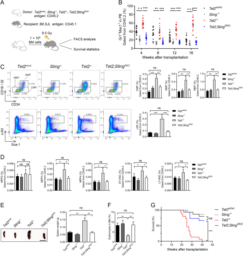
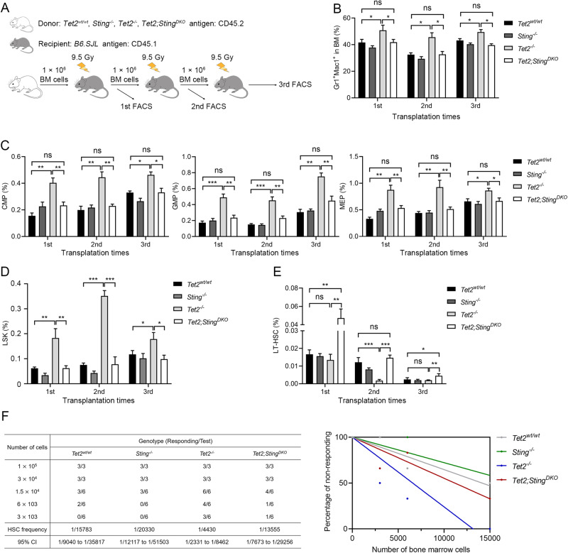
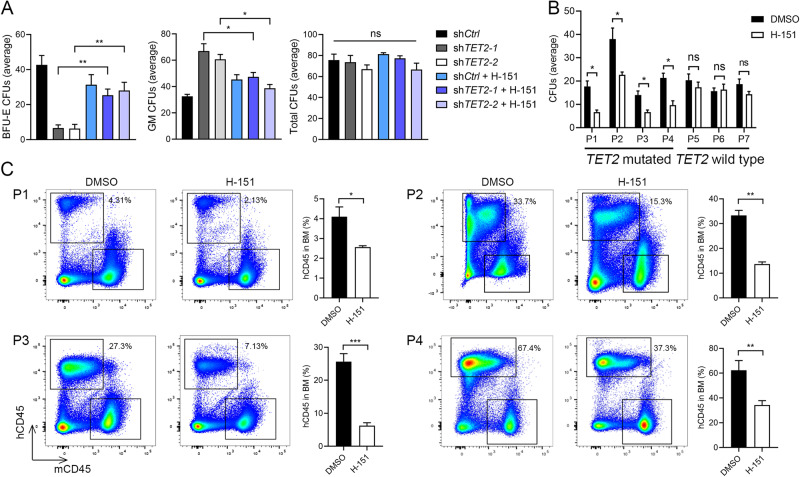
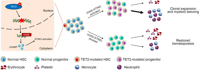
Somatic loss-of-function mutations of the dioxygenase Ten-eleven translocation-2 (TET2) occur frequently in individuals with clonal hematopoiesis (CH) and acute myeloid leukemia (AML). These common hematopoietic disorders can be recapitulated in mouse models. However, the underlying mechanisms by which the deficiency in TET2 promotes these disorders remain unclear. Here we show that the cyclic guanosine monophosphate-adenosine monophosphate synthase (cGAS)-stimulator of interferon genes (STING) pathway is activated to mediate the effect of TET2 deficiency in dysregulated hematopoiesis in mouse models. DNA damage arising in Tet2-deficient hematopoietic stem/progenitor cells (HSPCs) leads to activation of the cGAS-STING pathway which in turn promotes the enhanced self-renewal and development of CH. Notably, both pharmacological inhibition and genetic deletion of STING suppresses Tet2 mutation-induced aberrant hematopoiesis. In patient-derived xenograft (PDX) models, STING inhibition specifically attenuates the proliferation of leukemia cells from TET2-mutated individuals. These observations suggest that the development of CH associated with TET2 mutations is powered through chronic inflammation dependent on the activated cGAS-STING pathway and that STING may represent a potential target for intervention of relevant hematopoietic diseases.


Conflict of interest statement

The authors declare no competing interests.

Figures

Fig. 1
Fig. 1. cGAS-STING pathway is activated in Tet2−/− mouse HSPCs.
A Gene sets enrichment analysis of differentially expressed genes showing upregulation of type I interferon response in Tet2−/− hematopoietic stem cells (HSCs) compared to WT HSCs. There were 699 and 616 upregulated genes in Tet2-deficient LT-HSCs and ST-HSCs, respectively, compared to WT cells. DEGs were submitted to DAVID 6.7 (https://david.ncifcrf.gov) for gene ontology (GO) enrichment analyses (n = 4 mice, p < 0.05). B Heatmap showing activation of interferon- and inflammation-related genes in Tet2−/− LT-HSCs (P < 0.05). Scale bar denotes log2-transformed fold change. Four replicates were analyzed for each cell type. C Immunofluorescence of Tet2−/− LSK cells stained with γH2AX antibody (green). The quantification of γH2AX signal is shown to the right (MFI: Mean fluorescence intensity; scale bar, 5 μm). D cGAMP level in Tet2−/− BM-MNCs quantified by LC-MS (n = 5 mice). Statistical significance was assessed by t test (C, D). Data are mean ± s.e.m., *P < 0.05; ***P < 0.005.
Fig. 2
Fig. 2. Sting is required for the increased self-renewal and myeloid-skewed differentiation of Tet2−/− mouse HSPCs.
A Breeding strategy for inducible deletion of Tet2 in the hematopoietic system on the Sting−/− background. i.p., intraperitoneal injection. B Comparison of spleens from representative mice of indicated genotypes. Shown on the right is the quantification of spleen weight (n = 5 mice; scale bar, 1 cm). C Sting deletion attenuates the aberrant hematopoiesis caused by Tet2 deficiency. The percentages of LSK cells (top panel), oligopotent progenitor cells (CMP, GMP, and MEP, middle panel) and Mac1+Gr1+ myeloid cells (bottom panel) in the BM of indicated mice were determined by flow cytometric analysis. All mice were sacrificed for analysis 12 months after poly(I:C) injection. The quantification is shown to the right (n = 5 mice). D Deletion of Sting inhibits the expansion of MPP3 cell population and the enhanced self-renewal of HSCs in Tet2-deficient mice. The percentages of multipotent progenitor cells (MPP2/3/4), ST-HSCs, and LT-HSCs in the BM of indicated mice were measured by FACS (n = 8 mice). E PCA (Principal Component Analysis) plot showing the RNA-seq data of LT-HSCs derived from indicated genotypes of mice. Each dot represents an individual mouse (For Tet2−/−, n = 4; for other genotypes, n = 3). (F) Deletion of Sting attenuates the upregulation of innate immunity- and inflammation-related genes in LT-HSCs induced by Tet2 deficiency. Genes with log2(fold change) >1 and P < 0.05 are shown (For Tet2−/−, n = 4; for other genotypes, n = 3). LT-HSC, long term hematopoietic stem cell; ST-HSC, short term HSC; CMP, common myeloid progenitor; GMP, granulocyte macrophage progenitor; CLP, common lymphoid progenitor; MEP, megakaryocyte erythroid progenitor. Statistical significance was assessed by t test (BD). Data are mean ± s.e.m., *P < 0.05; **P < 0.01; “ns”: not significant.
Fig. 3
Fig. 3. Sting deletion abrogates Tet2 deficiency-induced skewed hematopoiesis in bone marrow transplantation models.
A Schematic of bone marrow transplantation assay. B Myeloid skewing of Tet2−/− cells after transplant (red dots) is corrected by Sting (blue dots) deletion. The scatter plot shows the quantification of donor-derived mature myeloid cells in the peripheral blood (PB) of primary recipients transplanted with indicated donor BM cells. C Sting deletion attenuates the expansion of Tet2−/− progenitor cells in recipient mice. Representative FACS plots of progenitor cells from the BM cells of indicated donor groups are shown on the left. Top row, CMP, GMP, MEP; Bottom row, LSK. Quantification is shown on the right. D Bar graphs showing the populations of MPP cells and HSCs in recipient mice analyzed by FACS. E Comparison of spleens from recipient mice of indicated genotypes. Shown to the right is the quantification of spleen weight. (n = 3 donors and 15 recipients for each genotype; scale bar, 1 cm). F Sting deletion restores normal erythropoiesis of Tet2−/− donors. The proportion of Ter119+ erythrocytes in the BM were analyzed by FACS. G Survival analysis of primary transplant recipients of indicated donor groups (n = 3 donors and 15 recipients for each genotype). In BM transplantation assays, all mice (n = 3 donors and 15 recipients for each genotype) were sacrificed and analyzed at 18 weeks after transplantation. Statistical significance was assessed with t-test (BF), and Mantel–Cox log-rank test (G). Data are mean ± s.e.m., *P < 0.05; **P < 0.01; ***P < 0.005; “ns”: not significant.
Fig. 4
Fig. 4. Blocking STING restores normal hematopoiesis of Tet2−/− BM cells in serial transplantation models.
A Schematic of serial transplantation assays. B Myeloid skewing of Tet2−/− BM donors is corrected by Sting deletion. C Sting deletion attenuates the expansion of transplanted Tet2−/− progenitor cells in recipient mice. D Sting deletion mitigates the abnormal expansion of Tet2−/− LSK cells in serial transplantation recipients. E Sting deletion increases the proportion of Tet2−/− donor LT-HSCs in serial transplantation recipients. F The frequencies of functional HSCs in each genotype of mice were measured by the limiting dilution assay. Table to the left displays the cell dose, responding rate, calculated HSC frequencies and 95% confidence interval (CI), while the Poisson statistical analysis is presented on the left, color lines indicate the best best-fit linear model. The frequencies and analysis were performed using the L-Calc software. All mice (n = 2 donors and 10 recipients for each genotype) in the serial transplantation assay were sacrificed and analyzed 18 weeks after transplantation. Statistical significance was assessed with two-way ANOVA (BD) and t test (E). Data are mean ± s.e.m., *P < 0.05; **P < 0.01; ***P < 0.005; “ns”: not significant.
Fig. 5
Fig. 5. Myeloid skewing of TET2-deficient human HSCs and leukemogenesis of TET2-mutated AML Patient cells are prevented by inhibiting STING.
A STING inhibitor H-151 reduces myeloid colony formation and restores erythroid colony formation of TET2-deficient cord blood CD34+ cells. CD34+ cells were infected with lentivirus carrying short hairpin RNA which targeted TET2 (shTET2-1 or shTET2-2) or scrambled control (shCtrl). Cells were cultured in methylcellulose medium for 14 days. B H-151 inhibits colony formation of TET2-mutated patient AML cells. P1-4 are AML patients with TET2 mutations, and P5-7 are those without TET2 mutation. C H-151 reduces the engraftment efficiency of TET2-mutated patient AML cells in B-NDG mice. The percentages of human CD45-positive cells (hCD45) in total BM are indicated. Statistical significance was assessed by t test (AC). Data are mean ± s.e.m., *P < 0.05; **P < 0.01; ***P < 0.005; “ns”: not significant.
Fig. 6
Fig. 6. Working model for the activation of STING in mediating TET2 deficiency-associated CH.
Cartoon summarizing our findings: HSPCs with mutated TET2 accumulate DNA damage, which in turn activates the cGAS-STING pathway leading to the production of inflammatory cytokines. This promotes clonal expansion and myeloid differentiation skewing of the mutant HSPCs. Blocking the cGAS-STING pathway suppresses the increased self-renewal and skewed differentiation potential of the mutant HSPCs, and thus restores normal hematopoiesis.

References

    1. He Y-F, Li B-Z, Li Z, Liu P, Wang Y, Tang Q, et al. Tet-mediated formation of 5-carboxylcytosine and its excision by TDG in mammalian DNA. Science. 2011;333:1303–7. doi: 10.1126/science.1210944. - DOI - PMC - PubMed
    1. Jaiswal S, Ebert BL. Clonal hematopoiesis in human aging and disease. Science. 2019;366:6465. doi: 10.1126/science.aan4673. - DOI - PMC - PubMed
    1. Xie M, Lu C, Wang J, McLellan MD, Johnson KJ, Wendl MC, et al. Age-related mutations associated with clonal hematopoietic expansion and malignancies. Nat Med. 2014;20:1472–8. doi: 10.1038/nm.3733. - DOI - PMC - PubMed
    1. Genovese G, Kahler AK, Handsaker RE, Lindberg J, Rose SA, Bakhoum SF, et al. Clonal hematopoiesis and blood-cancer risk inferred from blood DNA sequence. N. Engl J Med. 2014;371:2477–87. doi: 10.1056/NEJMoa1409405. - DOI - PMC - PubMed
    1. Busque L, Patel JP, Figueroa ME, Vasanthakumar A, Provost S, Hamilou Z, et al. Recurrent somatic TET2 mutations in normal elderly individuals with clonal hematopoiesis. Nat Genet. 2012;44:1179–81. doi: 10.1038/ng.2413. - DOI - PMC - PubMed

Publication types