. 2023 Oct 10;24(1):52.
doi: 10.1186/s12868-023-00822-3.

Effects of isoflurane and urethane anesthetics on glutamate neurotransmission in rat brain using in vivo amperometry

Affiliations

Effects of isoflurane and urethane anesthetics on glutamate neurotransmission in rat brain using in vivo amperometry

Joshua A Beitchman et al. BMC Neurosci. .

Abstract

Background: Aspects of glutamate neurotransmission implicated in normal and pathological conditions are predominantly evaluated using in vivo recording paradigms in rats anesthetized with isoflurane or urethane. Urethane and isoflurane anesthesia influence glutamate neurotransmission through different mechanisms; however, real-time outcome measures of potassium chloride (KCl)-evoked glutamate overflow and glutamate clearance kinetics have not been compared within and between regions of the brain. In order to maintain rigor and reproducibility within the literature between the two most common methods of anesthetized in vivo recording of glutamate, we compared glutamate signaling as a function of anesthesia and brain region in the rat strain most used in neuroscience.

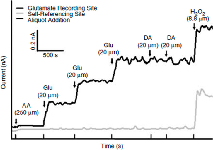
Methods: In the following experiments, in vivo amperometric recordings of KCl-evoked glutamate overflow and glutamate clearance kinetics (uptake rate and T80) in the cortex, hippocampus, and thalamus were performed using glutamate-selective microelectrode arrays (MEAs) in young adult male, Sprague-Dawley rats anesthetized with either isoflurane or urethane.

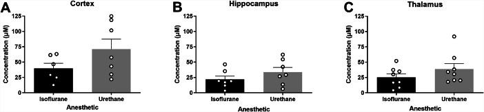
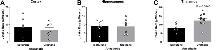
Results: Potassium chloride (KCl)-evoked glutamate overflow was similar under urethane and isoflurane anesthesia in all brain regions studied. Analysis of glutamate clearance determined that the uptake rate was significantly faster (53.2%, p < 0.05) within the thalamus under urethane compared to isoflurane, but no differences were measured in the cortex or hippocampus. Under urethane, glutamate clearance parameters were region-dependent, with significantly faster glutamate clearance in the thalamus compared to the cortex but not the hippocampus (p < 0.05). No region-dependent differences were measured for glutamate overflow using isoflurane.

Conclusions: These data support that amperometric recordings of KCl-evoked glutamate under isoflurane and urethane anesthesia result in similar and comparable data. However, certain parameters of glutamate clearance can vary based on choice of anesthesia and brain region. In these circumstances, special considerations are needed when comparing previous literature and planning future experiments.

Keywords: Amperometry; Cortex; Glutamate neurotransmission; Hippocampus; Isoflurane; Thalamus; Urethane.


Conflict of interest statement

The authors declare no competing interests.

Figures

Fig. 1
Fig. 1
The number of publications using urethane and isoflurane for neuroscience research between 1981–2020. A PubMed search indicates the use of urethane has decreased while the use of isoflurane has increased over the past 40 years
Fig. 2
Fig. 2
Representative calibration of a glutamate selective microelectrode array (MEA). Representative MEA calibration prior to in vivo recordings in which only one glutamate selective recording site and one self-referencing site are represented. Aliquots of 250 µM ascorbic acid (AA), 20 µM glutamate (Glu), 2 µM dopamine (DA), and 8.8 µM H2O2 are represented by vertical bars on the x-axis
Fig. 3
Fig. 3
Recording regions and amperometric calculations. (A) Anatomical regions of interest (ROI) in the rodent brain were the hippocampus (blue), thalamus (green), and cortex (yellow). Image modified from Paxinos and Watson (2007). * represents the tip of the MEA. (B) Representative peak, showing glutamate concentration (µM) as a function of time in seconds in response to local applications of 100 µM glutamate. Amperometric calculations used in the analysis (peak amplitude, uptake rate, and T80) are shown
Fig. 4
Fig. 4
Levels of evoked release of glutamate were similar in urethane and isoflurane-anesthetized rats. Local applications of volume-matched 120 mM potassium chloride (KCl) were made to the cortex, hippocampus, and thalamus. There was no significant difference between the glutamate overflow measured for rats anesthetized with isoflurane or urethane in the (A) cortex (t11 = 1.629, p = 0.131), (B) hippocampus (t12 = 1.247, p = 0.236), or (C) thalamus (t14 = 1.299, p = 0.214). Bar graphs represent mean + SEM. N = 6–8 per group
Fig. 5
Fig. 5
Glutamate uptake rates were similar in the cortex and hippocampus and different in the thalamus of urethane and isoflurane-anesthetized rats. Amplitude-matched signals from local applications of 100 µM exogenous glutamate compared for extracellular glutamate clearance in the cortex, hippocampus, and thalamus. No significant difference in the uptake rate between rats anesthetized with isoflurane or urethane in the (A) cortex (t11 = 0.544, p = 0.597) and (B) hippocampus (t12 = 0.148, p = 0.884). (C) Urethane administration was associated with a significantly faster uptake rate in thalamus than isoflurane (t13 = 2.817, p = 0.0145). Bar graphs represent mean + SEM. N = 6–9 per group
Fig. 6
Fig. 6
Extracellular clearance times (T80) were similar in urethane and isoflurane-anesthetized rats. Local application of 100 µM exogenous glutamate resulted in amplitude-matched signals to assess the T80 in the cortex, hippocampus, and thalamus. There was no significant difference in the time taken for 80% of the maximal amplitude to clear between rats anesthetized with isoflurane or urethane in the (A) cortex (t10 = 1.329, p = 0.213), (B) hippocampus (t9 = 0.525, p = 0.612), or (C) thalamus (t13 = 1.827, p = 0.090). Bar graphs represent mean + SEM. N = 4–9 per group
Fig. 7
Fig. 7
Glutamate clearance under urethane was capable of distinguishing region-dependent differences. (A-C) Under isoflurane, all outcome measures were similar between the cortex, hippocampus, and thalamus. (D) No significant differences were detected in KCl-evoked glutamate release across regions when using urethane. (E-F) Glutamate clearance kinetics under urethane changes as a function of the brain region when evaluating uptake rate (F2,20 = 4.41; p = 0.026) and T80 (F2,14 = 3.79; p = 0.044), where uptake rate was significantly faster and clearance time was significantly shorter in the thalamus compared to the cortex. Bar graphs represent mean + SEM. N = 6–9 per group

Update of

References

    1. Danbolt NC. Glutamate uptake. ProgNeurobiol. 2001;65(1):1–105. - PubMed
    1. Hascup KN, Findley CA, Britz J, Esperant-Hilaire N, Broderick SO, Delfino K, et al. Riluzole attenuates glutamatergic tone and cognitive decline in AβPP/PS1 mice. J Neurochem. 2021;156 4:513–23. doi: 10.1111/jnc.15224. - DOI - PMC - PubMed
    1. Blandini F, Porter RH, Greenamyre JT. Glutamate and Parkinson’s disease. Mol Neurobiol. 1996;12 1:73–94. doi: 10.1007/bf02740748. - DOI - PubMed
    1. Joyce JM, Mercier LJ, Stokoe M, La PL, Bell T, Batycky JM, et al. Glutamate, GABA and glutathione in adults with persistent post-concussive symptoms. Neuroimage Clin. 2022;36:103152. doi: 10.1016/j.nicl.2022.103152. - DOI - PMC - PubMed
    1. Meldrum BS. The role of glutamate in epilepsy and other CNS disorders. Neurology. 1994;44(11 Suppl 8):14–23. - PubMed

Publication types

LinkOut - more resources