

A hypervascular placental polyp after complete abortion: a case report
- PMID: 37817177
- PMCID: PMC10566062
- DOI: 10.1186/s12905-023-02672-x
A hypervascular placental polyp after complete abortion: a case report
Abstract
Background: Placental polyps are rare complications of delivery or abortion. They are thought to complicate less than 0.25% of all pregnancies, although the actual incidence is unknown. While they typically occur within four weeks of delivery or abortion, they can have a variable presentation, which can lead to a delay in care.
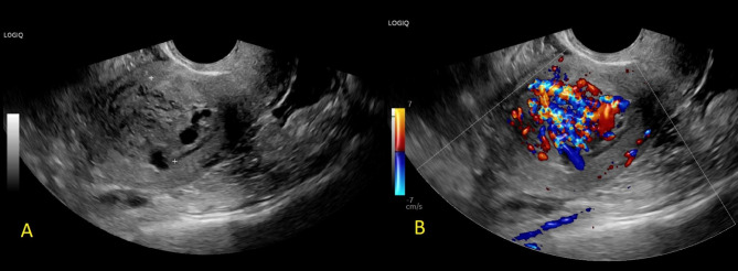
Case presentation: A 35-year-old G4P2012 patient presented at 9 weeks gestation for a medication abortion. Post-abortion ultrasound after one week confirmed the abortion was complete and her bleeding ceased. The patient then presented two months later with the new onset of worrisome bleeding. She was found on ultrasound to have a new hypervascular polypoidal mass in the endometrial cavity. She then underwent an in-office dilation and curettage with an electric vacuum aspirator, which was curative. A follow up ultrasound three months later demonstrated no recurrence.
Conclusions: Placental polyps are a rare complication following pregnancy and should be included in the differential when a patient presents with bleeding and a new mass in the endometrial cavity on ultrasound following a delivery or abortion, even when frankly retained products of conception had been ruled out at time of abortion.
Keywords: Abortion; Case report; Placental polyp; Puerperal disorder; Retained products of conception; Uterine bleeding.
© 2023. BioMed Central Ltd., part of Springer Nature.
Conflict of interest statement
The authors declare no competing interests.
Figures

Similar articles
-
Diagnostic and therapeutic decision-making with transvaginal sonography for first trimester spontaneous abortion, clinically thought to be incomplete or complete.Contraception. 1998 Jun;57(6):393-7. doi: 10.1016/s0010-7824(98)00046-8. Contraception. 1998. PMID: 9693399
-
Uterine preservation surgery for placental polyp.J Obstet Gynaecol Res. 2014 Jan;40(1):89-95. doi: 10.1111/jog.12128. Epub 2013 Aug 12. J Obstet Gynaecol Res. 2014. PMID: 23937267
-
Ultrasound of the postpartum uterus. Prediction of retained placental tissue.J Ultrasound Med. 1991 Aug;10(8):451-6. doi: 10.7863/jum.1991.10.8.451. J Ultrasound Med. 1991. PMID: 1942234
-
Differential diagnosis and management of placental polyp and uterine arteriovenous malformation: Case reports and review of the literature.Womens Health (Lond). 2016 Nov;12(6):538-543. doi: 10.1177/1745505717692590. Epub 2017 Feb 10. Womens Health (Lond). 2016. PMID: 29334028 Free PMC article. Review.
-
[Role of imaging in cases of bleeding after spontaneous or induced abortion].J Gynecol Obstet Biol Reprod (Paris). 2015 May;44(5):398-402. doi: 10.1016/j.jgyn.2014.10.013. Epub 2014 Oct 30. J Gynecol Obstet Biol Reprod (Paris). 2015. PMID: 25433565 Review. French.
Cited by
-
A Rare Case of a Hypervascular Placental Polyp Leading to Massive Postpartum Hemorrhage Requiring Hysterectomy.Case Rep Obstet Gynecol. 2025 Jun 25;2025:4120029. doi: 10.1155/crog/4120029. eCollection 2025. Case Rep Obstet Gynecol. 2025. PMID: 40606511 Free PMC article.
References
-
- Redline RW, Boyd TK, Roberts DJ. Placental and Gestational Pathology with Online Resource. Cambridge University Press; 2018.
-
- Watcharotone W, Leelaphatanadit C. Placental polyp: a case report. Siriraj Med J. 2005;57(9):391–3.
Publication types
MeSH terms
LinkOut - more resources
Full Text Sources
Medical
