

Surgical technique of spine-shortening vertebral osteotomy for adult tethered cord syndrome: a case report and review of the literature
- PMID: 37817238
- PMCID: PMC10566082
- DOI: 10.1186/s13256-023-04155-x
Surgical technique of spine-shortening vertebral osteotomy for adult tethered cord syndrome: a case report and review of the literature
Abstract
Background: Miyakoshi et al. reported three cases of tethered cord syndrome treated by spine-shortening vertebral osteotomy, which provided relief of the patients' symptoms with no complications. Although the details of these cases were described in a previous report, the surgical technique was not thoroughly explained. In the present report, we describe the details of our procedure with reference to a fourth case.
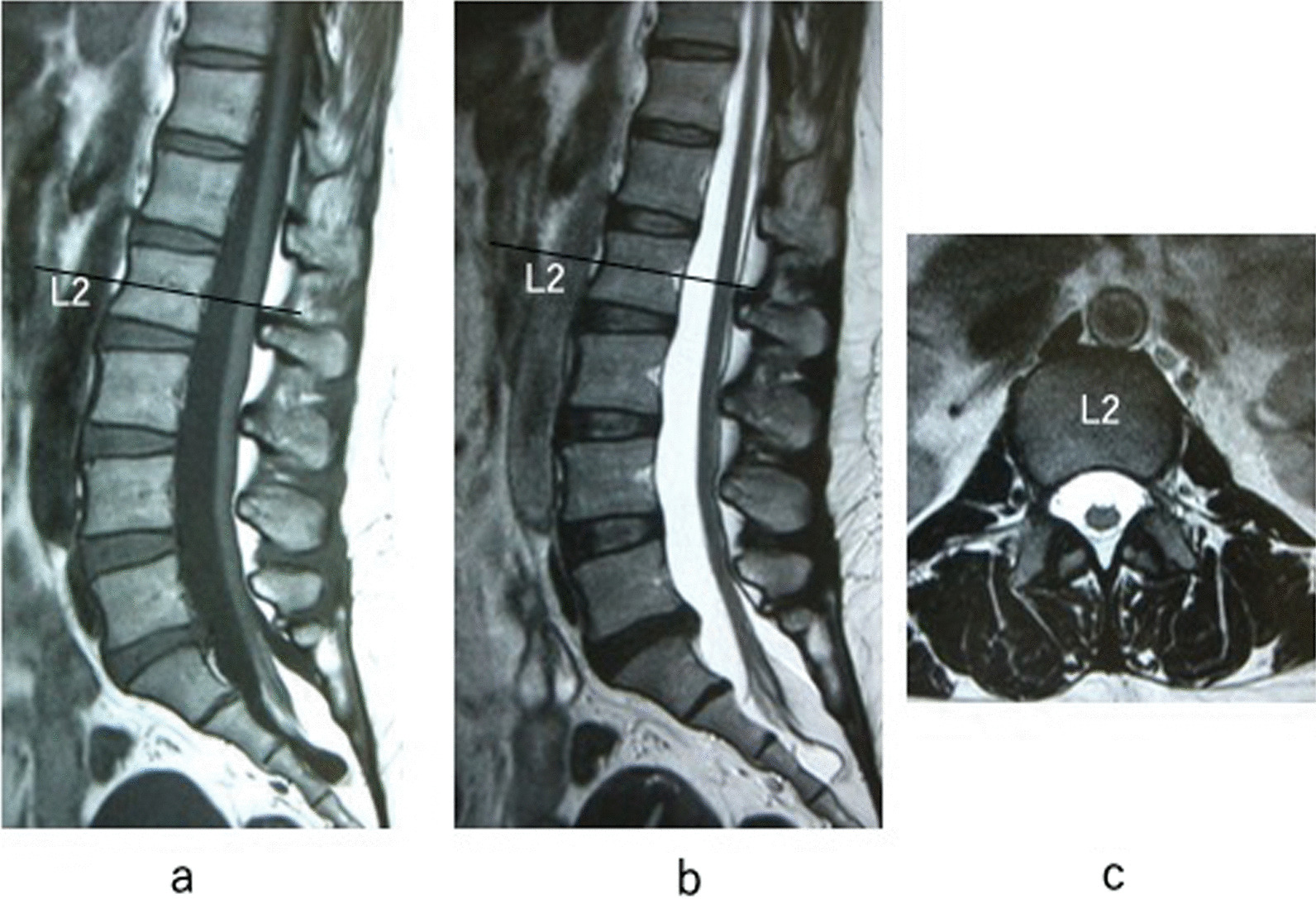
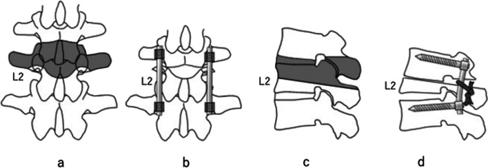
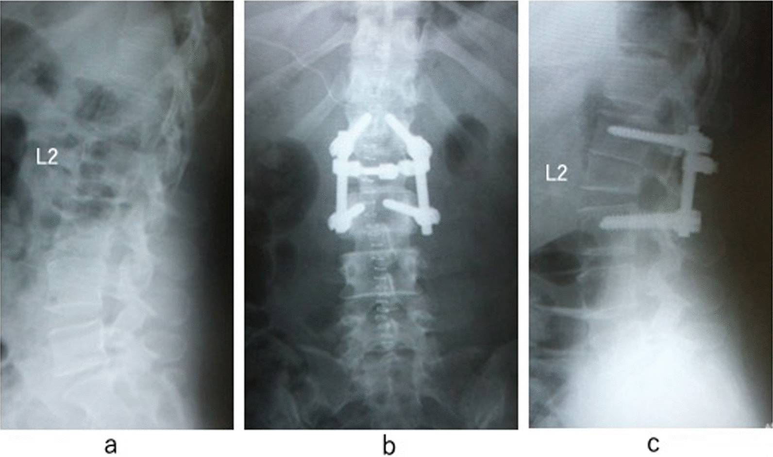
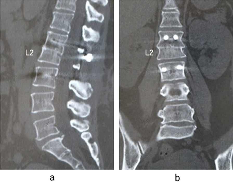
Case presentation: A 47-year-old Asian woman was admitted to our hospital with a 1-year history of worsening leg numbness and urinary dysfunction. Magnetic resonance imaging revealed a low-lying conus medullaris extending to the level of S2 and surrounded by fat tissue at that level. We diagnosed her condition as adult tethered cord syndrome, and spine-shortening vertebral osteotomy was planned. The target level for the osteotomy was L2. Bilateral pedicle screw implants were placed at L1 and L3 using an anterior-posterior image intensifier. In this procedure, it is essential to use monoaxial screws inserted exactly parallel to the rostral endplates of each vertebral body; this ensures appropriate alignment between the L1 caudal endplate and the L2 osteotomy surface. The upper one-third of the lamina of L2 was resected, and the bilateral two-thirds of the pedicle of L2 was removed with a surgical air drill. After exposure of the lateral side of the L1-2 disc, discectomy was performed with a knife and curette. Following complete discectomy of L1-2, the upper vertebral body of L2 was removed with a surgical air drill. After complete removal of the vertebral body, a straight rod was connected to two screws and applied pressure between the screws. Two polyethylene tapes were applied to the L2 lamina and bilateral rods.
Conclusion: Spine-shortening osteotomy that preserves the caudal one-third of the pedicle and lamina with one-above and one-below instrumentation successfully reduced the spinal cord tension without causing neural damage.
Keywords: Adult tethered cord syndrome; Case report; Spine-shortening vertebral osteotomy; Surgical technique.
© 2023. BioMed Central Ltd., part of Springer Nature.
Conflict of interest statement
The authors declare that they have no competing interests.
Figures
References
-
- Zhang C, Chang CC, Mummaneni PV, Yuan C, Dhall S, Jian F, et al. Spinal column shortening versus revision detethering for recurrent adult tethered cord syndrome: a preliminary comparison of perioperative and clinical outcomes. J Neurosurg Spine. 2020;7:1–7. - PubMed
Publication types
MeSH terms
LinkOut - more resources
Full Text Sources
Medical
