Review
. 2024 Oct;20(41):e2303682.
doi: 10.1002/smll.202303682. Epub 2023 Oct 10.

Nanomedicine for Maternal and Fetal Health

Affiliations
Review

Nanomedicine for Maternal and Fetal Health

Davell Carter et al. Small. 2024 Oct.

Abstract

Conception, pregnancy, and childbirth are complex processes that affect both mother and fetus. Thus, it is perhaps not surprising that in the United States alone, roughly 11% of women struggle with infertility and 16% of pregnancies involve some sort of complication. This presents a clear need to develop safe and effective treatment options, though the development of therapeutics for use in women's health and particularly in pregnancy is relatively limited. Physiological and biological changes during the menstrual cycle and pregnancy impact biodistribution, pharmacokinetics, and efficacy, further complicating the process of administration and delivery of therapeutics. In addition to the complex pharmacodynamics, there is also the challenge of overcoming physiological barriers that impact various routes of local and systemic administration, including the blood-follicle barrier and the placenta. Nanomedicine presents a unique opportunity to target and sustain drug delivery to the reproductive tract and other relevant organs in the mother and fetus, as well as improve the safety profile and minimize side effects. Nanomedicine-based approaches have the potential to improve the management and treatment of infertility, obstetric complications, and fetal conditions.

Keywords: infertility; obstetrics; pregnancy; vaginal drug delivery; women's health.


Conflict of interest statement

Conflict of Interest

The mucus-penetrating particle technology is licensed and in clinical development for ocular indications by Kala Pharmaceuticals. L.M.E and Johns Hopkins own company stock. Under a licensing agreement between Kala Pharmaceuticals and the Johns Hopkins University, L.M.E. and the University are entitled to royalty distributions related to the technology. These arrangements have been reviewed and approved by the Johns Hopkins University in accordance with its conflict of interest policies. All authors contributed to the manuscript, assisted in revisions, read, and approved the submitted version.

Figures
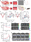
Figure 1.
Figure 1.. Biodistribution of nanoparticles to the reproductive system during the mouse estrous cycle.
(a). During the estrus stage there is increased blood supply to the ovary to support preovulatory follicles. After ovulation, a dense blood network termed the corpus luteum is observed. A higher density of blood vessels around the follicle results in a higher accumulation of nanoparticles (blue) in the reproductive system. (b) Contrarily, there are fewer blood vessels in the ovary and around the follicles specifically during the diestrus stage. (c, d) 80 nm Gd-loaded PEGylated liposomes (Gd-lipo) were imaged using cryo-transmission electron microscopy (c, scale bar 100 nm) and sized using dynamic light scattering (DLS) in d. (e) Gd-lipo were injected intravenously to female mice at different stages of the menstrual cycle. (f) NP accumulation 24 h post administration was quantified using elemental analysis for Gd or by mRNA expression. Results are shown as the injected %Gd normalized to the organ weight. 1.8-fold more liposomes reached the ovaries at the estrus stage (n = 8) compared to the diestrus stage (n = 8, red and blue represent two independent experiments). (g) 2.5-fold more liposomes reached the uterus at the estrus stage (n = 8) compared to at the diestrus stage (n = 7). (h) Biodistribution of liposomes to ovarian and breast cancer tumors and the efficacy of cancer treatment were affected by the female mouse cycle. For ovarian cancer, treatment efficacy of doxorubicin-loaded liposome was evaluated using IVIS imaging, enabled by the luciferase-expressing cells, during the estrus (n = 5 (days 0 and 7), n = 2 (day 14)) and diestrus stages (n = 5 (days 0 and 7), n = 3 (day 14)). Arrows indicate treatment times. (i) IVIS images of luminescent ovarian cancer tumor (left panel, treatment during diestrus; right panel, treatment during estrus). (j) In mice bearing orthotopic ovarian cancer, the efficacy of doxorubicin-loaded liposomes was negatively affected by dosing in the estrus phase. (k) IVIS images of 4T1 mCherry breast cancer tumor (top, treatment during estrus; bottom, treatment during diestrus). Three representative mice from each group are shown. Adapted with permission from (16).
Figure 2.
Figure 2.. Biological barriers to nanomedicines targeting in the female reproductive tract.
(a)Systemically administered NPs decorated with tissue-specific ligands can accumulate in the uterus. Alternatively, vaginally administered drugs such as progesterone can distribute to the uterus before pooling into the bloodstream in a phenomenon recognized as the uterine first pass effect. (b) In pregnancy, the placenta is the interface that connects the fetal and maternal blood. Intravenous administration of NPs in the maternal blood can lead to their crossing the placenta into the fetus. Several studies have identified particle characteristics amenable to transplacental crossing, and small and neutral or anionic NPs have shown preferential transport to the fetus. (c) The cervicovaginal mucus is the first line of defense covering the vaginal mucosa. PEGylated muco-inert NPs with a size smaller than the average pore size between the mucin proteins penetrate the mucus and reach the underlining epithelial layers. (d) Molecules entry to the ovarian follicle is strictly regulated by the blood-follicle barrier, which shows preference towards smaller particles with a cationic surface charge. A surge in ovulatory hormones, such as luteinizing hormone, could enhance the permeation of macromolecules into the follicle.
Figure 3.
Figure 3.. Nanomedicine for the management of female infertility.
(a) Overview of GnRH (6-D-Phe) nanostructures and fibril formation. In the presence of zinc ions (Zn2+), GnRH precipitates to form nanostructures that self-assemble to form fibrils. (b) In vitro release profiles Zn2+: GnRH [6-D-Phe] and GnRH [6-D-Phe] oil depot formulations. The complex was formed by incubating GnRH and zinc ions in Tris buffer at pH 7.8, while the in-situ complex was formulated in purified water. Oil vehicle (OV) 1 was composed medium chain triglyceride (MCT), 3% (w/w) aluminium distearate, 1% phospholipon 90 H, and 5% kolliphor ELP. OV2 consisted of castor oil: MCT 50:50% (w/w). (c) In vitro drug release profile of progesterone (Prog) from Prog solution, liposome (Lipo/Prog), and chitosan-coated liposomes (CS-Lipo/Prog) in phosphate-buffered saline (pH 7.4). (d) The plasma concentration-time curve of Prog after oral administration of Prog, Lipo/Prog, CS-Lipo/Prog (18 mg/kg, Prog) in Wistar rats (mean ± SD, n = 5), and the area under the curve (AUC0-infinite) and relative bioavailability of different formulations after oral administration. (e) Microinjection into the ovary. (f) Functional assessment of the BFB. FITC-dextran (green) was injected interstitially in the mouse ovary. Samples were recovered 30 min after microinjection and immune-stained with anti-Müllerian hormone (AMH), which is produced by granulosa cells. (g) Appearance of ovaries 1 week after AAV-mCherry injection. Arrowheads indicate cells expressing the red fluorescent protein. (h) Microinjecting of AAVs into the ovarian stroma penetrates the BFB and achieves long-term gene expression. Introduction of an AAV carrying the mouse Kitl gene restores oogenesis in congenitally infertile KitlSl-t/KitlSl-t mutant mouse ovaries, which lack Kitl expression but contain only primordial follicles. Healthy offspring without AAV integration were born by natural mating. (a, b) Adapted with permission from (76), (c, d) from (83), (e-h) from (59).
Figure 4.
Figure 4.. Nanomedicine applications in obstetric conditions.
(a) Preparation of trophoblast-targeted NPs encapsulating Nrf2 and sFTL1 siRNA. NP loaded with Nrf2 and sFTL1 siRNA was constructed from carboxyl- polyethylene glycol-poly (D,L-lactide) (COOH-PEG5K-PLA8K), cationic lipid DOTAP, and siNrf2 and sisFTL1 using the double emulsion method. Trophoblast-targeted nanoparticles were fabricated by conjugating peptides, which could specifically bind with the chondroitin sulfate. (b) A Tumor-homing peptides bind to the placenta. Pregnant mice were intravenously injected with phage bearing the surface peptides CGKRK or iRGD or the control sequence G7 (1.5 × 10¹⁰ colony-forming units per mouse). After 30 min, mice were subjected to cardiac perfusion; phage were recovered from individual organs and quantified; results are expressed as fold titers relative to those of the control sequence G7. (c) CGKRK binds to membrane-associated calreticulin. Human first trimester placenta incubated with carboxyfluorescein-CGKRK and immune-stained with an antibody to calreticulin (red). Blue, DAPI (nuclei). Scale bars, 10 μm. (d) Schematic of maleimide-based conjugation of bioactive nanoparticles to the ECFC surface’s free thiols followed by in situ PEGylation. Continuous pseudo-autocrine stimulation of GDM-ECFCs with SB-431542 improves their clinical potential for therapeutic vasculogenesis. (e) Real-time RT-PCR quantification of TAGLN expression in normal ECFC and GDM-ECFC under Vehicle (DMSO) control or treatment with 5 μM SB-431542 (SB) for 72 h (four biological replicates, n = 4; mean ± s.d.). TAGLN expression is normalized to ECFC. (f) The number of migrating cells after 4 h were quantified. Four biological replicates (n = 4; mean ± s.d.) were used for normal ECFCs (black data dots) and GDM-ECFCs (red data dots). SB-NPs significantly improve cell migration of GDM-ECFCs (*p = 0.039), but not normal ECFCs (p = 0.955). (g) Illustration of indomethacin (IND) liposome decorated with oxytocin receptor antagonist (ORA, Atosiban). (h) IND concentrations in the maternal uterus and fetus. IND was administered either in free form or within LIP-IND-ORA by IV injection to pregnant mice. IND concentrations in the maternal uterus and fetus were determined by LC-MS/MS analysis. Mean ± SEM, n = 6. **p-value < 0.01 to the levels of IND when the drug was administered via LIP-IND-ORA (g). PTB prevention using muco-inert P4/TSA nanosuspensions. (i) Pregnancy survival curves showing the percentage of mice remaining pregnant after double distal uterine injections of 20 μg of LPS on E15. Daily vaginal administration of P4 NS (1 mg, n = 20; repeated from Fig. 2B), TSA (15 μg, n = 20), or P4/TSA NS (1 mg/15 μg, n = 20) compared to LPS alone (n = 30) (**p = 0.003, Mantel-Cox test). (j) Daily vaginal administration of P4/TSA (n = 8) or intraperitoneal (i.p.) injection of P4/TSA NS (1 mg/15 μg, n = 8) compared to LPS alone (n = 10). Vaginal administration significantly reduced PTB rates compared to intraperitoneal injection (*p = 0.048) and LPS alone (p = 0.024). (a) Adapted with permission from (136), (b, c) from (141), (d-f) from (164), (g, h) from (32), (h, i) from (55).
Figure 5.
Figure 5.. Nanomedicine applications in fetal health.
(a) Fetal brain uptake of intra-amniotically delivered dendrimers in a mouse model of intrauterine inflammation and preterm birth. Non-treated fetal brain. (b) Distribution of Cy5 dendrimer in brains of LPS-induced neuroinflammation in fetuses at 24 h post dendrimer administration. Blue: DAPI staining of nucleus; red: dendrimer-Cy5. (c) Higher magnification of untreated brains immune-stained for microglia//macrophages (Iba1, green), and (d) higher magnifications of fetal brain 24 h post injection of Cy5-dendrimers showing co-localization between Cy5-dendrimer and Iba1. (e) Biodistribution of poly(lactic-co-glycolic acid) (PLGA) nanoparticles (NPs) in fetal organs after vitelline vein (IV) or intra-amniotic (IA) delivery. (f) LNP-mediated intra-amniotic mRNA delivery. IVIS image of dam and exposed uterine horn with pups in the four left sacs receiving PBS control and pups in the five right sacs receiving A4 LNP injections. Right - strong luciferase expression in the uterine horn where pups received A12 LNP injections, other than one sac (denoted with white arrow) that instead received PBS as a control injection. (g) IVIS images (left) of the highest luciferase expression in each organ for all conditions. Quantification (right) of fetal organ bioluminescence following dissection. There were no significant differences in the normalized total flux for A12 LNP compared to A4 LNP or PBS control across four organs shown. (h) Intra-vitelline vein injection in mice. (i) LNP-mediated mRNA delivery to fetuses. IVIS images (left) and quantification (right) of luciferase signal from livers of fetuses injected with LNPs. Each fetus was injected via the vitelline vein, extracted, and imaged by IVIS 4 hours after injection. Quantifications are the normalized total flux calculated by dividing the luminescence from the area of interest by the background from each individual image. The normalized total flux was averaged across injected fetuses. *p < 0.001 by one-way analysis of variance (ANOVA) with post hoc Tukey-Kramer compared to all other treatment groups, unless indicated otherwise, and outliers were detected using Grubbs’ test and removed from analysis; minimum n = 3 per treatment group; error bars represent SEM. (a-d) Adapted with permission from (183), (e) from (65), (f, g) from (64), (h, i) from (61).

Similar articles

Cited by

References

    1. Ybot-Gonzalez P, Greene NDE, Copp AJ, Henderson DJ, Massa V, Editorial: Maternal-foetal crosstalk impacts on offspring development. Front Cell Dev Biol 11, 1133159 (2023). - PMC - PubMed
    1. Hoekzema E et al., Pregnancy leads to long-lasting changes in human brain structure. Nat Neurosci 20, 287–296 (2017). - PubMed
    1. Soma-Pillay P, Nelson-Piercy C, Tolppanen H, Mebazaa A, Physiological changes in pregnancy. Cardiovasc J Afr 27, 89–94 (2016). - PMC - PubMed
    1. Chandra A, Copen CE, Stephen EH, Infertility and impaired fecundity in the United States, 1982–2010: data from the National Survey of Family Growth. Natl Health Stat Report, 1–18, 11 p following 19 (2013). - PubMed
    1. Ventura SJ, Curtin SC, Abma JC, Henshaw SK, Estimated pregnancy rates and rates of pregnancy outcomes for the United States, 1990–2008. Natl Vital Stat Rep 60, 1–21 (2012). - PubMed

Publication types

LinkOut - more resources