

Fetal circulatory physiology and brain development in complex congenital heart disease: A multi-modal imaging study
- PMID: 37817395
- PMCID: PMC11004088
- DOI: 10.1002/pd.6450
Fetal circulatory physiology and brain development in complex congenital heart disease: A multi-modal imaging study
Abstract
Objective: Fetuses with complex congenital heart disease have altered physiology, contributing to abnormal neurodevelopment. The effects of altered physiology on brain development have not been well studied. We used multi-modal imaging to study fetal circulatory physiology and brain development in hypoplastic left heart syndrome (HLHS) and d-transposition of the great arteries (TGA).
Methods: This prospective, cross-sectional study investigated individuals with fetal congenital heart disease and controls undergoing fetal echocardiography and fetal brain MRI. MRI measured total brain volume and cerebral oxygenation by the MRI quantification method T2*. Indexed cardiac outputs (CCOi) and vascular impedances were calculated by fetal echocardiography. Descriptive statistics assessed MRI and echocardiogram measurement relationships by physiology.
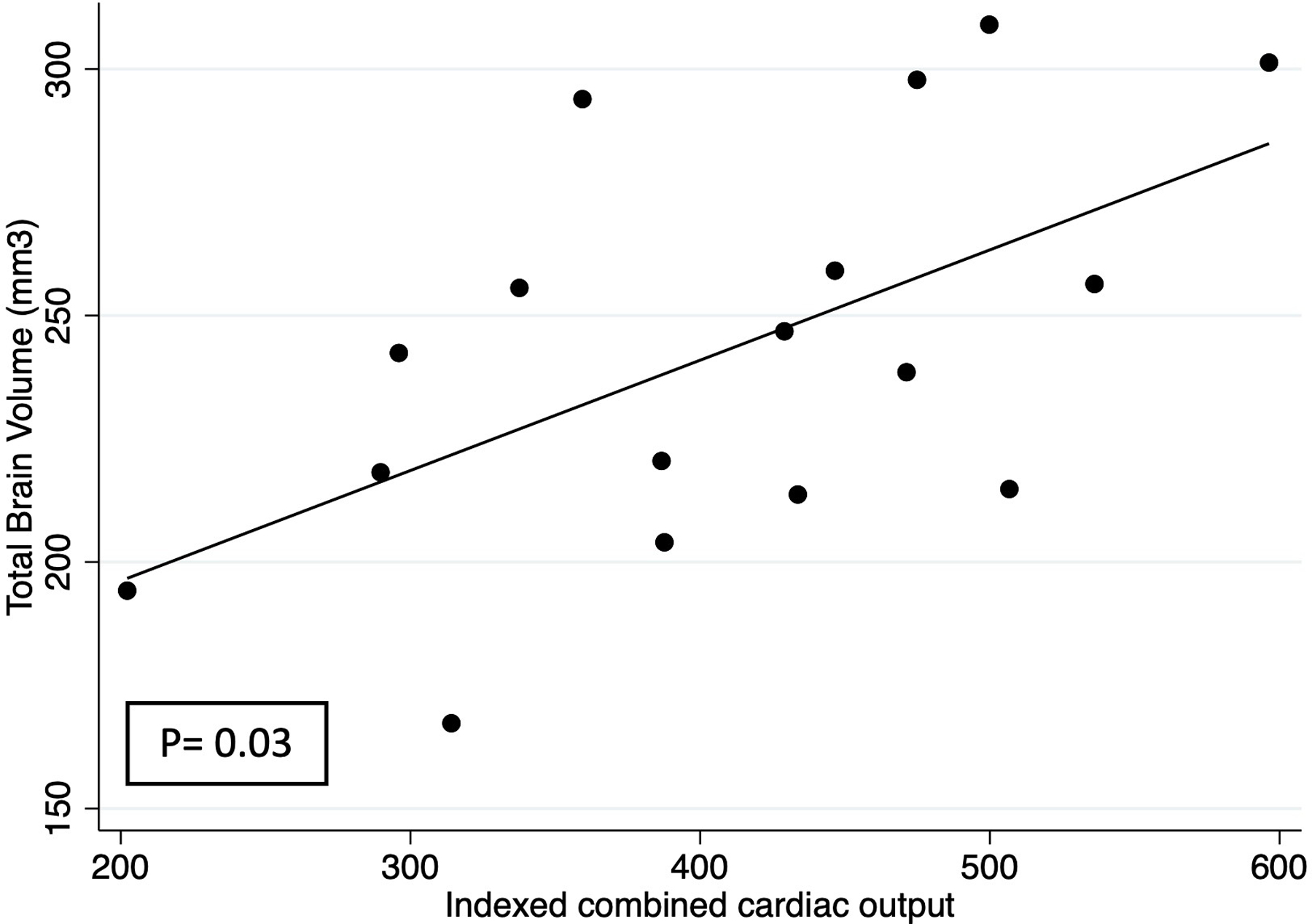
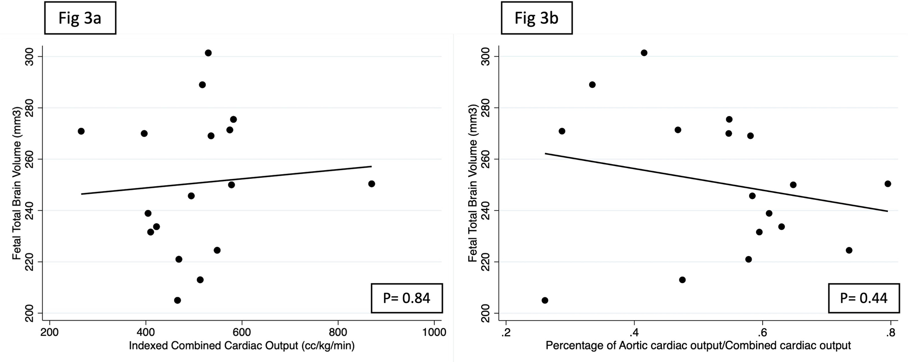
Results: Sixty-six participants enrolled (control = 20; HLHS = 25; TGA = 21), mean gestational age 33.8 weeks (95% CI: 33.3-34.2). Total brain volume and T2* were significantly lower in fetuses with cardiac disease. CCOi was lower in HLHS, correlating with total brain volume - for every 10% CCOi increase, volume increased 8 mm3 (95% CI: 1.78-14.1; p = 0.012). Echocardiography parameters and cerebral oxygenation showed no correlation. TGA showed no CCOi or aortic output correlation with MRI measures.
Conclusions: In HLHS, lower cardiac output is deleterious to brain development. Our findings provide insight into the role of fetal cardiovascular physiology in brain health.
© 2023 The Authors. Prenatal Diagnosis published by John Wiley & Sons Ltd.
Conflict of interest statement
Conflict of Interest Statement
All authors have no conflicts to disclose
Figures

References
-
- Limperopoulos C, Majnemer A, Shevell MI, et al. Neurodevelopmental status of newborns and infants with congenital heart defects before and after open heart surgery. J Pediatr 2000. Nov 1;137(5):638–45. - PubMed
-
- Limperopoulos C, Majnemer A, Shevell MI, et al. Functional limitations in young children with congenital heart defects after cardiac surgery. Pediatrics 2001. Dec 1;108(6):1325–31. - PubMed
-
- Carmant LS, Boucoiran I, Mathe M, et al. Prenatal markers of atypical neurodevelopment in children with congenital heart defects. J Matern Fetal Neonatal Med 2021. Apr 13;0(0):1–5. - PubMed
-
- Majnemer A, Limperopoulos C, Shevell M, et al. Long-term neuromotor outcome at school entry of infants with congenital heart defects requiring open-heart surgery. J Pediatr 2006. Jan 1;148(1):72–7. - PubMed
-
- Massaro AN, Glass P, Brown J, et al. Neurobehavioral abnormalities in newborns with congenital heart disease requiring open-heart surgery. J Pediatr 2011. Apr 1;158(4):678–681.e2. - PubMed
Publication types
MeSH terms
Grants and funding
LinkOut - more resources
Full Text Sources
Medical
