. 2023 Oct 31;42(10):113240.
doi: 10.1016/j.celrep.2023.113240. Epub 2023 Oct 17.

Toll-like receptor 4 and CD11b expressed on microglia coordinate eradication of Candida albicans cerebral mycosis

Affiliations

Toll-like receptor 4 and CD11b expressed on microglia coordinate eradication of Candida albicans cerebral mycosis

Yifan Wu et al. Cell Rep. .

Abstract

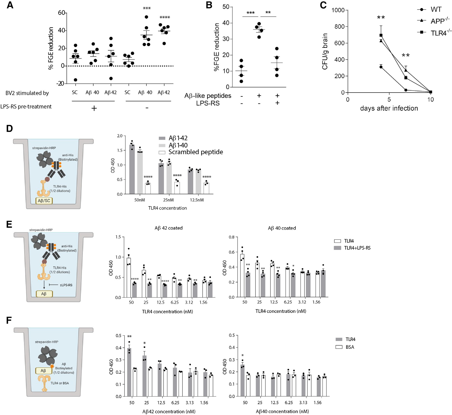
The fungal pathogen Candida albicans is linked to chronic brain diseases such as Alzheimer's disease (AD), but the molecular basis of brain anti-Candida immunity remains unknown. We show that C. albicans enters the mouse brain from the blood and induces two neuroimmune sensing mechanisms involving secreted aspartic proteinases (Saps) and candidalysin. Saps disrupt tight junction proteins of the blood-brain barrier (BBB) to permit fungal brain invasion. Saps also hydrolyze amyloid precursor protein (APP) into amyloid β (Aβ)-like peptides that bind to Toll-like receptor 4 (TLR4) and promote fungal killing in vitro while candidalysin engages the integrin CD11b (Mac-1) on microglia. Recognition of Aβ-like peptides and candidalysin promotes fungal clearance from the brain, and disruption of candidalysin recognition through CD11b markedly prolongs C. albicans cerebral mycosis. Thus, C. albicans is cleared from the brain through innate immune mechanisms involving Saps, Aβ, candidalysin, and CD11b.

Keywords: Alzheimer’s disease; CD11b; CP: Immunology; CP: Neuroscience; Candida albicans; Toll-like Receptor 4; amyloid beta; blood-brain barrier; candidalysin; cerebral mycosis; microglia; secreted aspartic proteinase.


Conflict of interest statement

Declaration of interests The authors declare no competing interests.

Figures

Figure 1.
Figure 1.. Saps mediate C. albicans cerebral invasion
Wild-type mice were challenged i.v. with wild-type parental strain or Sap-deficient C. albicans at indicated doses. (A and B) Cerebral fungal burdens were assessed 4 days post-infection as colony-forming units (CFU) expressed per gram of brain post-challenge with (A) sap1–3Δ/Δ, sap4–6Δ/Δ and (B) sap2Δ/Δ C. albicans. (C) Brain fungal burdens post-i.v. challenge with sap2Δ/Δ C. albicans from mice pretreated with pertussis toxin (PT) or vehicle. (D) Mice were challenged i.v. with 100,000 viable cells of wild-type and sap2Δ/Δ C. albicans. After 2 h, TRITC-dextran were injected i.v., and 2 h later, brains were perfused and homogenized, and the MFI of TRITC was quantitated. (E) Transendothelial electrical resistance (TEER) of monolayers of HBEC-5i cells was quantitated over time after Sap2 incubation. n ≥ 4, mean ± SEM, *p < 0.05, **p < 0.01, ***p < 0.001, and ****p < 0.0001 using one-way ANOVA followed by Tukey’s test for multiple comparison. Data are representative of two independent experiments.
Figure 2.
Figure 2.. Saps hydrolyze APP to generate Aβ peptides
(A) N2a cells were incubated with indicated amounts of recombinant Saps, after which Aβ40- and Aβ42-like peptides were quantitated from supernatants by ELISA. (B) Recombinant human APP was incubated with recombinant Saps at pH 3.5 or 6, and Aβ40- and Aβ42-like peptides were quantitated by ELISA. (C) Recombinant human APP was incubated with a mixture of recombinant Saps, and Aβ-like peptides were visualized by western blotting (SDS-PAGE) for Aβ peptides. (D) BACE1−/− mice were challenged with C. albicans and Aβ40- or Aβ42-like peptides were quantitated from brain homogenates by ELISA. (E) Immunofluorescence staining of FIGGs revealing IBA1-expressing microglial cells, Aβ, and DAPI+ nuclei from C. albicans-challenged BACE1−/− or APP−/− mice. n ≥ 4, mean ± SEM, **p < 0.01 and ****p < 0.0001 using one-way ANOVA followed by Dunnett’s test for multiple comparison (B) or two-tailed Student’s t test (A and D). Data are representative of three independent experiments.
Figure 3.
Figure 3.. Aβ peptides enhance the anti-fungal activity of BV-2 cells via TLR4
(A and B) Fungistasis assays in which BV-2 cells were pretreated with LPS-RS before activation by (A) Aβ peptides or scrambled control (SC) peptide or (B) Aβ-like peptides derived from the Sap-dependent cleavage of APP and addition of viable C. albicans. (C) Brain fungal burdens of wild-type (WT), TLR4−/−, and APP−/− mice assessed between days 4 and 10 after i.v. injection of 25,000 viable WT C. albicans cells. (D–F) Schematic diagrams of modified ELISAs assessing binding between Aβ peptides and TLR4 with capture and detecting reagent configurations with and without addition of LPS-RS and aggregate data as indicated. n ≥ 4. Mean ± SEM. *p < 0.05, **p < 0.01, ***p < 0.001, and ****p < 0.0001 using one-way ANOVA followed by Dunnett’s test for multiple comparison or two-tailed Student’s t test. Data are representative of two independent experiments.
Figure 4.
Figure 4.. Candidalysin enhances cerebral clearance of C. albicans
(A) WT mice were challenged i.v. with 25,000 viable cells of WT or ece1Δ/Δ C. albicans, and cerebral clearance was assessed over 60 days. (B) Immunofluorescence staining of FIGGs for DAPI, IBA-1, Aβ, and GFAP 21 days post-challenge. (C) Schematic diagram of fungistasis assay and aggregate data of BV-2 cells challenged with WT or ece1Δ/Δ C. albicans after stimulation with either SC or Aβ42 peptides. (D) BV-2 cells were incubated with synthetic candidalysin (10 μM) overnight, and secretion of IL-1, IL-6, TNF, and IFN-β was assessed by ELISA. n ≥ 4, mean ± SEM, *p < 0.05, **p < 0.01, ***p < 0.001, and ****p < 0.0001 using two-way ANOVA (C) or two tailed Student’s t test (D–F). Data are representative of two independent experiments.
Figure 5.
Figure 5.. Candidalysin activates microglial anti-fungal responses through CD11b
(A) WT or CD11b−/− mice were challenged i.v. with 25,000 viable cells of WT or ece1Δ/Δ C. albicans, and cerebral clearance was assessed over 20 days. (B–D) Schematic diagram of fungistasis assay (B) and aggregate data of candidalysin-activated BV-2 cells challenged with either WT or ece1Δ/Δ C. albicans with and without anti-CD11b or NIF blockade of CD11b (C and D). (E and F) Fungistasis assay of candidalysin-activated (E) primary microglia or (F) primary astrocytes from WT mice with WT C. albicans with and without anti-CD11b or NIF blockade of CD11b. (G) Pull-down assays using CD11-expressing CHO cells. CHO-CD11a/b/c cell lysates (prey) were incubated with biotinylated candidalysin (Bio-Clys; bait) or biotinylated serine (Bio-serine; control), and the mixtures were loaded onto prewashed streptavidin beads. Bait-prey complexes were eluted from the beads and loaded onto SDS-PAGE gels to detect CD11a, CD11b, and CD11c via western blotting. (H–J) Schematic diagrams and aggregate data depicting in vitro assays in which the dose-dependent binding of plate-bound (H) CD11b extracellular domain or (I) candidalysin/SC peptide to the other reagent or (J) CD11b or CD11c extracellular domains binding to plate-bound candidalysin were determined colorimetrically (OD, optical density). (K) Flow cytometric analysis of splenocytes from WT or CD11b−/− mice incubated with AF647-conjugated CL (10 μM). n = 4, mean ± SEM, *p < 0.05, **p < 0.01, ***p < 0.001, and ****p < 0.0001 using one-way ANOVA followed by Tukey’s test for multiple comparison (A–F), two tailed Student’s t test (H–J), or two-way ANOVA (K). Data are representative of two or three independent experiments.

References

    1. Brown GD, Denning DW, Gow NAR, Levitz SM, Netea MG, and White TC (2012). Hidden killers: human fungal infections. Sci. Transl. Med. 4, 165rv13–165rv113. - PubMed
    1. Eggimann P, Garbino J, and Pittet D (2003). Epidemiology of Candida species infections in critically ill non-immunosuppressed patients. Lancet Infect. Dis. 3, 685–702. - PubMed
    1. Parker JC Jr., McCloskey JJ, and Lee RS (1978). The emergence of candidosis. The dominant postmortem cerebral mycosis. Am. J. Clin. Pathol. 70, 31–36. - PubMed
    1. McCarthy MW, Kalasauskas D, Petraitis V, Petraitiene R, and Walsh TJ (2017). Fungal Infections of the Central Nervous System in Children. J. Pediatric Infect. Dis. Soc. 6, e123–e133. 10.1093/jpids/pix059. - DOI - PubMed
    1. Pendlebury WW, Perl DP, and Munoz DG (1989). Multiple microabscesses in the central nervous system: a clinicopathologic study. J. Neuropathol. Exp. Neurol. 48, 290–300. - PubMed

Publication types

LinkOut - more resources