. 2023 Oct 11;8(1):156.
doi: 10.1038/s41541-023-00751-6.

Effect of mRNA-LNP components of two globally-marketed COVID-19 vaccines on efficacy and stability

Affiliations

Effect of mRNA-LNP components of two globally-marketed COVID-19 vaccines on efficacy and stability

Lizhou Zhang et al. NPJ Vaccines. .

Abstract

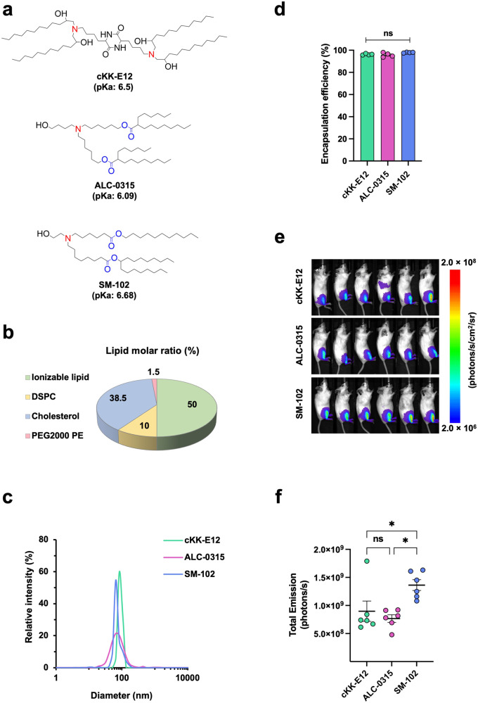
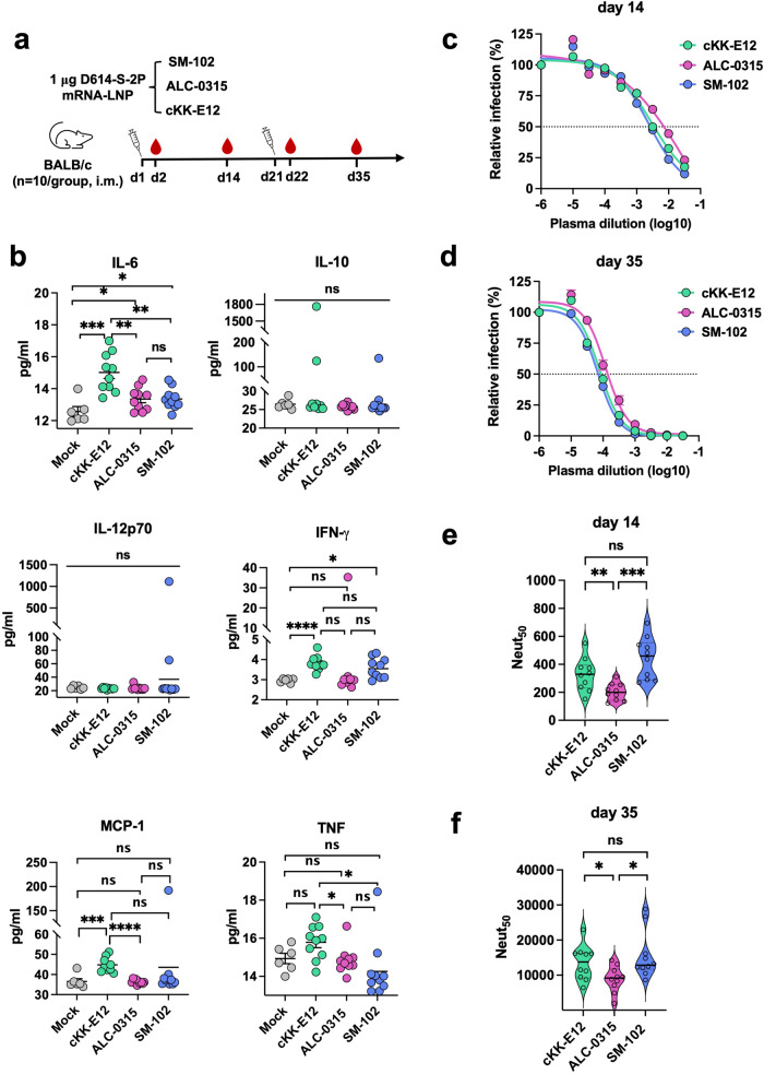
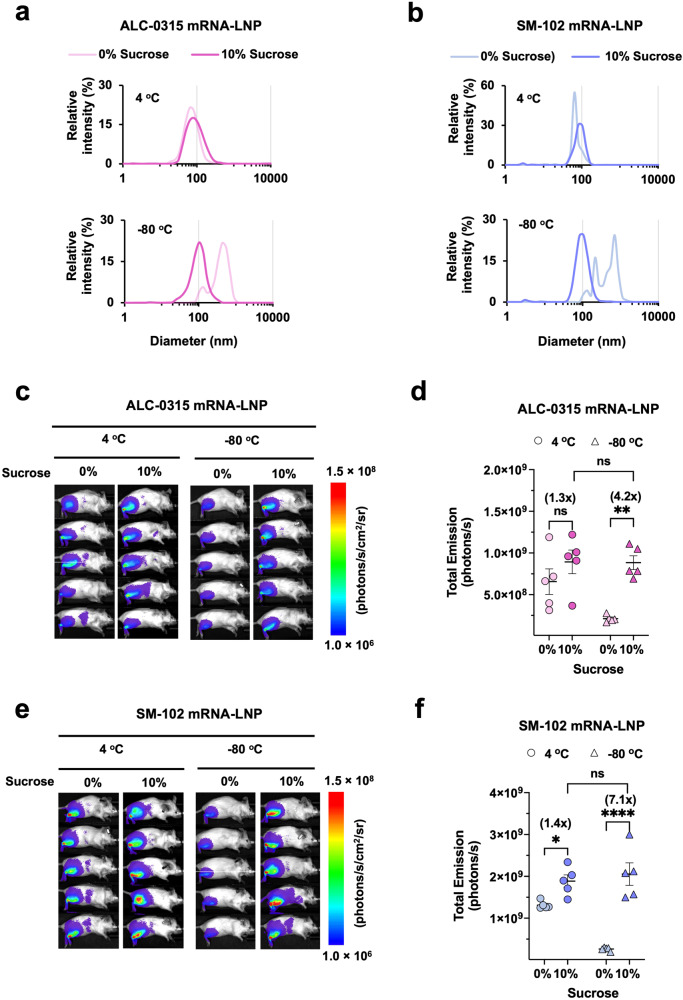
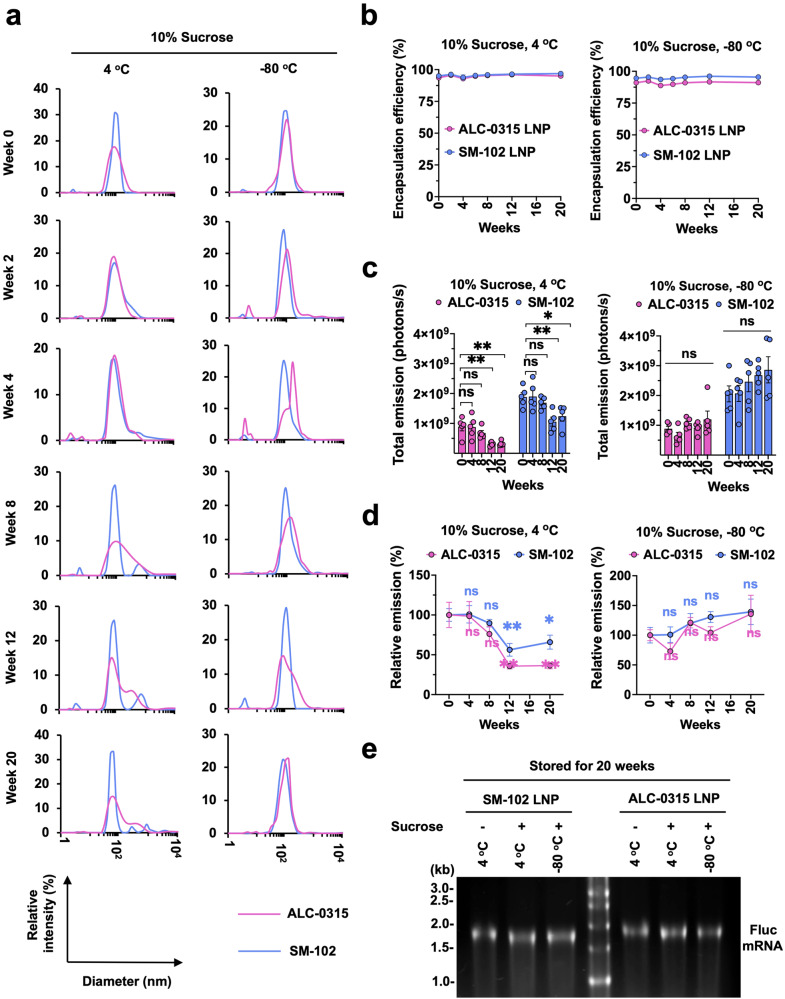
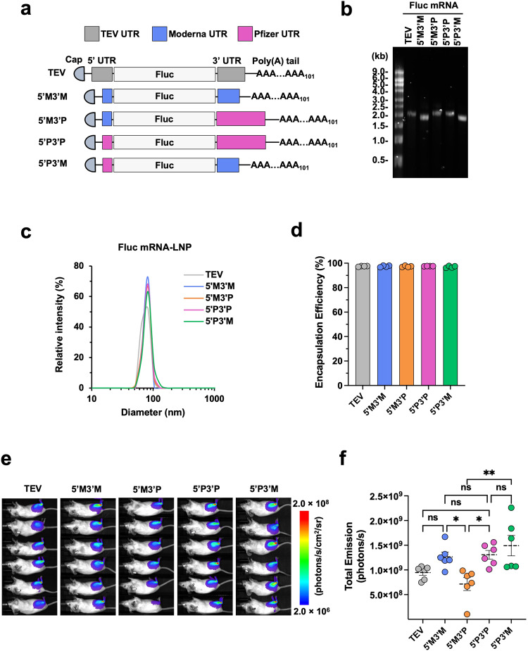
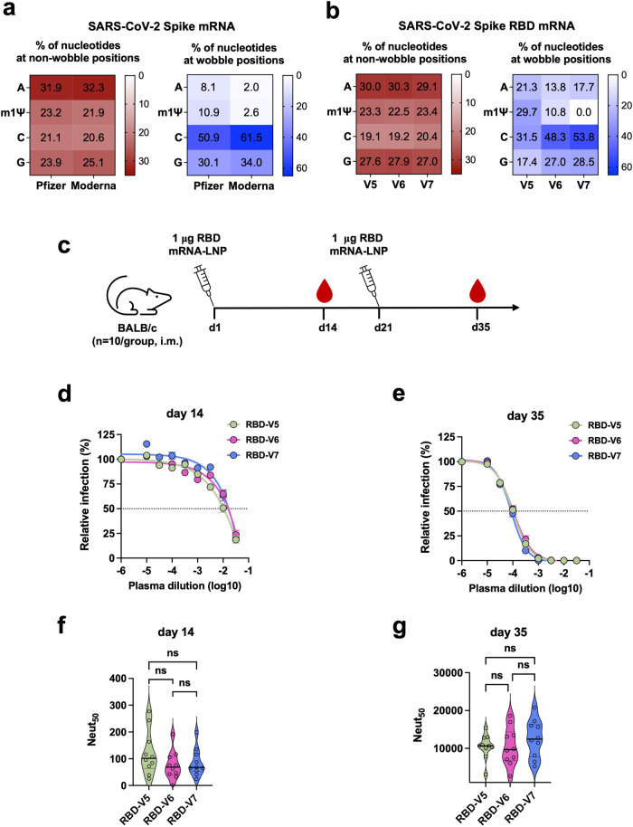
During the COVID-19 pandemic, Pfizer-BioNTech and Moderna successfully developed nucleoside-modified mRNA lipid nanoparticle (LNP) vaccines. SARS-CoV-2 spike protein expressed by those vaccines are identical in amino acid sequence, but several key components are distinct. Here, we compared the effect of ionizable lipids, untranslated regions (UTRs), and nucleotide composition of the two vaccines, focusing on mRNA delivery, antibody generation, and long-term stability. We found that the ionizable lipid, SM-102, in Moderna's vaccine performs better than ALC-0315 in Pfizer-BioNTech's vaccine for intramuscular delivery of mRNA and antibody production in mice and long-term stability at 4 °C. Moreover, Pfizer-BioNTech's 5' UTR and Moderna's 3' UTR outperform their counterparts in their contribution to transgene expression in mice. We further found that varying N1-methylpseudouridine content at the wobble position of mRNA has little effect on vaccine efficacy. These findings may contribute to the further improvement of nucleoside-modified mRNA-LNP vaccines and therapeutics.


Conflict of interest statement

The authors declare no competing interests.

Figures

Fig. 1
Fig. 1. SM-102 ionizable lipid is moderately more efficient than ALC-0315 in intramuscular delivery of mRNA in mice.
a Chemical structures of ionizable lipids cKK-E12, ALC-0315, and SM-102. Protonatable nitrogens are indicated in red, and biodegradable ester bonds in blue. Shown pKa values are apparent pKa. b A pie chart showing the molar ratio of lipids for LNP formulation. DSPC: Distearoylphosphatidylcholine; PEG2000 PE: 1,2-dioleoyl-sn-glycero-3-phosphoethanolamine-N-[methoxy(polyethylene glycol)-2000]. c Size distribution of Fluc mRNA-LNPs was measured by dynamic light scattering (DLS) and plotted with particle size on the X axis and relative intensity of scattered light on the Y axis. Plots shown are representative of two independent experiments, each conducted with two independent Fluc mRNA-LNP preparations. d Encapsulation efficiency of Fluc mRNA-LNPs was determined by Quant-iT™ RiboGreen RNA Assay Kit. Encapsulation Efficiency (%) = [(Fluorescence)total mRNA – (Fluorescence)outside mRNA] / (Fluorescence)total mRNA × 100%. e Images of mice at 24 h after injected with 1 μg Fluc mRNA-LNP. Bioluminescence intensity is presented in radiance (photons/s/cm2/sr) in a rainbow scale. f Bioluminescence shown in e was quantified as total emission in the region of interest. Each dot represents an individual mouse. Two independent preparations of mRNA-LNPs were used for each group. Data are presented as Mean ± SEM of n = 6 mice per group. Statistical significance among the groups was analyzed by one-way ANOVA with Tukey’s multiple comparisons test (ns, not significant; *p < 0.05).
Fig. 2
Fig. 2. SM-102 elicits comparable inflammatory response but higher antibody production in mice compared to ALC-0315.
a Timeline of mouse immunization and bleeding. Ten BALB/c mice per group were intramuscularly vaccinated at the indicated days, each time with 1 μg SARS-CoV-2 spike mRNA-LNP prepared with the indicated ionizable lipid. Blood was collected at the indicated time points to measure cytokines and neutralizing antibodies. b Pro-inflammatory cytokines/chemokines in the plasma collected at day 2 were detected using the murine inflammation kit and analyzed by Accuri Flow cytometer. Data are presented as Mean ± SEM of n = 10 mice per group. Cytokine levels in the d22 plasma are presented in Supplementary Fig. 1. c, d SARS-CoV-2 pseudovirus (PV) was preincubated with or without (presented at x = − 6) serially diluted plasma collected at day 14 c or 35 d. H1299-hACE2 cells were incubated with these preincubated mixes and analyzed 24 h later by measuring luciferase activity. Entry of SARS-CoV-2 PV in the presence of immune plasma relative to that in the absence of plasma is shown. Each dot on the curve represents the average value from ten mice. The dashed lines in the figures indicate 50% neutralization. e, f Violin plots show the Neut50 value of individual mouse plasmas (n = 10 per group) collected at day 14 e or 35 f. The central thick lines indicate the median of ten Neut50 values per group. Statistical significance among the groups was analyzed by one-way ANOVA with Tukey’s multiple comparisons test (ns, not significant; *p < 0.05, **p < 0.01, ***p < 0.001, and ****p < 0.0001).
Fig. 3
Fig. 3. Sucrose enhances intramuscular delivery efficiency of SM-102 and ALC-0315 mRNA-LNP in mice.
a, b The particle size distribution of ALC-0315 a or SM-102 b Fluc mRNA-LNP stored for 1 h at 4 °C (upper panel) or −80 °C (lower panel) with or without 10% sucrose was measured by DLS. Particle size is plotted on the X axis with relative intensity of scattered light on the Y axis. Plots shown are representative of two independent experiments, each conducted with two independent Fluc mRNA-LNP preparations. c, e Images of mice at 24 h post intramuscular injection of 1 μg Fluc mRNA-LNP formulated with either ALC-0315 c or SM-102 e. Bioluminescence intensity is shown in radiance (photons/s/cm2/sr) in a rainbow scale. d, f Bioluminescence imaged in c, e, respectively, was quantified as total emission in the region of interest. Each dot represents an individual mouse. Data are presented as Mean ± SEM of n = 5 mice per group. Statistical significance among the groups was analyzed by two-way ANOVA with Sidak’s multiple comparisons test (ns, not significant; *p < 0.05, **p < 0.01, and ****p < 0.0001).
Fig. 4
Fig. 4. ALC-0315 mRNA-LNP is moderately less stable than SM-102 mRNA-LNP at 4 °C.
a Particle size of ALC-0315 or SM-102 Fluc mRNA-LNP stored at 4 °C and −80 °C was measured by DLS at indicated time points. Particle size is plotted on the X axis with relative intensity of scattered light on the Y axis. Plots shown are representative of two independent experiments, each conducted with two independent Fluc mRNA-LNP preparations. b Encapsulation efficiency of the Fluc mRNA-LNP shown in a was measured at the indicated time points, using Quant-iTTM RiboGreen RNA Reagent. c Bioluminescence from the mice was quantified as total emission in the region of interest at 24 h after the injection of the Fluc mRNA-LNP at the indicated time points. d Bioluminescence values shown in c were normalized to that measured at week 0. Data are presented as Mean ± SEM of n = 5 mice per group. Statistical significance between week 0 and other time points within the same group was analyzed by one-way ANOVA with Dunnett’s multiple comparisons test. (ns, not significant; *p < 0.05 and **p < 0.01). e Encapsulated mRNA in LNP after 20 weeks of storage at 4 °C or −80 °C. 4 μl of 50 μg/ml mRNA-LNP was loaded per lane. Image shown is the representative of two independent mRNA-LNP preparations.
Fig. 5
Fig. 5. UTRs from Moderna and Pfizer-BioNTech mRNA differentially contribute to translation.
a Schematic diagrams of the Fluc mRNAs with different UTRs but with the same 5’ cap and 3’ poly(A) tail of 101 nucleotides. b Size and purity of Fluc mRNAs were checked by RNA gel electrophoresis. c Size distribution of the LNPs shown in b was measured by DLS. Particle size is plotted on X axis and relative intensity of scattered light on Y axis. Presented plots are the representative of two independent experiments, each conducted with two independent Fluc mRNA-LNP preparations. d Encapsulation efficiency of the same Fluc mRNA-LNPs. e Bioluminescence images of mice at 24 h post intramuscular injection of 1 μg Fluc mRNA-LNP containing the indicated UTRs. Bioluminescence intensity is presented in radiance (photons/s/cm2/sr) in a rainbow scale. f Bioluminescence shown in e was quantified as total emission in the region of interest. Each dot represents an individual mouse. Data are presented as Mean ± SEM of n = 6 mice per group. Statistical significance among the groups was analyzed by one-way ANOVA with Tukey’s multiple comparisons test (ns, not significant; *p < 0.05 and **p < 0.01).
Fig. 6
Fig. 6. m1Ψ content at the wobble position of RBD mRNA does not significantly contribute to vaccine efficacy in mice.
a Nucleotide composition of the spike mRNA of Pfizer-BioNTech and Moderna COVID-19 vaccines. The numbers shown in the boxes are the percentage of the indicated nucleotide in those at non-wobble positions (left panel) or at the wobble position (right panel). b Nucleotide composition of three spike RBD mRNAs designed to contain varying level of wobble m1Ψ content. RBD-V6 and RBD-V7 mRNA have wobble m1Ψ content close to that of the Pfizer-BioNTech and Moderna COVID-19 vaccines, respectively. Otherwise, these RBD mRNAs encode the identical protein and share the same 5′ cap, UTRs, and poly(A) tail. Their LNPs were formulated with the same lipids. c Timeline of mouse immunization and bleeding. d, e SARS-CoV-2 PV neutralization assays conducted with plasmas collected at days 14 d and 35 e. Each dot on the curves represents the average value from ten mouse plasmas. Dashed lines indicate 50% neutralization. f, g Violin plots show the Neut50 values of mouse immune plasmas (n = 10 per group) shown in d, e, respectively. Each dot represents an individual mouse. The thick horizontal lines and error bars indicate Mean ± SEM of n = 10 mice per group. Statistical significance among the groups was analyzed by one-way ANOVA with Tukey’s multiple comparisons test (ns, not significant).

Similar articles

Cited by

References

    1. Vogel AB, et al. BNT162b vaccines protect rhesus macaques from SARS-CoV-2. Nature. 2021;592:283–289. - PubMed
    1. Corbett KS, et al. SARS-CoV-2 mRNA vaccine design enabled by prototype pathogen preparedness. Nature. 2020;586:567–571. - PMC - PubMed
    1. Rijkers, G. T. et al. Antigen Presentation of mRNA-Based and Virus-Vectored SARS-CoV-2 Vaccines. Vaccines (Basel)9, 10.3390/vaccines9080848 (2021). - PMC - PubMed
    1. Laczkó D, et al. A single immunization with nucleoside-modified mRNA vaccines elicits strong cellular and humoral immune responses against SARS-CoV-2 in mice. Immunity. 2020;53:724–732.e727. - PMC - PubMed
    1. Pardi N, Hogan MJ, Porter FW, Weissman D. mRNA vaccines - a new era in vaccinology. Nat Rev Drug Discov. 2018;17:261–279. - PMC - PubMed