. 2023 Oct 11;13(1):17224.
doi: 10.1038/s41598-023-44427-6.

The role of intrathecal free light chains kappa for the detection of autoimmune encephalitis in subacute onset neuropsychiatric syndromes

Affiliations

The role of intrathecal free light chains kappa for the detection of autoimmune encephalitis in subacute onset neuropsychiatric syndromes

Dominic Bertram et al. Sci Rep. .

Abstract

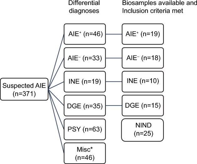
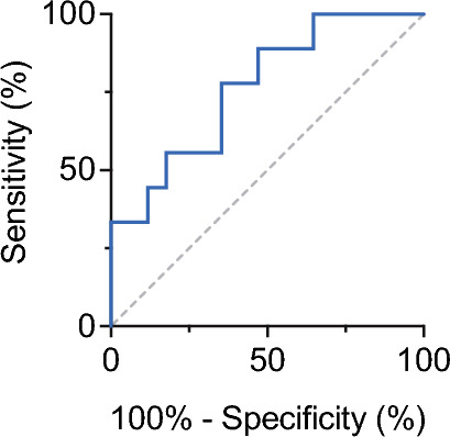
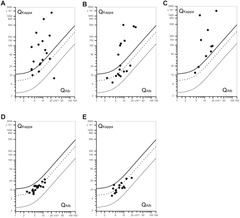
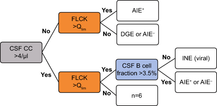
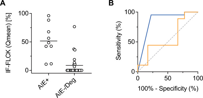
Intrathecal synthesis of free light chains kappa (FLCK) is increasingly recognized as a marker of inflammatory CNS pathologies. Here, we tested the performance of FLCK in differentiating autoimmune encephalitis (AIE) from non-inflammatory etiologies in subacute onset neuropsychiatric syndromes. Patients undergoing diagnostic work-up for suspected autoimmune encephalitis at our department between 2015 and 2020 were retrospectively assessed for definitive diagnosis, available CSF and blood samples, as well as complete clinical records. Intrathecal FLCK was measured along with established CSF markers of CNS inflammation. The study cohort consisted of 19 patients with antibody-mediated AIE (AIE+), 18 patients with suspected AIE but without detectable autoantibodies (AIE-), 10 patients with infectious (viral) encephalitis (INE), and 15 patients with degenerative encephalopathies (DGE). 25 age- and sex-matched patients with non-inflammatory neurological diseases (NIND) were used as a control group. All AIE+ patients exhibited intrathecal synthesis of FLCK compared to only 39% of AIE- patients and 81% of patients in the INE group. No intrathecal synthesis of FLCK was found in DGE and NIND patients. While intrathecal FLCK was equally specific for an inflammatory etiology as oligoclonal bands (OCB) in the cerebrospinal fluid (CSF), the sensitivity of intrathecal FLCK for any inflammatory intrathecal process was higher than that of OCB (83% vs. 38%). Intrathecal FLCK synthesis was found to discriminate AIE+ from non-inflammatory encephalopathies and AIE- when the CSF cell count was normal [receiver operating characteristic (ROC) analysis area under the curve (AUC): 0.867, p = 0.002], while it failed to differentiate between AIE+ and INE in the presence of CSF pleocytosis (AUC: 0.561, p = 0.607). In conclusion, in the absence of CSF pleocytosis, intrathecal FLCK discriminated AIE+ from competing diagnoses in our cohort of subacute onset neuropsychiatric syndromes. In addition to established markers of CSF inflammation, intrathecal FLCK might support clinical decision-making and contribute to selecting patients for (repeated) antibody testing.


Conflict of interest statement

The authors declare no competing interests.

Figures

Figure 1
Figure 1
Graphical representation of the study cohort. Between 2015 and 2020, 371 patients underwent diagnostic work-up for suspected AIE. Diagnostic categories were AIE+ (antibody-positive autoimmune encephalitis), AIE (antibody-negative suspected autoimmune encephalitis), INE (infectious encephalitis), DGE (degenerative encephalopathy), PSY (psychiatric diagnoses). A large number of cases (n = 175) could not be assigned to any of the categories. After the application of exclusion criteria (see “Methods”) and screening for complete (blood and CSF) biosamples, 62 patients were included in the further analysis [87 individuals, including the control group of 25 patients with NIND (non-inflammatory neurologic disease)].
Figure 2
Figure 2
Receiver operating characteristics for the CSF fraction of B cells in the differentiation of AIE+ and INE under conditions of CSF pleocytosis and intrathecal FLCK synthesis > Qlim. AUC: 0.765, Cutoff 3.5%, p = 0.02.
Figure 3
Figure 3
Double logarithmic quotient-diagrams (Reiber diagrams) of QFLCK (QKappa), i.e. FLCK CSF/serum ratio, set against QAlb, i.e. Albumin CSF/serum ratio. (A) AIE+, 78% (n = 15/19) of QKappa > Qlim; (B) AIE, 39% (n = 7/18) of QKappa > Qlim; (C) INE, 81% (n = 9/11) of QKappa > Qlim; (D) DGE and (E) NIND, all QKappa < Qlim. Symbols above Qlim indicate intrathecal FLCK synthesis. Qlim thick upper line, Qmean dotted middle line, Qlow thin lower line.
Figure 4
Figure 4
Sequential diagnostic approach applicable to this cohort using CSF white cell count, intrathecal FLCK synthesis, and CSF B cell fraction. Pre-stratification of the cohort into CSF pleocytosis (CC > 4/μL) and normocytosis. In the upper arm (CSF normocytosis), intrathecal FLCK synthesis discriminated AIE+ from other diagnoses. In the lower arm (CSF pleocytosis), QFLCK > Qlim indicated either infectious or autoimmune encephalitis. In the event of CSF pleocytosis and QFLCK > Qlim, the intrathecal B cell fraction (< 3.5%) distinguished infectious from autoimmune encephalitis with AUC of 0.75.
Figure 5
Figure 5
Test performance of IF-FLCK. (A) Individual IF-FLCK values for the CSF-normocytic subgroup (AIE+, AIE, and DGE, for visualization displayed with reference to Qmean), (B) receiver operating characteristics with respect to QFLCK > Qlim after stratification of the cohort into CSF pleocytosis and CSF normocytosis. In CSF normocytosis, the intrathecal fraction of FLCK (IF-FLCK) distinguished AIE+ from the rest (DGE, AIE) with a sensitivity of 95.2% and a specificity of 77.8% (AUC 0.867, p = 0.002, blue line). In CSF pleocytosis, the capacity of IF-FLCK to discriminate AIE+ or AIE from INE was poor (AUC 0.561, p = 0.607, orange line).

References

    1. Titulaer MJ, et al. Treatment and prognostic factors for long-term outcome in patients with anti-NMDA receptor encephalitis: An observational cohort study. Lancet Neurol. 2013;12:157–165. doi: 10.1016/S1474-4422(12)70310-1. - DOI - PMC - PubMed
    1. Dubey D, et al. Autoimmune encephalitis epidemiology and a comparison to infectious encephalitis. Ann. Neurol. 2018;83:166–177. doi: 10.1002/ana.25131. - DOI - PMC - PubMed
    1. Graus F, et al. A clinical approach to diagnosis of autoimmune encephalitis. Lancet Neurol. 2016;15:391–404. doi: 10.1016/S1474-4422(15)00401-9. - DOI - PMC - PubMed
    1. Ruiz-Garcia R, et al. Limitations of a commercial assay as diagnostic test of autoimmune encephalitis. Front. Immunol. 2021;12:691536. doi: 10.3389/fimmu.2021.691536. - DOI - PMC - PubMed
    1. Thaler FS, et al. Rituximab treatment and long-term outcome of patients with autoimmune encephalitis: Real-world evidence from the GENERATE registry. Neurol. Neuroimmunol. Neuroinflamm. 2021 doi: 10.1212/NXI.0000000000001088. - DOI - PMC - PubMed

Publication types

Substances

Supplementary concepts