. 2023 Oct;622(7982):393-401.
doi: 10.1038/s41586-023-06594-4. Epub 2023 Oct 11.

Design and testing of a humanized porcine donor for xenotransplantation

Affiliations

Design and testing of a humanized porcine donor for xenotransplantation

Ranjith P Anand et al. Nature. 2023 Oct.

Abstract

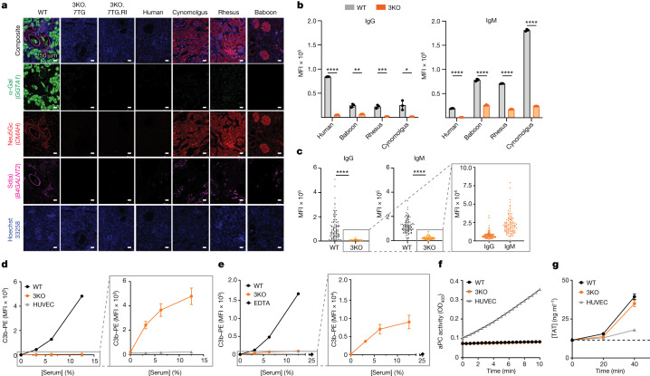
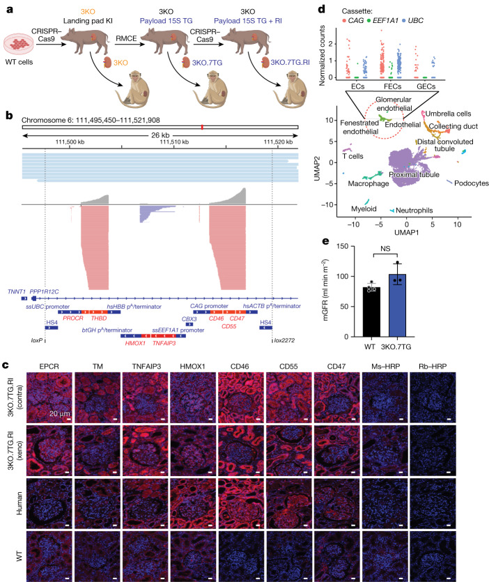
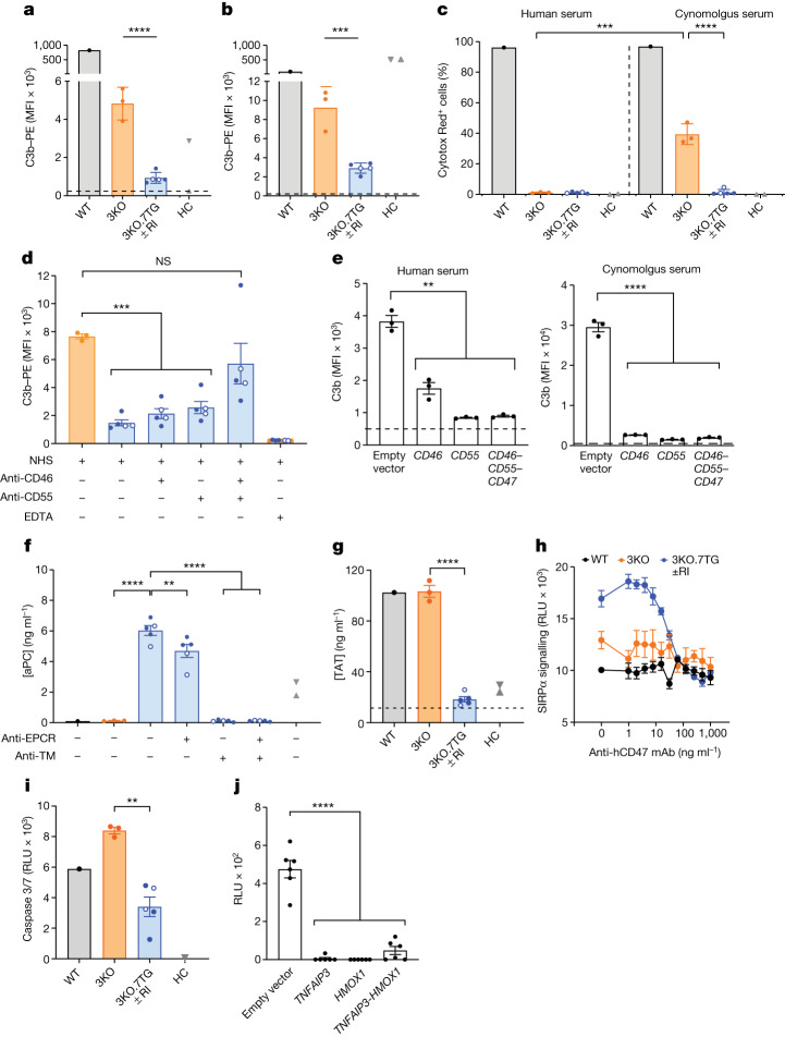
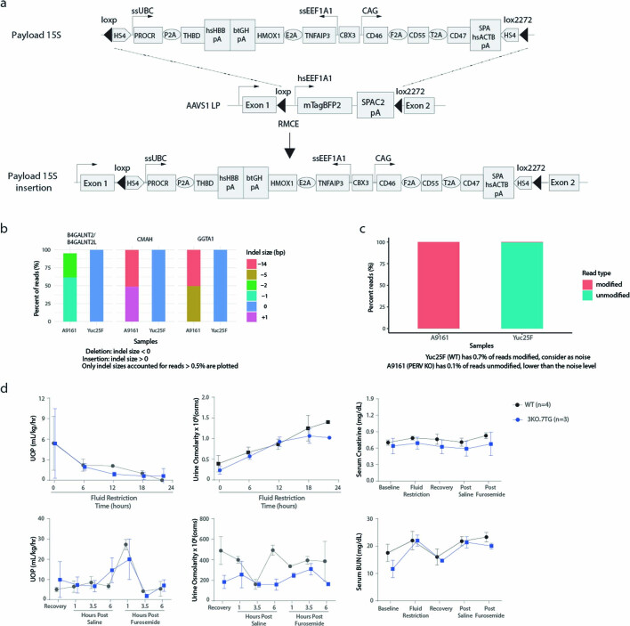
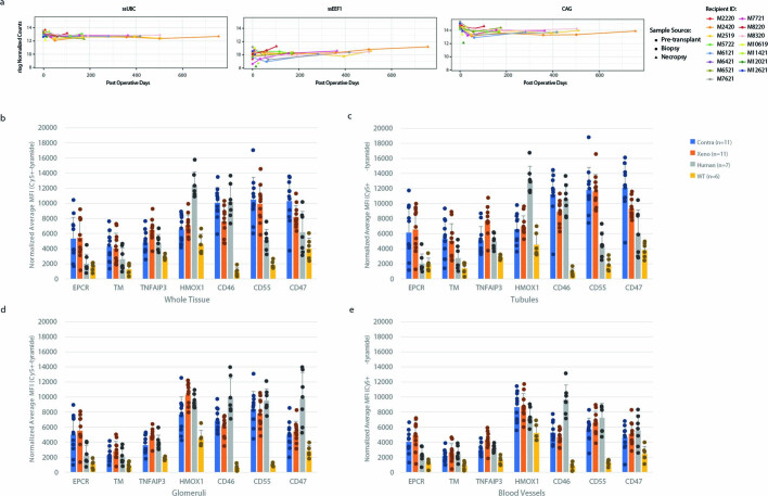
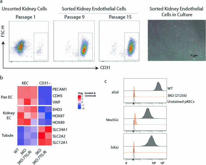
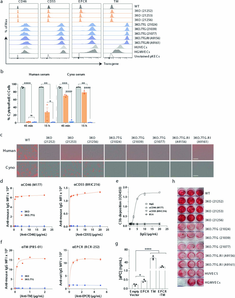
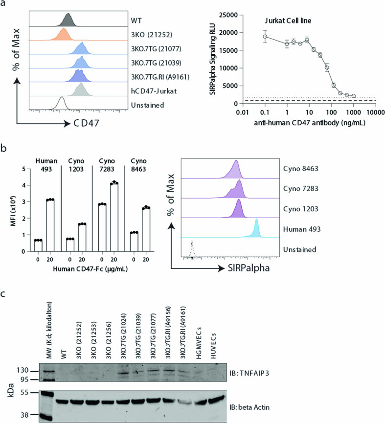
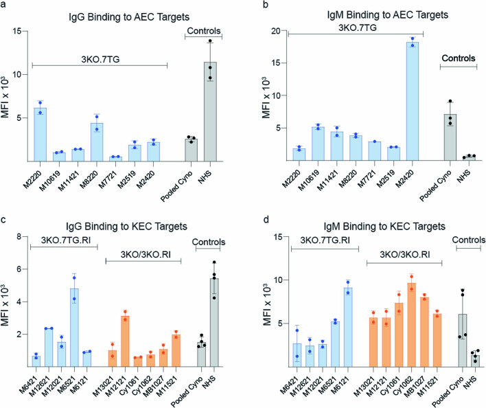
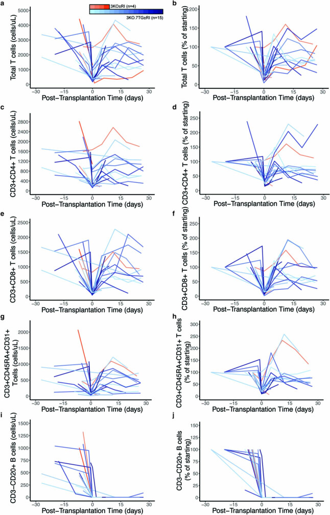
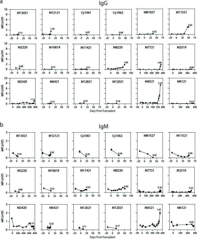
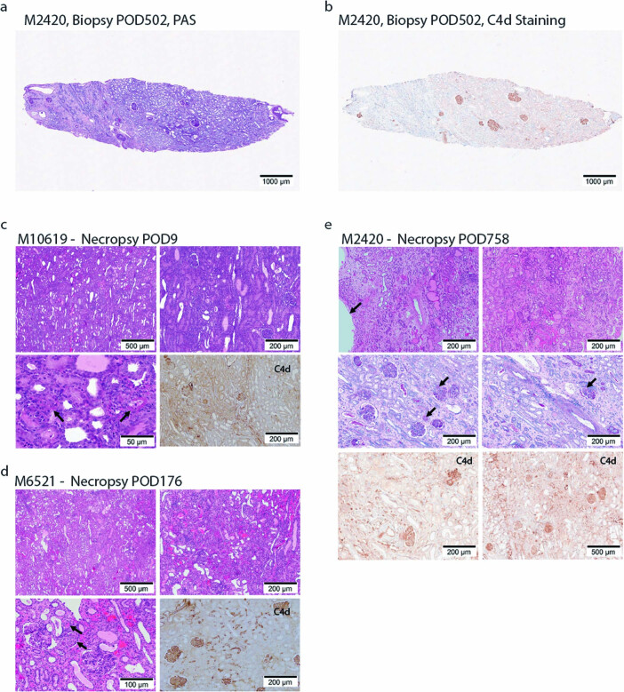
Recent human decedent model studies1,2 and compassionate xenograft use3 have explored the promise of porcine organs for human transplantation. To proceed to human studies, a clinically ready porcine donor must be engineered and its xenograft successfully tested in nonhuman primates. Here we describe the design, creation and long-term life-supporting function of kidney grafts from a genetically engineered porcine donor transplanted into a cynomolgus monkey model. The porcine donor was engineered to carry 69 genomic edits, eliminating glycan antigens, overexpressing human transgenes and inactivating porcine endogenous retroviruses. In vitro functional analyses showed that the edited kidney endothelial cells modulated inflammation to an extent that was indistinguishable from that of human endothelial cells, suggesting that these edited cells acquired a high level of human immune compatibility. When transplanted into cynomolgus monkeys, the kidneys with three glycan antigen knockouts alone experienced poor graft survival, whereas those with glycan antigen knockouts and human transgene expression demonstrated significantly longer survival time, suggesting the benefit of human transgene expression in vivo. These results show that preclinical studies of renal xenotransplantation could be successfully conducted in nonhuman primates and bring us closer to clinical trials of genetically engineered porcine renal grafts.


Conflict of interest statement

eGenesis has filed multiple patent applications covering the subject matter of this paper. R.P.A., J.V.L., D.H., A.A., D.A.-A, J.C.C., S. Chhangawala, R.J.E., N.E., K.G., A.K.G., X.G., K.C.H., P.H., S.H., N.H., L.A.K., Y.K., T.L., F.L., M.L., S.C.L., C.N., M.P., V.B.P., R.A.P., R.P., L.P., L.Q., W.T.S., D. Stevens, K.S., O.D.S., Y.X., S.Y., G.E.Z., M.C., M.E.Y. and W.Q. contributed to this work as employees of eGenesis and may have an equity interest, in the form of stock options, in eGenesis. J.N.C. and X.T. contributed to this work as employees of eGenesis. D.J.F. was partially supported by eGenesis. R.B.C., D.J.F., T.K. and G.L. are consultants for eGenesis. J.F.M. serves on the eGenesis Scientific Advisory Board. G.M.C. is co-founder and scientific advisor to eGenesis.

Figures

Fig. 1
Fig. 1. Functional incompatibilities exist between porcine donor and primate recipient.
a, IHC confirmed that the three glycan antigens are expressed in the WT porcine kidney, but have been eliminated from the 3KO.7TG (donor ID 21077) and the 3KO.7TG.RI (donor ID A9161) kidneys, a pattern similar to that of the human kidney. In comparison, OWMs express the Neu5Gc antigen. IHC analyses of 3KO were performed for all contralateral kidney samples included in this study and 3KO phenotypes confirmed for all. b, WT porcine KECs bound human and NHP preformed antibodies, whereas 3KO porcine KECs showed markedly reduced antibody binding. Each sample was run with three technical replicates. Error bars are s.d. c, The 3KO AECs bound significantly less IgG and IgM than the WT AECs, when incubated with 96 individual cynomolgus monkey serum samples. Note that 3KO AECs retained a substantial level of IgG and IgM binding. Statistical analysis was performed using Wilcoxon matched-pairs signed rank test. d, WT KECs showed significant C3b deposition in human serum, compared with human umbilical vein endothelial cells (HUVECs). Although markedly reduced, the 3KO KECs retained some level of C3b deposition (right panel). Error bars are s.e.m. e, WT KECs showed C3b deposition and 3KO KECs showed less deposition when incubated in the cynomolgus monkey serum. Error bars are s.e.m. f, Porcine KECs (WT or 3KO) did not produce aPC, whereas HUVECs readily produced aPC. For df, WT KEC (n = 1), 3KO KEC (n = 3) and HUVEC (n = 1). OD405, optical density at 405 nm. g, When incubated with human whole blood, WT (n = 1) and 3KO (n = 1) porcine KECs triggered coagulation, measured as TAT formation. Error bars are s.e.m. For dg, each point is a biological replicate examined over at least two independent experiments. With the exception of c, statistical analyses were performed using two-tailed unpaired Student’s t-tests. ****P < 0.0001, ***P < 0.001, **P < 0.01, *P < 0.05. Source Data
Fig. 2
Fig. 2. Yucatan porcine donor is engineered to carry 69 genomic edits.
a, The porcine donor kidney, 3KO.7TG.RI, was engineered to eliminate three glycan antigens (3KO), overexpress seven human transgenes (PL15S) and inactivate PERV elements (RI) through three rounds of editing and cloning. The donor kidney, 3KO.7TG, carries 3KO and PL15S, without RI. KI, knock in; RMCE, recombinase-mediated cassette exchange. b, Reads from Nanopore long-read whole-genome sequencing of the 3KO.7TG.RI donor, A9161, were aligned to a custom chromosome carrying PL15S inserted at the AAVS1 genomic safe harbour site (top). Reads from Nanopore direct RNA-seq of A9161 kidney mRNA were aligned to the custom chromosome (bottom). All three transcription units were transcribed. c, All seven human transgenic proteins were detected in the contralateral (contra) kidney of A9161 recovered at transplantation (row 1), and the xenograft (xeno) kidney harvested at necropsy on post-transplantation day 176 (row 2). HRP, horseradish peroxidase; TM, thrombomodulin; Ms–HRP, goat anti-mouse secondary antibody conjugated with HRP; Rb–HRP, goat anti-rabbit secondary antibody conjugated with HRP. d, Three endothelial cell types were identified from kidney dissociated cell populations by single-cell RNA-seq, including endothelial cells (ECs) (PECAM1+PLVAPEHD3GATA5), glomerular endothelial cells (GECs) (PECAM1+EHD3+GATA5+) or fenestrated endothelial cells (FECs) (PECAM1+PLVAP+) (bottom), and transgene expression was examined among the three endothelial cell types (top). Mean log2-normalized unique molecular identifier counts were plotted against the three PL15S transcription units (ssUBC, ssEEF1A1 and CAG). UMAP, uniform manifold approximation and projection. e, The 3KO.7TG porcine donors (n = 3) showed normal measured glomerular filtration rate (mGFR) compared with age-matched WT Yucatan pigs (n = 4). Unpaired two-tailed Student’s t-test; error bars are s.e.m. Points are biological replicates and data are from one experiment. Source Data
Fig. 3
Fig. 3. Human transgenes confer protection.
a,b, In human (a) or cynomolgus monkey (b) serum, deposition of C3b on 3KO.7TG ± RI KECs was further reduced. The dashed lines indicate average C3b deposition (serum + EDTA). c, In human or cynomolgus monkey serum, 3KO.7TG ± RI KECs were protected from complement-dependent cytotoxicity. 3KO KECs showed residual complement-dependent cytotoxicity in cynomolgus monkey serum. d, The 3KO.7TG ± RI KECs mitigated C3b deposition when human CD46, CD55 or both were not blocked by antibodies. NHS, normal human serum. e, 3KO KECs carrying CD46, CD55 or CD46CD55CD47 polycistronic genes regulated C3b deposition in human or cynomolgus monkey serum. The dashed lines indicate average C3b deposition from EDTA-treated serum. f, The 3KO.7TG ± RI KECs readily generated aPC. aPC production was reduced when EPCR or thrombomodulin (TM) was blocked with antibodies, with blocking of thrombomodulin being more effective. The dashed line indicates average aPC production without cells. g, 3KO.7TG ± RI KECs regulated TAT formation in whole blood. The dashed line indicates baseline TAT in donor blood. h, Human CD47 (hCD47) on 3KO.7TG ± RI KECs signalled through SIRPα on human Jurkat cells. WT KECs (n = 1), 3KO KECs (n = 1), 3KO.7TG ± RI (n = 2 for 3KO.7TG and n = 1 for 3KO.7TG.RI). mAb, monoclonal antibody; RLU, relative light unit. i, 3KO.7TG ± RI KECs were protected from human TNF-induced caspase 3/7 activation. j, 3KO kidney cortex-derived cells expressing TNFAIP3, HMOX1 or TNFAIP3HMOX1 polycistronic genes were protected from human TNF-induced caspase 3/7 activation. In ad,fi, WT (n = 1), 3KO (n = 3), 3KO.7TG (n = 3, solid blue circles), 3KO.7TG.RI (n = 2, open circles), human controls (HC) and human umbilical vein endothelial cells (n = 1, grey triangle). In ad,f,g, human glomerular microvascular endothelial cells (n = 1, grey inverted triangle). For ad,fi, data are from biological replicate, repeated at least twice. For e,j, data are from a technical replicate. Error bars indicate s.e.m. Statistical analyses were two-tailed, paired (3KO.7TG ± RI with versus without blocking antibodies (f)) and unpaired (all other comparisons) Student’s t-tests. ****P < 0.0001, ***P < 0.001, **P < 0.01; NS, not significant. Source Data
Fig. 4
Fig. 4. Engineered porcine kidneys support life in cynomolgus monkeys.
a, Significantly improved survival probability (P = 0.026) for recipients transplanted with 3KO.7TG ± RI renal grafts (n = 15) compared with 3KO ± RI (n = 6). Statistical analysis was performed using a log-rank test. b, Serum creatinine levels generally remained within normal range, except when associated with graft failure. Creatinine levels above 6 mg dl−1 meet criteria for euthanasia. Dashed lines indicate normal range of serum creatinine levels. c, Periodic acid–Schiff staining of a kidney biopsy sample derived from recipient M2420, carrying a 3KO.7TG renal graft, at POD 502, appears normal. d,e, Blood vessels (d) and glomerulus (e) of M2420 renal graft biopsy at POD 502 appear normal. f, C4d-positive staining in glomeruli, but not in peritubular capillaries, was observed in M2420 renal graft biopsy at POD 502. Source Data
Extended Data Fig. 1
Extended Data Fig. 1. Yucatan porcine donor is engineered to carry 69 genomic edits.
a, Payload 15 S was inserted into the AAVS1 genomic safe harbor site by recombinase mediated cassette exchange (RMCE). It carries three transcription units, with the ssUBC promoter expressing the PROCR and the THBD cDNAs, the ssEEF1A1 promoter expressing the TNFAIP3 and the HMOX1 cDNAs, and the CAG promoter expressing the CD46, CD55, and CD47 cDNAs. The transgenic construct is flanked by the loxP/lox2272 sequences and replaced the landing pad sequence flanked by the same lox sites, in an RMCE reaction. b, Indel patterns for the B4GALNT2/B4GALNT2L, CMAH, and GGTA1 genes carried in the porcine donor A9161, analyzed from Amplicon sequencing. c, Region encompassing the catalytic core of the reverse transcriptase (RT) was amplified by a PCR reaction and sequenced, from the porcine endogenous retroviral sequences of donor A9161. Compared with the PCR product amplified from the wild type Yuc25F cells, those amplified from A9161 had been modified and predicted to obliterate the RT activity. d, Kidneys from 3KO.7TG donors (n = 3) responded to fluid challenges in vivo, including fluid restriction, saline bolus challenge, and furosemide (1 mgkg−1), similar to WT Yucatan (n = 4) kidneys. Data are from one independent experiment. Source Data
Extended Data Fig. 2
Extended Data Fig. 2. Human transgenes are expressed.
a, RNA was extracted from contralateral, biopsy, and necropsy kidney samples (3KO.7TG, n = 8; 3KO.7TG.RI, n = 7) and sequencing performed on the Illumina platform. Transgene expression from each of the three transcription units, ssUBC, ssEEF1A1, and CAG, was analyzed. b, Human transgenic protein expression, as detected by IHC, was quantitated from contralateral and necropsy kidney samples for completed NHP transplant studies, except for NHP recipient M7721 (porcine donor 21405), for which a contralateral kidney sample was not available (3KO.7TG, n = 6; 3KO.7TG.RI, n = 5). c–e, Transgenic protein expression was quantitated from tubular cells, glomeruli, and blood vessels, respectively. Each dot represents data from an independent kidney sample. Source Data
Extended Data Fig. 3
Extended Data Fig. 3. Porcine kidney endothelial cells are derived and characterized.
a, Kidney endothelial cells (KECs) were enriched by sorting for CD31+ cells twice. Dissociated cells from a kidney preparation contained a low percentage of CD31+ cells (2%). A subsequent second sort substantially enriched the percentage of CD31+ cells. CD31 expression was maintained over passaging as shown with passages 9 and 15 cells. KECs were isolated and enriched CD31 expression was assessed for lines from  41 independent kidneys. (Right Panel) A 40X phase-contrast image of double sorted KECs is provided. b, CD31+ KECs expressed endothelial marker genes, analyzed by RNA-seq. c, Compared to WT KECs, 3KO KECs lacked the three glycans, analyzed by flow cytometry.
Extended Data Fig. 4
Extended Data Fig. 4. Human CD46, CD55, EPCR, and TM proteins are expressed and functional.
a, Human CD46, CD55, EPCR, and TM proteins were detected on the surface of 3KO.7TG ± RI KECs. b, WT (n = 3) and 3KO (n = 3) KECs were lysed, whereas the 3KO.7TG KECs (n = 3) showed significantly reduced complement dependent cytotoxicity (CDC), when incubated with human or cynomolgus monkey serum over an extended period of time. c, Representative images from 3b, collected from an Incucyte after fluorescent dye staining. Scale bar (in white): 400 μm. d, Anti-human CD46 (clone M177, left) and CD55 (clone BRIC216, right) antibodies specifically bound human CD46 or CD55, as demonstrated by the lack of binding to 3KO KECs (n = 1, blue line), compared to the 3KO.7TG KECs (n = 1, orange line). e, M177 and BRIC216 antibodies did not activate complement fixation compared to human IgG (hIgG). f, Similarly, anti-human TM (clone PBS-01) and EPCR (clone RCR-252) antibodies are human specific. (3KO, n = 1; 3KO.7TG, n = 1). For d-f, average of 2 technical replicates. g, EPCR, TM, or both expressed in KECs contributed to aPC production, with TM showing more significant effect. Points are technical replicates. h, Extensive clotting was observed when WT (n = 1) and 3KO (n = 3) KECs were incubated with human whole blood, while 3KO.7TG (n = 3) or 3KO.7TG.RI KECs (n = 2) showed minimal clotting, similar to human umbilical vein and human glomerular microvascular endothelial cells (HUVECs and HGMVECs). Samples in the same row are technical replicates. Pictures were taken after plasma or nonclotted blood was removed from wells. For b,g, ****P < 0.0001, ***P < 0.001, **P < 0.01, * P < 0.05 by unpaired, two-tailed Student’s t-tests. Source Data
Extended Data Fig. 5
Extended Data Fig. 5. Human CD47 and TNFAIP3 proteins are expressed.
a, (Left) Human CD47 was detected on 3KO.7TG ± RI KECs and on Jurkat T cells overexpressing human CD47 (hCD47-Jurkat). (Right) In the SIRPα reporter assay, hCD47-Jurkat cells were incubated with SIRPα expressing Jurkat signaling cells, and engagement of hCD47 with human SIRPα produced an activation signal, which was blocked with increasing anti-human CD47 antibodies. Dashed lines: signals of cells alone. Error bars represent standard deviation from mean. b, (Left) Recombinant human CD47-Fc fusion protein bound to human (n = 1) or cynomolgus monkey (cyno) (n = 3) monocytes. Each point represents technical replicates. (Right) SIRPα expression on the monocytes used in the binding assay is shown. Data are representative of two independent experiments. c, 3KO.7TG ± RI KECs expressed TNFAIP3 protein as analyzed by Western blot. Uncropped images are provided in Supplementary Fig. 3. Representative data from two independent experiments. Source Data
Extended Data Fig. 6
Extended Data Fig. 6. Cynomolgus monkeys were examined for preformed IgG and IgM antibodies.
a, Pre-transplant serum samples from NHP recipients that received 3KO.7TG renal grafts were incubated with aortic endothelial cells (AECs) derived from a 3KO porcine donor and bound IgG measured. Displayed data were compiled from n = 3 independent experiments, with reference controls run in each experiment. For a given sample, each point represents a technical replicate, while points for the two reference controls represent experimental replicates. b, Same as in a but anti-porcine IgM binding was measured. c, Pre-transplant serum samples from NHP recipients that received 3KO.7TG.RI or 3KO ± RI renal grafts were incubated with KECs derived from a 3KO porcine donor and bound IgG measured. Displayed data were compiled from n = 4 independent experiments, with reference controls run in each experiment. For a given sample, each point represents a technical replicate, while points for the two reference controls represent experimental replicates. d, Same as in c but anti-porcine IgM antibody binding was measured. NHS, normal human serum; Pooled Cyno, serum pooled from 96 cynomolgus monkeys. Source Data
Extended Data Fig. 7
Extended Data Fig. 7. Lymphocytes were depleted following induction immunosuppression.
Flow cytometry demonstrated depletion of circulating immune cells with rhATG and anti-CD20 mAb. Absolute counts of immune cells pre-transplant (left panels) and relative changes as compared to the first pre-transplant measurement (right panels) for total T cells (a,b), CD3 + CD4 + T cells (c,d), CD3 + CD8 + T cells (e,f), CD3 + CD45RA + CD31+ recent thymic emigrant T cells (g,h), and CD3-CD20 + B cells (i,j). Source Data
Extended Data Fig. 8
Extended Data Fig. 8. Development of de novo anti-porcine IgG and IgM antibodies.
a, Pre- and post-transplant serum samples from NHP recipients that received 3KO ± RI or 3KO.7TG ± RI renal grafts were incubated with aortic or kidney derived endothelial cells isolated from a 3KO porcine donor and bound IgG measured across 2 technical replicates for each sample. b, Same as in a but anti-porcine IgM binding was measured. For a and b, data are compiled from seven independent experiments, where all samples from a given recipient were screened in the same experiment. Source Data
Extended Data Fig. 9
Extended Data Fig. 9. Histological evaluation of transplanted kidney samples.
a & b, whole kidney biospy section for recipient M2420 at post-operative day (POD) 502 (see related high power images in Fig. 4f–i). Whole slide scan of a PAS (periodic acid-Schiff) stained section demonstrates a focal scar at left with preserved parenchyma at middle and right (a) and stained with anti-C4d antibody and visualized with horse-radish peroxidase shows predominantly glomerular C4d and peritubular capillary C4d score of 0 by Banff (b). c, d, & e: Histopathologic photomicrographs from necropsy samples of xenografts isolated from M10619 (POD9), M6521 (POD176), and M2420 (POD758) respectively. Kidney sections were stained with H&E (haematoxylin and eosin) and C4d (c, d) or H&E, PAS, and C4d (e) and photomicrographs of various magnifications taken. The C4d Banff scores for the 3 xenokidneys are 2, 3, and 3 respectively.

Comment in

References

    1. Montgomery RA, et al. Results of two cases of pig-to-human kidney xenotransplantation. N. Engl. J. Med. 2022;386:1889–1898. doi: 10.1056/NEJMoa2120238. - DOI - PubMed
    1. Porrett PM, et al. First clinical‐grade porcine kidney xenotransplant using a human decedent model. Am. J. Transplant. 2022;22:1037–1053. doi: 10.1111/ajt.16930. - DOI - PubMed
    1. Griffith BP, et al. Genetically modified porcine-to-human cardiac xenotransplantation. N. Engl. J. Med. 2022;387:35–44. doi: 10.1056/NEJMoa2201422. - DOI - PMC - PubMed
    1. Adams AB, et al. Anti-C5 antibody tesidolumab reduces early antibody-mediated rejection and prolongs survival in renal xenotransplantation. Ann. Surg. 2021;274:473–480. doi: 10.1097/SLA.0000000000004996. - DOI - PMC - PubMed
    1. Iwase H, et al. Immunological and physiological observations in baboons with life-supporting genetically engineered pig kidney grafts. Xenotransplantation. 2017;24:e12293. doi: 10.1111/xen.12293. - DOI - PMC - PubMed

Publication types

Substances